| Links: | ESBL Programs , ESBL Databases |


This database reports results from proteomic profiling of subcellular fractions in cultured kidney collecting duct cells. The database was created by Chin-Rang Yang, Pumipat Tongyoo, and Mark A. Knepper in the Epithelial Systems Biology Laboratory (ESBL) in the Division of Intramural Research (DIR) of the National Heart, Lung and Blood Institute (NHLBI).
Methods: We performed large-scale quantification of proteins using LC-MS/MS of subcellular
fractions from differential centrifugation in vasopressin- and vehicle-treated mouse mpkCCD cells. Quantification was done
by SILAC. Cells treated with the vasopressin analog dDAVP (heavy) or vehicle (light) were mixed 1:1 prior to homogenization
and fractionation by differential centrifugation. Fractions were subjected to SDS-PAGE, trypsinization and quantification by
LC-MS/MS. Differential centrifugation fractions: 1000 Xg pellet (1K), 4000 Xg pellet (4K), 17,000 Xg pellet (17K), 200,000 Xg
pellet (200Kp), and 200,000 Xg supernatant (200Ks). 8608 proteins were quantified. Abundance Levels in Fraction (x1000):
The sum of light(ctrl)-channel intensities (area-under-curve) from the reconstructed ion chromatograms (MS1) of all peptides
mapped to a given protein. The intensities were scaled as total proteins from each fraction were loaded to Mass Spectrometry.The median Log2 ratio of Heavy(dDAVP)/Light(Ctrl) channels from all peptides mapped to a given protein. The values of intensities were median-normalized in each channel before calculating ratios.
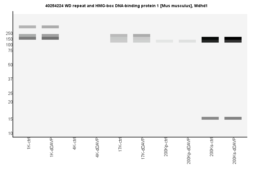
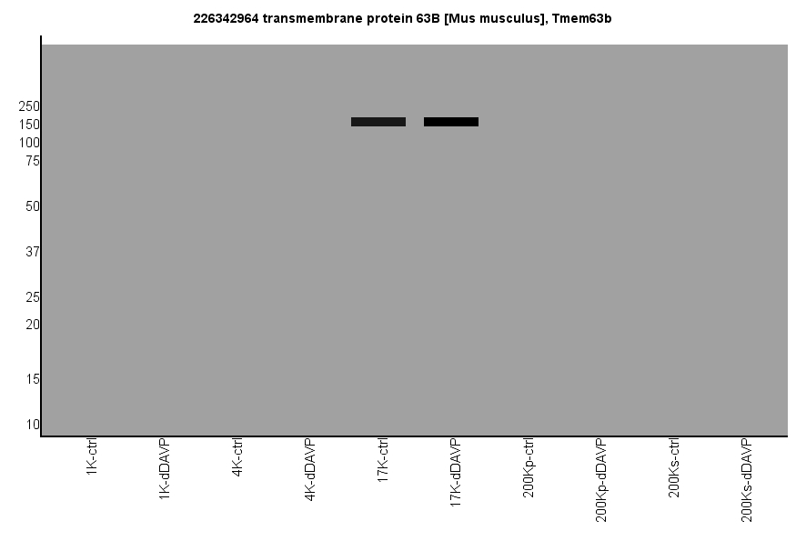
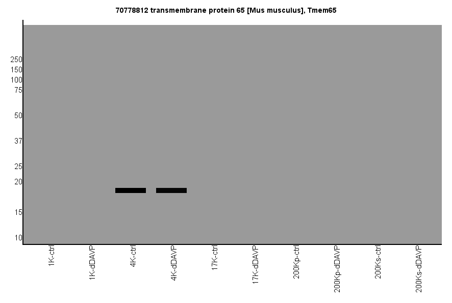
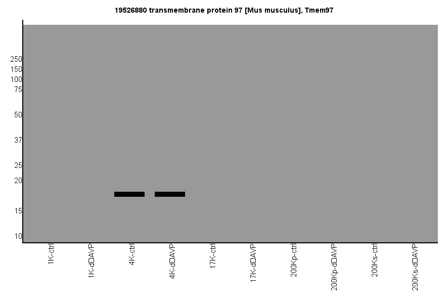
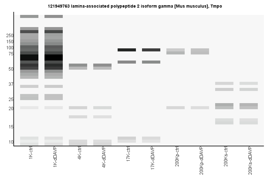
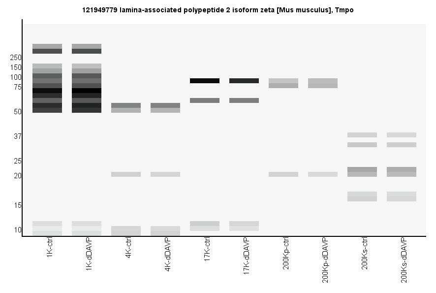
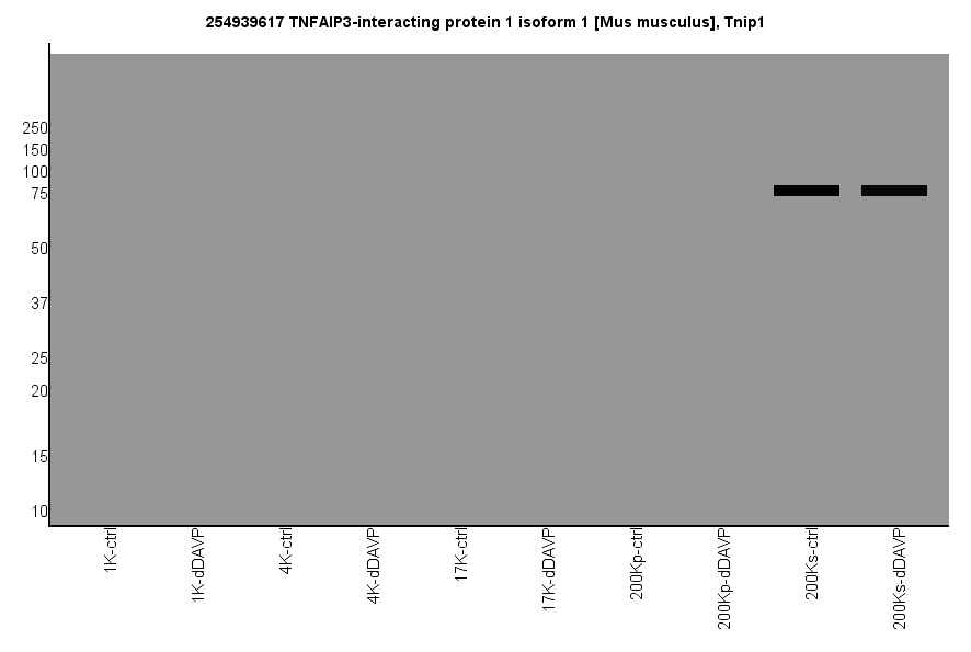
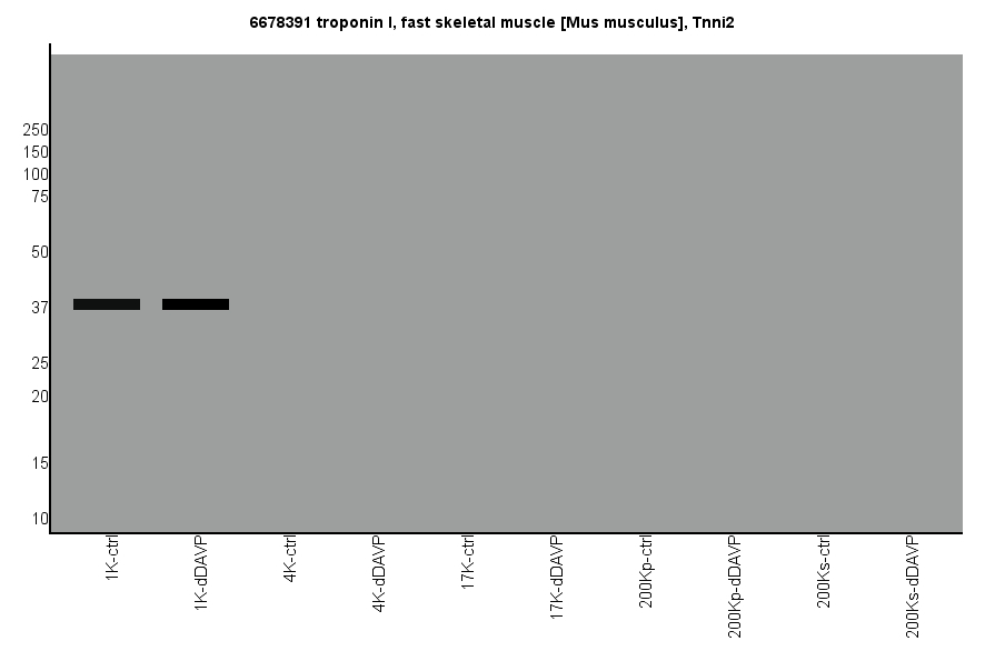
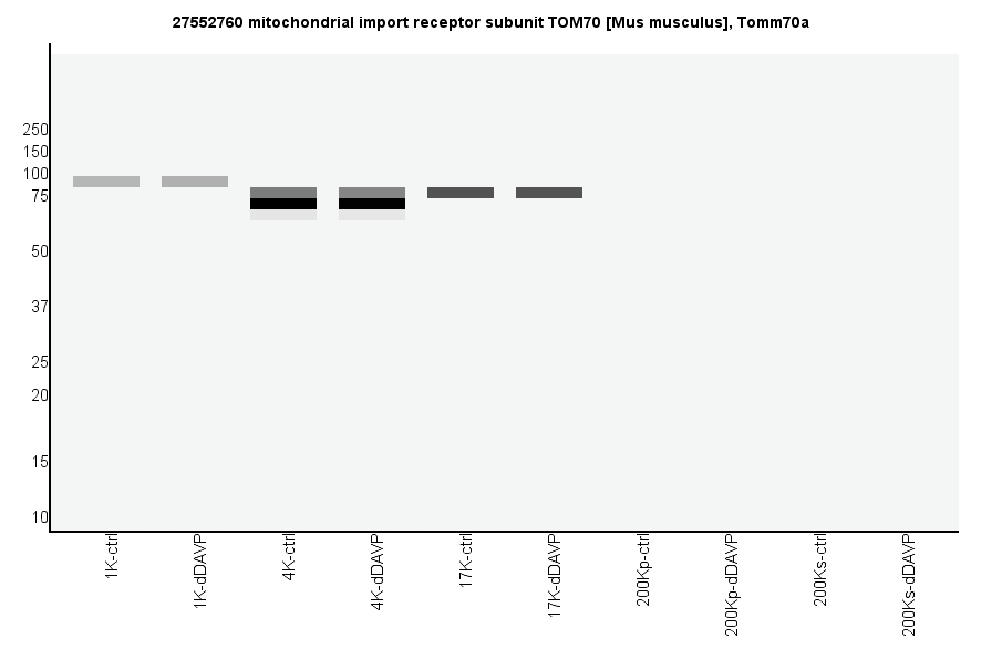
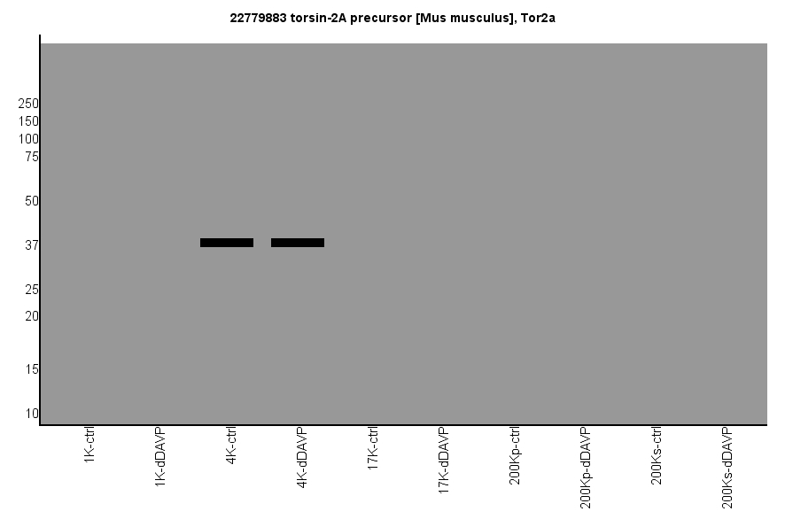
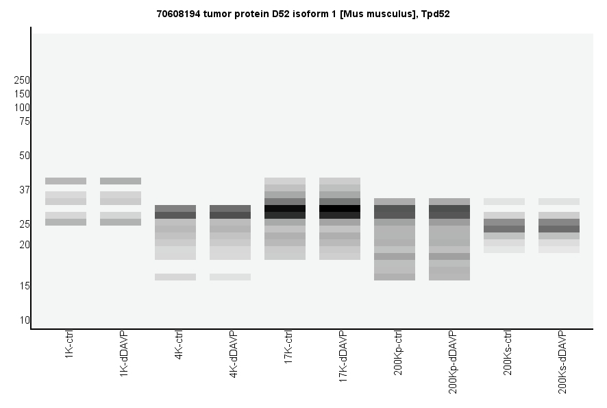
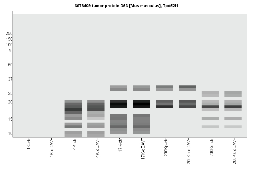
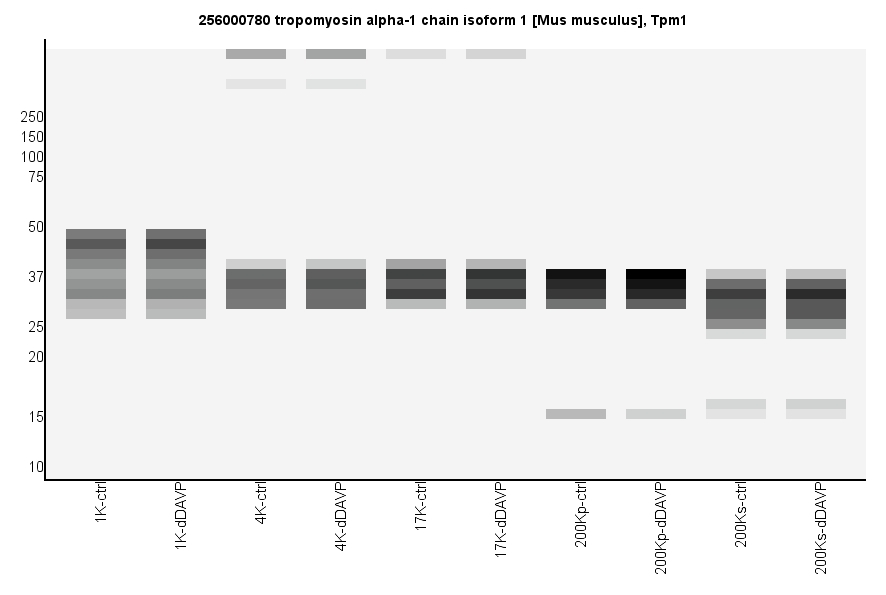
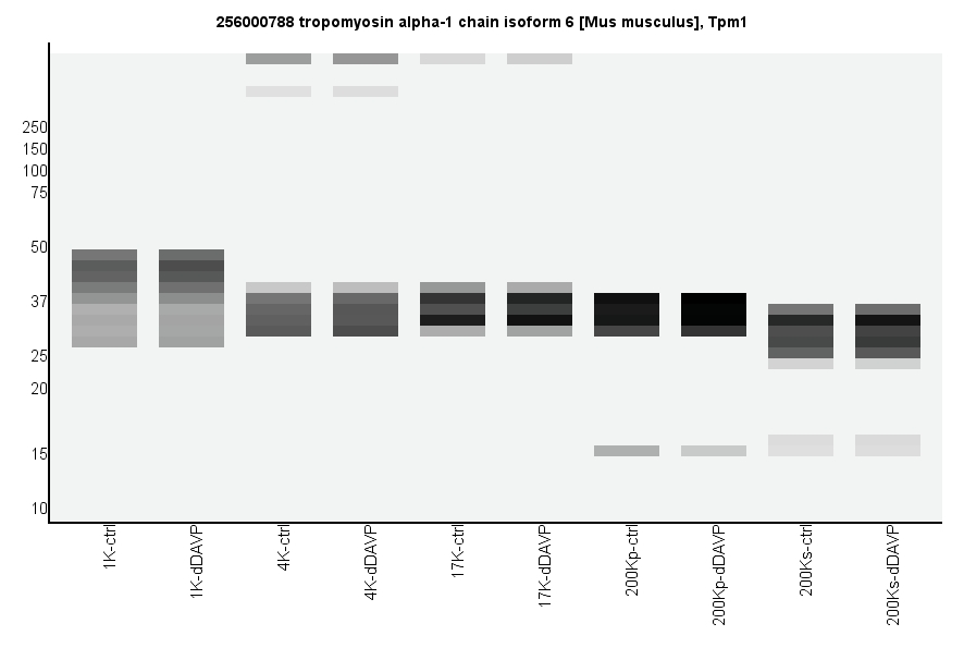
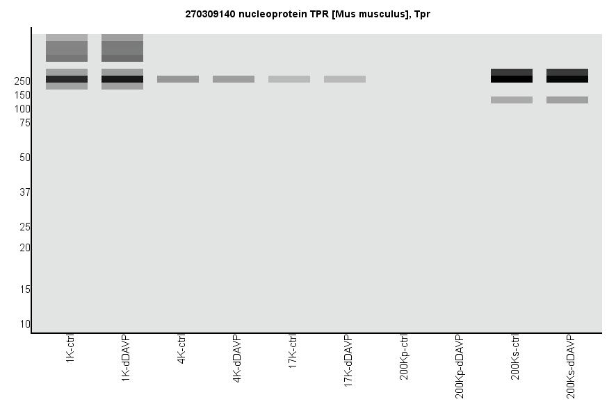
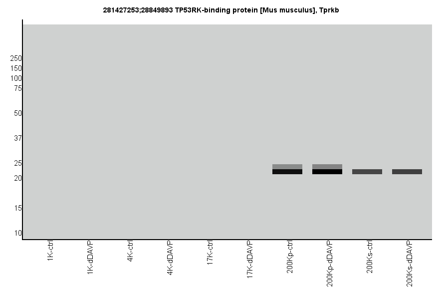
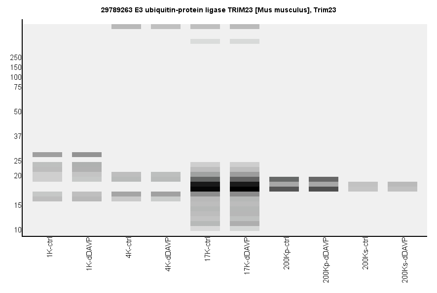
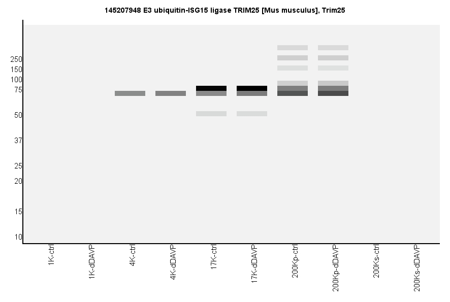
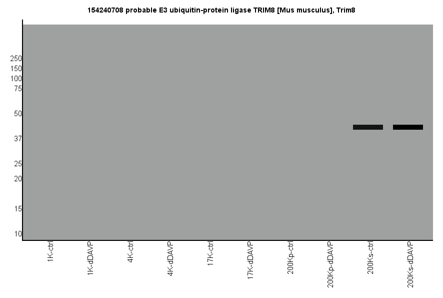
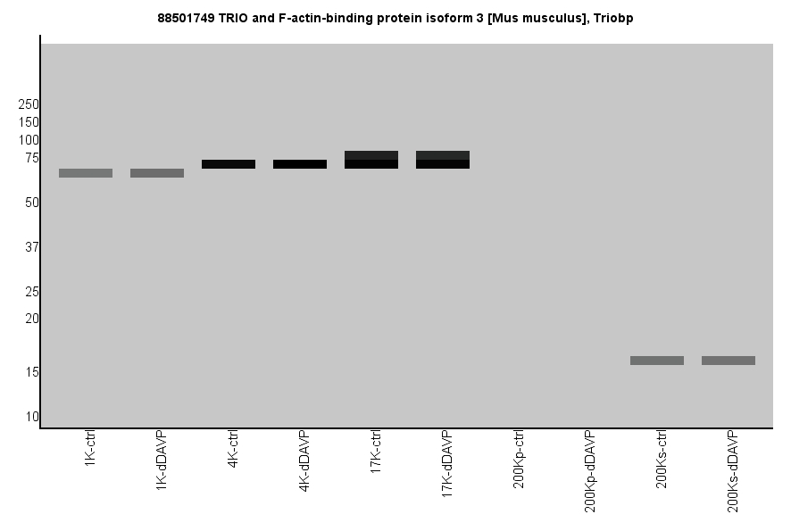
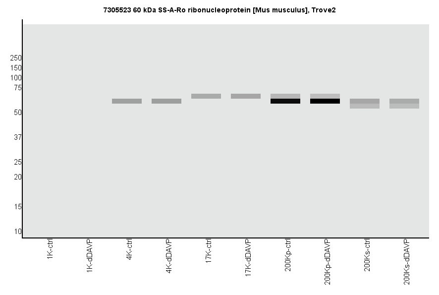
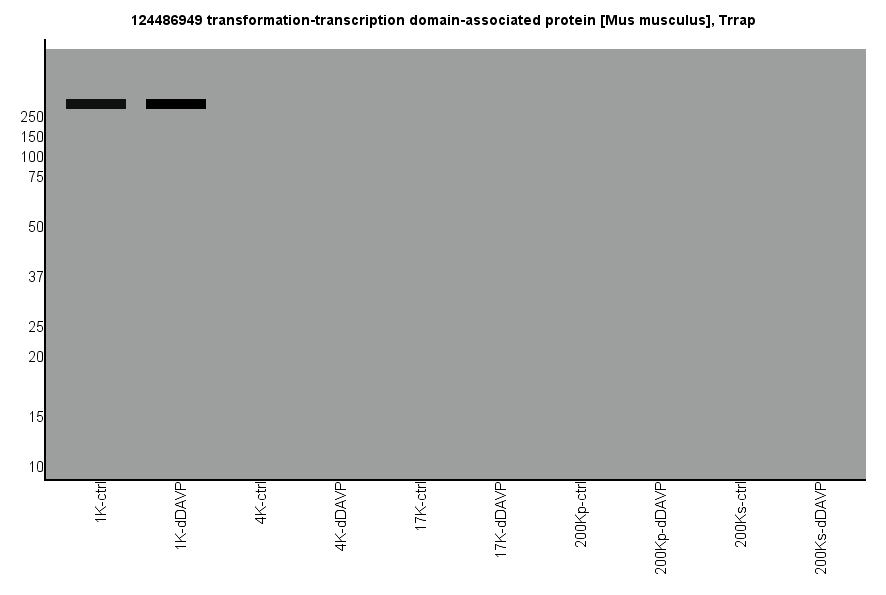
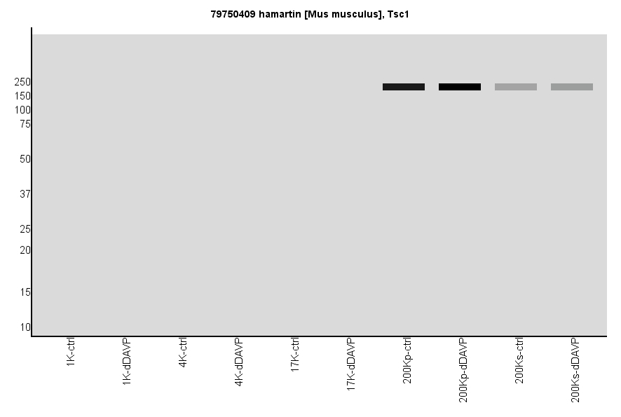
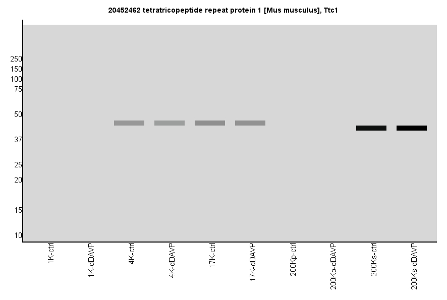
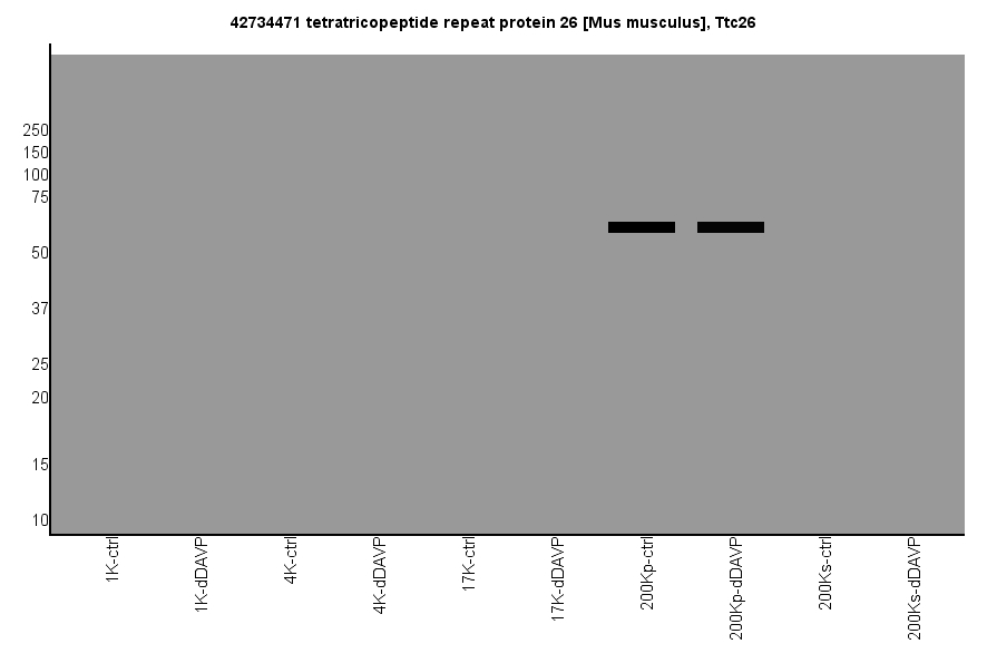
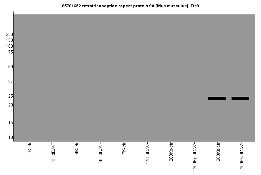
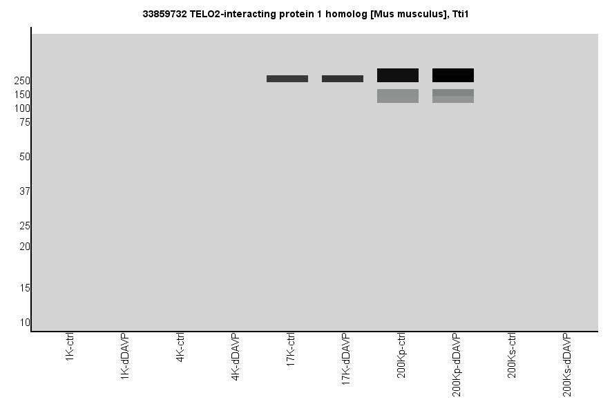
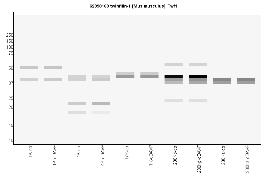
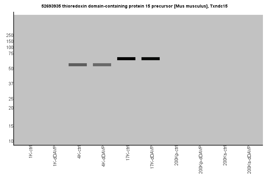
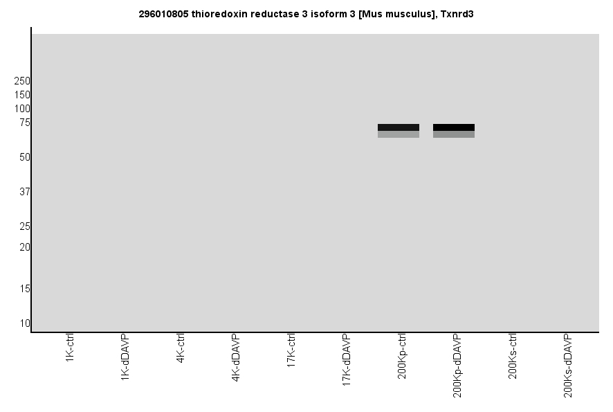
Virtual Western Blot: The data can be viewed as virtual western blots that map the peptide intensities to individual
slices from the SDS-PAGE for a given protein. The intensities were scaled as 100ug proteins were loaded equally in each lane.
The virtual blots can be viewed by mouse over the GI Number at the left. Clicking on the Official Gene Symbol for a
given protein will reveal its NCBI RefSeq record. The virtual western blot images show data normalized using a logarithmic conversion scale to map
peptide intensities to gray-scale levels. Using this conversion method, low-abundance peptides are generally visible in the
blot images. Switch back to the linear conversion scale
database page.
For relative protein abundances in this cell line, please visit Relative Protein Abundances in Mouse mpkCCD Cells database page.
For apparent vs. calculated MW ratios in this cell line, please visit Apparent / Calculated MW Ratios in Mouse mpkCCD Cells database page.
Reference I: Deep Proteomic Profiling of Vasopressin-Sensitive Collecting Duct Cells. I. Virtual Western Blots and Molecular Weight Distributions.
Yang, et al. (2015), PMID: 26310816
Reference II: Deep Proteomic Profiling of Vasopressin-Sensitive Collecting Duct Cells. II. Bioinformatic Analysis of Vasopressin Signaling.
Yang et al. (2015), PMID: 26310817
(Mouse over to see the table color scale)
Download full database
| Abundance Levels in Fractions (x1000) | Median Log2 (dDAVP/Ctrl) Ratio | |||||||||||||||
| GI Number | Gene Symbol | 1K | 4K | 17K | 200Kp | 200Ks | 1K | 4K | 17K | 200Kp | 200Ks | Annotation | ||||
| 124487289 | A1cf | 0 | 0 | 2526 | 1375 | 0 | n.v. | n.v. | 0.27 | 0.08 | n.v. | APOBEC1 complementation factor | ||||
| 283483963 | A4galt | 0 | 0 | 154 | 0 | 0 | n.v. | n.v. | 0.07 | n.v. | n.v. | lactosylceramide 4-alpha-galactosyltransferase | ||||
| 241982696 | Aaas | 970 | 1397 | 225 | 0 | 0 | 0.06 | 0.03 | -0.21 | n.v. | n.v. | aladin | ||||
| 21313520 | Aacs | 0 | 2776 | 4520 | 819 | 79368 | n.v. | 0.24 | 0.16 | 0.3 | 0.09 | acetoacetyl-CoA synthetase | ||||
| 110625673 | Aagab | 0 | 15 | 54 | 57 | 975 | n.v. | 0.22 | -0.04 | 0.19 | -0.01 | alpha- and gamma-adaptin-binding protein p34 | ||||
| 73695877 | Aak1 | 0 | 90 | 231 | 1211 | 5815 | n.v. | 0.39 | 0.06 | 0.24 | 0.11 | AP2 associated kinase 1 isoform 2 | ||||
| 91992157 | Aak1 | 0 | 90 | 231 | 1211 | 5815 | n.v. | 0.39 | 0.06 | 0.24 | 0.11 | AP2 associated kinase 1 isoform 1 | ||||
| 295789038 | Aamdc | 0 | 0 | 25 | 0 | 10131 | n.v. | n.v. | 0.4 | n.v. | 0.04 | UPF0366 protein C11orf67 homolog isoform 1 | ||||
| 295789042 | Aamdc | 0 | 0 | 25 | 0 | 6019 | n.v. | n.v. | 0.4 | n.v. | 0.01 | UPF0366 protein C11orf67 homolog isoform 3 | ||||
| 295789044 | Aamdc | 0 | 0 | 25 | 0 | 6019 | n.v. | n.v. | 0.4 | n.v. | 0.01 | UPF0366 protein C11orf67 homolog isoform 4 | ||||
| 40254393 | Aamdc | 0 | 0 | 25 | 0 | 10131 | n.v. | n.v. | 0.4 | n.v. | 0.04 | UPF0366 protein C11orf67 homolog isoform 2 | ||||
| 299473737 | Aamp | 0 | 211 | 206 | 25 | 12434 | n.v. | 0.08 | -0.13 | -0.12 | 0.03 | angio-associated migratory cell protein isoform 1 | ||||
| 82524294 | Aamp | 0 | 211 | 206 | 25 | 12434 | n.v. | 0.08 | -0.13 | -0.12 | 0.03 | angio-associated migratory cell protein isoform 2 | ||||
| 258679441 | Aar2 | 0 | 0 | 0 | 3332 | 0 | n.v. | n.v. | n.v. | -0.08 | n.v. | protein AAR2 homolog | ||||
| 34610207 | Aars | 183 | 1363 | 17299 | 11959 | 316626 | -0.09 | 0.13 | 0.13 | 0.47 | 0.14 | alanine--tRNA ligase, cytoplasmic | ||||
| 52630311 | Aars2 | 0 | 405 | 0 | 0 | 0 | n.v. | -0.26 | n.v. | n.v. | n.v. | alanine--tRNA ligase, mitochondrial precursor | ||||
| 21450213 | Aarsd1 | 0 | 0 | 1013 | 16 | 59780 | n.v. | n.v. | -0.1 | 0.55 | 0.09 | alanyl-tRNA editing protein Aarsd1 | ||||
| 227496837 | Aasdhppt | 0 | 0 | 0 | 0 | 214 | n.v. | n.v. | n.v. | n.v. | 0.37 | L-aminoadipate-semialdehyde dehydrogenase-phosphopantetheinyl transferase | ||||
| 9790013 | Aatf | 0 | 0 | 0 | 573 | 0 | n.v. | n.v. | n.v. | 0.14 | n.v. | protein AATF | ||||
| 283483966 | Abat | 0 | 328 | 549 | 0 | 0 | n.v. | 0.03 | -0.2 | n.v. | n.v. | 4-aminobutyrate aminotransferase, mitochondrial isoform 2 precursor | ||||
| 37202121 | Abat | 0 | 328 | 549 | 0 | 0 | n.v. | 0.03 | -0.2 | n.v. | n.v. | 4-aminobutyrate aminotransferase, mitochondrial isoform 1 precursor | ||||
| 116292744 | Abca13 | 0 | 148 | 0 | 1244 | 0 | n.v. | 0.21 | n.v. | 0.01 | n.v. | ATP-binding cassette sub-family A member 13 | ||||
| 110225379 | Abca2 | 0 | 0 | 124 | 0 | 0 | n.v. | n.v. | -0.1 | n.v. | n.v. | ATP-binding cassette sub-family A member 2 | ||||
| 88759350 | Abca3 | 0 | 0 | 0 | 0 | 3106 | n.v. | n.v. | n.v. | n.v. | -0.15 | ATP-binding cassette sub-family A member 3 | ||||
| 6671495 | Abca4 | 0 | 0 | 275 | 0 | 0 | n.v. | n.v. | 0.13 | n.v. | n.v. | retinal-specific ATP-binding cassette transporter | ||||
| 146134400 | Abca5 | 0 | 0 | 711 | 0 | 0 | n.v. | n.v. | -0.38 | n.v. | n.v. | ATP-binding cassette sub-family A member 5 | ||||
| 15451840 | Abca7 | 0 | 6 | 7 | 0 | 0 | n.v. | 0 | 0.3 | n.v. | n.v. | ATP-binding cassette sub-family A member 7 | ||||
| 153792543 | Abca9 | 0 | 825 | 0 | 0 | 0 | n.v. | -0.35 | n.v. | n.v. | n.v. | ATP-binding cassette sub-family A member 9 | ||||
| 9506367 | Abcb10 | 0 | 466 | 0 | 0 | 0 | n.v. | -0.16 | n.v. | n.v. | n.v. | ATP-binding cassette sub-family B member 10, mitochondrial | ||||
| 17647117 | Abcb6 | 0 | 737 | 61 | 0 | 0 | n.v. | -0.66 | -0.13 | n.v. | n.v. | ATP-binding cassette sub-family B member 6, mitochondrial | ||||
| 169234938 | Abcb7 | 0 | 357 | 0 | 0 | 0 | n.v. | -0.1 | n.v. | n.v. | n.v. | ATP-binding cassette sub-family B member 7, mitochondrial | ||||
| 27753995 | Abcb8 | 0 | 438 | 0 | 0 | 0 | n.v. | -0.11 | n.v. | n.v. | n.v. | ATP-binding cassette sub-family B member 8, mitochondrial precursor | ||||
| 6678848 | Abcc1 | 805 | 4920 | 4087 | 0 | 0 | 0.15 | -0.14 | -0.24 | n.v. | n.v. | multidrug resistance-associated protein 1 | ||||
| 116063566 | Abcc2 | 1501 | 0 | 0 | 0 | 0 | 0.01 | n.v. | n.v. | n.v. | n.v. | canalicular multispecific organic anion transporter 1 | ||||
| 90403595 | Abcc3 | 0 | 470 | 40 | 0 | 0 | n.v. | -0.25 | -0.6 | n.v. | n.v. | canalicular multispecific organic anion transporter 2 | ||||
| 255683320 | Abcc4 | 751 | 3073 | 3907 | 90 | 0 | -0.39 | 0.1 | -0.01 | -0.58 | n.v. | ATP-binding cassette, sub-family C (CFTR/MRP), member 4 isoform 1 | ||||
| 255683324 | Abcc4 | 751 | 3073 | 3907 | 90 | 0 | -0.39 | 0.1 | -0.01 | -0.58 | n.v. | ATP-binding cassette, sub-family C (CFTR/MRP), member 4 isoform 2 | ||||
| 255683328 | Abcc4 | 751 | 3073 | 3907 | 90 | 0 | -0.39 | 0.1 | -0.01 | -0.58 | n.v. | ATP-binding cassette, sub-family C (CFTR/MRP), member 4 isoform 3 | ||||
| 6671497 | Abcd1 | 0 | 50 | 582 | 0 | 0 | n.v. | 0.14 | -0.54 | n.v. | n.v. | ATP-binding cassette sub-family D member 1 | ||||
| 60218877 | Abcd3 | 137972 | 13356 | 28472 | 0 | 0 | 0.07 | -0.07 | -0.2 | n.v. | n.v. | ATP-binding cassette sub-family D member 3 | ||||
| 226052788 | Abcd4 | 0 | 58 | 148 | 0 | 0 | n.v. | -0.28 | -0.74 | n.v. | n.v. | ATP-binding cassette sub-family D member 4 | ||||
| 114205431 | Abce1 | 0 | 3513 | 47314 | 151204 | 3245 | n.v. | 0.12 | 0.05 | 0.08 | 0.25 | ATP-binding cassette sub-family E member 1 | ||||
| 39930335 | Abcf1 | 1886 | 674 | 61489 | 56660 | 0 | 0.42 | -0.05 | -0.03 | -0.06 | n.v. | ATP-binding cassette sub-family F member 1 | ||||
| 23956078 | Abcf2 | 0 | 6215 | 37683 | 144115 | 0 | n.v. | 0.13 | 0.05 | 0.05 | n.v. | ATP-binding cassette sub-family F member 2 isoform 1 | ||||
| 299473734 | Abcf2 | 0 | 6215 | 33633 | 130764 | 0 | n.v. | 0.13 | 0.06 | 0.05 | n.v. | ATP-binding cassette sub-family F member 2 isoform 2 | ||||
| 29789050 | Abcf3 | 0 | 0 | 104 | 3585 | 9189 | n.v. | n.v. | -0.04 | 0.1 | 0.01 | ATP-binding cassette sub-family F member 3 | ||||
| 6752944 | Abcg2 | 0 | 0 | 600 | 3233 | 0 | n.v. | n.v. | -0.27 | -0.19 | n.v. | ATP-binding cassette sub-family G member 2 | ||||
| 269784760 | Abhd10 | 0 | 1330 | 0 | 0 | 0 | n.v. | -0.24 | n.v. | n.v. | n.v. | abhydrolase domain-containing protein 10, mitochondrial precursor | ||||
| 440918693 | Abhd10 | 0 | 1330 | 0 | 0 | 0 | n.v. | -0.24 | n.v. | n.v. | n.v. | mycophenolic acid acyl-glucuronide esterase, mitochondrial isoform 2 precursor | ||||
| 159110817 | Abhd12 | 20375 | 3219 | 651 | 0 | 0 | 0.07 | -0.33 | -0.32 | n.v. | n.v. | monoacylglycerol lipase ABHD12 | ||||
| 124487441 | Abhd13 | 0 | 186 | 51 | 0 | 0 | n.v. | -0.7 | -0.1 | n.v. | n.v. | abhydrolase domain-containing protein 13 | ||||
| 171460960 | Abhd14b | 6151 | 14569 | 13837 | 3495 | 200300 | 0.38 | 0.25 | 0.09 | 0.14 | 0.15 | alpha/beta hydrolase domain-containing protein 14B | ||||
| 30519896 | Abhd16a | 0 | 1273 | 2112 | 0 | 0 | n.v. | -0.23 | -0.32 | n.v. | n.v. | abhydrolase domain-containing protein 16A | ||||
| 38142456 | Abhd17b | 0 | 0 | 66 | 0 | 0 | n.v. | n.v. | -0.4 | n.v. | n.v. | abhydrolase domain-containing protein FAM108B1 precursor | ||||
| 326937491 | Abhd4 | 0 | 0 | 186 | 9119 | 0 | n.v. | n.v. | -0.1 | 0.06 | n.v. | abhydrolase domain-containing protein 4 isoform 1 | ||||
| 326937494 | Abhd4 | 0 | 0 | 186 | 9119 | 0 | n.v. | n.v. | -0.1 | 0.06 | n.v. | abhydrolase domain-containing protein 4 isoform 2 | ||||
| 13385690 | Abhd5 | 0 | 1432 | 439 | 1506 | 0 | n.v. | 0.33 | 0.15 | 0.14 | n.v. | 1-acylglycerol-3-phosphate O-acyltransferase ABHD5 | ||||
| 31560264 | Abhd6 | 1831 | 5622 | 686 | 0 | 0 | -0.2 | -0.3 | -0.59 | n.v. | n.v. | monoacylglycerol lipase ABHD6 | ||||
| 116089341 | Abi1 | 0 | 3290 | 4223 | 2955 | 814 | n.v. | -0.01 | -0.07 | -0.29 | -0.63 | abl interactor 1 isoform 1 | ||||
| 116089343 | Abi1 | 0 | 3290 | 4223 | 2955 | 814 | n.v. | -0.01 | -0.07 | -0.29 | -0.63 | abl interactor 1 isoform 4 | ||||
| 116089345 | Abi1 | 0 | 3290 | 4223 | 2955 | 814 | n.v. | -0.01 | -0.07 | -0.29 | -0.63 | abl interactor 1 isoform 2 | ||||
| 116089347 | Abi1 | 0 | 3290 | 4223 | 2955 | 814 | n.v. | -0.01 | -0.07 | -0.29 | -0.63 | abl interactor 1 isoform 3 | ||||
| 116089351 | Abi1 | 0 | 3290 | 4223 | 2955 | 814 | n.v. | -0.01 | -0.07 | -0.29 | -0.63 | abl interactor 1 isoform 5 | ||||
| 162951865 | Abl1 | 0 | 0 | 1 | 0 | 0 | n.v. | n.v. | 0.36 | n.v. | n.v. | tyrosine-protein kinase ABL1 isoform b | ||||
| 162951870 | Abl1 | 0 | 0 | 1 | 0 | 0 | n.v. | n.v. | 0.36 | n.v. | n.v. | tyrosine-protein kinase ABL1 isoform a | ||||
| 157057145 | Ablim1 | 0 | 0 | 0 | 0 | 1981 | n.v. | n.v. | n.v. | n.v. | 0.81 | actin-binding LIM protein 1 isoform 1 | ||||
| 157057174 | Ablim1 | 0 | 0 | 0 | 0 | 1981 | n.v. | n.v. | n.v. | n.v. | 0.81 | actin-binding LIM protein 1 isoform 2 | ||||
| 157057554 | Ablim1 | 0 | 0 | 0 | 0 | 1981 | n.v. | n.v. | n.v. | n.v. | 0.81 | actin-binding LIM protein 1 isoform 3 | ||||
| 37574113 | Abr | 0 | 23 | 18 | 0 | 0 | n.v. | 0.01 | -0.25 | n.v. | n.v. | active breakpoint cluster region-related protein isoform 1 | ||||
| 38683820 | Abr | 0 | 23 | 18 | 0 | 0 | n.v. | 0.01 | -0.25 | n.v. | n.v. | active breakpoint cluster region-related protein isoform 2 | ||||
| 38683822 | Abr | 0 | 23 | 18 | 0 | 0 | n.v. | 0.01 | -0.25 | n.v. | n.v. | active breakpoint cluster region-related protein isoform 3 | ||||
| 153791468 | Abracl | 3442 | 1939 | 1803 | 1653 | 664 | 0.13 | 0.17 | -0.01 | 0.08 | 0.39 | costars family protein ABRACL | ||||
| 49402267 | Abtb2 | 0 | 165 | 0 | 0 | 0 | n.v. | -0.03 | n.v. | n.v. | n.v. | ankyrin repeat and BTB/POZ domain-containing protein 2 | ||||
| 18700004 | Acaa1a | 75567 | 8488 | 33302 | 0 | 0 | 0.05 | -0.17 | -0.24 | n.v. | n.v. | 3-ketoacyl-CoA thiolase A, peroxisomal precursor | ||||
| 22122797 | Acaa1b | 38699 | 4027 | 13973 | 0 | 0 | 0.04 | -0.2 | -0.24 | n.v. | n.v. | 3-ketoacyl-CoA thiolase B, peroxisomal precursor | ||||
| 29126205 | Acaa2 | 694915 | 124712 | 10371 | 29484 | 54929 | -0.03 | -0.12 | -0.41 | -0.2 | -0.13 | 3-ketoacyl-CoA thiolase, mitochondrial | ||||
| 125656173 | Acaca | 0 | 1010 | 1810 | 28224 | 18602 | n.v. | -0.05 | -0.01 | 0.24 | -0.09 | acetyl-CoA carboxylase 1 | ||||
| 157042798 | Acacb | 0 | 0 | 865 | 4551 | 8343 | n.v. | n.v. | 0.03 | 0.27 | 0.01 | acetyl-Coenzyme A carboxylase beta precursor | ||||
| 156255157 | Acad10 | 622 | 3881 | 0 | 0 | 0 | -0.02 | -0.38 | n.v. | n.v. | n.v. | acyl-CoA dehydrogenase family member 10 | ||||
| 74271799 | Acad11 | 9611 | 430 | 11278 | 0 | 0 | -0.06 | -0.18 | -0.23 | n.v. | n.v. | acyl-CoA dehydrogenase family member 11 | ||||
| 257467675 | Acad12 | 0 | 3881 | 0 | 0 | 0 | n.v. | -0.38 | n.v. | n.v. | n.v. | acyl-Coenzyme A dehydrogenase family, member 10-like | ||||
| 118403322 | Acad8 | 5126 | 205 | 0 | 0 | 0 | -0.12 | -0.17 | n.v. | n.v. | n.v. | isobutyryl-CoA dehydrogenase, mitochondrial precursor | ||||
| 100817933 | Acad9 | 44648 | 16015 | 622 | 0 | 0 | -0.01 | -0.05 | -0.37 | n.v. | n.v. | acyl-CoA dehydrogenase family member 9, mitochondrial | ||||
| 31982520 | Acadl | 326927 | 79582 | 2254 | 4731 | 10999 | 0.01 | -0.16 | -0.49 | -0.2 | -0.29 | long-chain specific acyl-CoA dehydrogenase, mitochondrial precursor | ||||
| 6680618 | Acadm | 172736 | 54161 | 471 | 1997 | 8996 | 0.06 | -0.12 | -0.07 | -0.2 | -0.34 | medium-chain specific acyl-CoA dehydrogenase, mitochondrial precursor | ||||
| 31982522 | Acads | 50891 | 1198 | 0 | 4207 | 6768 | -0.06 | -0.21 | n.v. | -0.04 | -0.33 | short-chain specific acyl-CoA dehydrogenase, mitochondrial precursor | ||||
| 17647119 | Acadsb | 824 | 246 | 0 | 0 | 0 | 0.29 | -0.36 | n.v. | n.v. | n.v. | short/branched chain specific acyl-CoA dehydrogenase, mitochondrial | ||||
| 23956084 | Acadvl | 89004 | 43583 | 3616 | 1555 | 0 | -0.03 | -0.07 | -0.22 | -1.17 | n.v. | very long-chain specific acyl-CoA dehydrogenase, mitochondrial precursor | ||||
| 62079289 | Acap2 | 0 | 372 | 819 | 2136 | 14972 | n.v. | 0.29 | 0.01 | -0.58 | -0.05 | arf-GAP with coiled-coil, ANK repeat and PH domain-containing protein 2 | ||||
| 21450129 | Acat1 | 80546 | 36172 | 896 | 2636 | 1810 | -0.01 | -0.19 | -0.41 | -0.22 | -0.99 | acetyl-CoA acetyltransferase, mitochondrial precursor | ||||
| 148747461 | Acat2 | 0 | 42 | 1204 | 43989 | 30179 | n.v. | 0.18 | 0.03 | 0.29 | 0.07 | acetyl-CoA acetyltransferase, cytosolic | ||||
| 110625948 | Acat3 | 0 | 0 | 795 | 22109 | 19898 | n.v. | n.v. | 0.03 | 0.32 | 0.08 | acetyl-Coenzyme A acetyltransferase 3 | ||||
| 229608928 | Acbd3 | 2837 | 10394 | 1803 | 2079 | 675 | 0.12 | 0.08 | 0.2 | 0.02 | 0.85 | Golgi resident protein GCP60 | ||||
| 156255161 | Acbd5 | 1391 | 2622 | 1196 | 0 | 11 | 0.1 | -0.06 | -0.16 | n.v. | -2.23 | acyl-CoA-binding domain-containing protein 5 isoform d | ||||
| 156255163 | Acbd5 | 1391 | 2622 | 1196 | 0 | 11 | 0.1 | -0.06 | -0.16 | n.v. | -2.23 | acyl-CoA-binding domain-containing protein 5 isoform a precursor | ||||
| 156255165 | Acbd5 | 1391 | 2622 | 1196 | 0 | 11 | 0.1 | -0.06 | -0.16 | n.v. | -2.23 | acyl-CoA-binding domain-containing protein 5 isoform b precursor | ||||
| 27229192 | Acbd5 | 1391 | 2622 | 1196 | 0 | 11 | 0.1 | -0.06 | -0.16 | n.v. | -2.23 | acyl-CoA-binding domain-containing protein 5 isoform c | ||||
| 224809391 | Acbd6 | 0 | 0 | 0 | 0 | 129 | n.v. | n.v. | n.v. | n.v. | 0.06 | acyl-CoA-binding domain-containing protein 6 isoform 2 | ||||
| 224809393 | Acbd6 | 0 | 0 | 0 | 0 | 129 | n.v. | n.v. | n.v. | n.v. | 0.06 | acyl-CoA-binding domain-containing protein 6 isoform 3 | ||||
| 224809389 | Acbd6 | 0 | 0 | 34 | 0 | 3074 | n.v. | n.v. | 0.14 | n.v. | 0.09 | acyl-CoA-binding domain-containing protein 6 isoform 1 | ||||
| 60593059 | Acd | 0 | 0 | 0 | 475 | 203 | n.v. | n.v. | n.v. | -0.29 | -0.08 | adrenocortical dysplasia protein | ||||
| 194394192 | Acin1 | 26287 | 0 | 45 | 0 | 0 | -0.13 | n.v. | 0.17 | n.v. | n.v. | apoptotic chromatin condensation inducer in the nucleus isoform 1 | ||||
| 194394195 | Acin1 | 26287 | 0 | 45 | 0 | 0 | -0.13 | n.v. | 0.17 | n.v. | n.v. | apoptotic chromatin condensation inducer in the nucleus isoform 3 | ||||
| 336176080 | Acin1 | 26287 | 0 | 45 | 0 | 0 | -0.13 | n.v. | 0.17 | n.v. | n.v. | apoptotic chromatin condensation inducer in the nucleus isoform 5 | ||||
| 336176082 | Acin1 | 26287 | 0 | 45 | 0 | 0 | -0.13 | n.v. | 0.17 | n.v. | n.v. | apoptotic chromatin condensation inducer in the nucleus isoform 6 | ||||
| 146231985 | Acin1 | 26287 | 307 | 45 | 8 | 4284 | -0.13 | 0.13 | 0.17 | -0.04 | -0.1 | apoptotic chromatin condensation inducer in the nucleus isoform 2 | ||||
| 194394197 | Acin1 | 26287 | 307 | 45 | 8 | 4284 | -0.13 | 0.13 | 0.17 | -0.04 | -0.1 | apoptotic chromatin condensation inducer in the nucleus isoform 4 | ||||
| 29293809 | Acly | 0 | 843 | 24622 | 526495 | 227877 | n.v. | 0.02 | -0.05 | 0.1 | -0.07 | ATP-citrate synthase isoform 2 | ||||
| 313151222 | Acly | 0 | 843 | 24622 | 542998 | 227877 | n.v. | 0.02 | -0.05 | 0.1 | -0.07 | ATP-citrate synthase isoform 1 | ||||
| 110347487 | Aco1 | 0 | 2067 | 5002 | 8079 | 78297 | n.v. | 0.25 | 0.09 | 0.44 | 0.16 | cytoplasmic aconitate hydratase | ||||
| 18079339 | Aco2 | 1252388 | 119059 | 14815 | 1195 | 42153 | -0.01 | -0.14 | -0.34 | -0.17 | -0.25 | aconitate hydratase, mitochondrial precursor | ||||
| 6753550 | Acot1 | 152031 | 16974 | 565 | 0 | 6340 | -0.13 | -0.23 | -0.4 | n.v. | -0.32 | acyl-coenzyme A thioesterase 1 | ||||
| 154426268 | Acot10 | 0 | 3707 | 0 | 0 | 0 | n.v. | -0.07 | n.v. | n.v. | n.v. | acyl-coenzyme A thioesterase 10, mitochondrial precursor | ||||
| 18482377 | Acot12 | 0 | 163 | 0 | 0 | 0 | n.v. | 0.67 | n.v. | n.v. | n.v. | acyl-coenzyme A thioesterase 12 | ||||
| 13385260 | Acot13 | 7414 | 4476 | 401 | 0 | 1842 | -0.03 | -0.03 | -0.3 | n.v. | -0.14 | acyl-coenzyme A thioesterase 13 | ||||
| 238624114 | Acot2 | 223626 | 21495 | 565 | 0 | 6340 | -0.06 | -0.29 | -0.4 | n.v. | -0.32 | acyl-coenzyme A thioesterase 2, mitochondrial precursor | ||||
| 19527406 | Acot3 | 0 | 0 | 0 | 0 | 2774 | n.v. | n.v. | n.v. | n.v. | -0.32 | acyl-coenzyme A thioesterase 3 | ||||
| 269308227 | Acot4 | 52081 | 7958 | 0 | 0 | 4605 | -0.18 | -0.25 | n.v. | n.v. | -0.45 | acyl-coenzyme A thioesterase 4 | ||||
| 238550185 | Acot5 | 0 | 0 | 0 | 0 | 2774 | n.v. | n.v. | n.v. | n.v. | -0.32 | acyl-coenzyme A thioesterase 5 | ||||
| 110626167 | Acot6 | 52081 | 7958 | 0 | 0 | 4605 | -0.18 | -0.25 | n.v. | n.v. | -0.45 | acyl-coenzyme A thioesterase 6 | ||||
| 225690614 | Acot7 | 0 | 119 | 565 | 377 | 21987 | n.v. | 0.36 | -0.09 | 0.27 | 0.39 | cytosolic acyl coenzyme A thioester hydrolase isoform 2 | ||||
| 225690616 | Acot7 | 0 | 119 | 565 | 377 | 21987 | n.v. | 0.36 | -0.09 | 0.27 | 0.39 | cytosolic acyl coenzyme A thioester hydrolase isoform 1 | ||||
| 225690618 | Acot7 | 0 | 79 | 565 | 377 | 21987 | n.v. | 1.28 | -0.09 | 0.27 | 0.39 | cytosolic acyl coenzyme A thioester hydrolase isoform 3 | ||||
| 254587964 | Acot8 | 476 | 7770 | 3080 | 0 | 0 | 0.49 | -0.09 | -0.26 | n.v. | n.v. | acyl-coenzyme A thioesterase 8 | ||||
| 31980998 | Acot9 | 7872 | 4718 | 0 | 0 | 0 | -0.06 | -0.11 | n.v. | n.v. | n.v. | acyl-coenzyme A thioesterase 9, mitochondrial | ||||
| 429484484 | Acox1 | 16457 | 26417 | 26297 | 0 | 535 | -0.13 | -0.02 | -0.2 | n.v. | -1.38 | peroxisomal acyl-coenzyme A oxidase 1 isoform 2 | ||||
| 66793429 | Acox1 | 16457 | 26417 | 26297 | 0 | 535 | -0.13 | -0.02 | -0.2 | n.v. | -1.38 | peroxisomal acyl-coenzyme A oxidase 1 isoform 1 | ||||
| 34328334 | Acox3 | 0 | 2127 | 449 | 0 | 0 | n.v. | -0.04 | -0.02 | n.v. | n.v. | peroxisomal acyl-coenzyme A oxidase 3 | ||||
| 159032062 | Acp1 | 13675 | 8598 | 12841 | 22813 | 15465 | -0.01 | 0.18 | 0.01 | 0.01 | 0.09 | low molecular weight phosphotyrosine protein phosphatase isoform 1 | ||||
| 29150253 | Acp2 | 11295 | 5912 | 3731 | 647 | 0 | 0.02 | -0.13 | -0.23 | -0.41 | n.v. | lysosomal acid phosphatase precursor | ||||
| 66773165 | Acp6 | 0 | 7782 | 0 | 0 | 793 | n.v. | -0.24 | n.v. | n.v. | 0.55 | lysophosphatidic acid phosphatase type 6 precursor | ||||
| 24418933 | Acsf2 | 18690 | 14483 | 275 | 0 | 0 | -0.02 | -0.09 | -0.99 | n.v. | n.v. | acyl-CoA synthetase family member 2, mitochondrial precursor | ||||
| 113199775 | Acsf3 | 1501 | 1067 | 0 | 0 | 0 | -0.16 | -0.26 | n.v. | n.v. | n.v. | acyl-CoA synthetase family member 3, mitochondrial precursor | ||||
| 31560705 | Acsl1 | 0 | 7256 | 2731 | 0 | 0 | n.v. | -0.22 | -0.28 | n.v. | n.v. | long-chain-fatty-acid--CoA ligase 1 | ||||
| 209977076 | Acsl3 | 5840 | 2749 | 10207 | 0 | 0 | 0.37 | -0.14 | -0.17 | n.v. | n.v. | long-chain-fatty-acid--CoA ligase 3 isoform b | ||||
| 75992920 | Acsl3 | 5840 | 3067 | 11096 | 0 | 0 | 0.37 | -0.04 | -0.16 | n.v. | n.v. | long-chain-fatty-acid--CoA ligase 3 isoform a | ||||
| 46518528 | Acsl4 | 4760 | 4471 | 7605 | 4338 | 268 | -0.03 | 0.05 | 0.03 | 0.03 | -0.39 | long-chain-fatty-acid--CoA ligase 4 isoform 1 | ||||
| 75992925 | Acsl4 | 4760 | 4471 | 7605 | 4338 | 268 | -0.03 | 0.05 | 0.03 | 0.03 | -0.39 | long-chain-fatty-acid--CoA ligase 4 isoform 2 | ||||
| 58218988 | Acsl5 | 3863 | 15212 | 5553 | 0 | 0 | 0.26 | -0.3 | -0.37 | n.v. | n.v. | long-chain-fatty-acid--CoA ligase 5 | ||||
| 31980996 | Acss2 | 0 | 0 | 0 | 0 | 2024 | n.v. | n.v. | n.v. | n.v. | -0.36 | acetyl-coenzyme A synthetase, cytoplasmic | ||||
| 33563240 | Acta1 | 5646106 | 506364 | 954028 | 1091070 | 3808375 | 0 | 0.18 | -0.04 | -0.04 | 0.02 | actin, alpha skeletal muscle | ||||
| 6671507 | Acta2 | 4870901 | 471728 | 862002 | 1034770 | 3713317 | 0 | 0.18 | -0.04 | -0.06 | 0.02 | actin, aortic smooth muscle | ||||
| 6671509 | Actb | 9565955 | 664316 | 1289302 | 2003867 | 6630832 | 0.02 | 0.19 | -0.02 | 0 | 0.02 | actin, cytoplasmic 1 | ||||
| 30425250 | Actbl2 | 2546275 | 280349 | 352546 | 605396 | 2762795 | 0 | 0.14 | -0.05 | -0.08 | 0.04 | beta-actin-like protein 2 | ||||
| 14192922 | Actc1 | 5646106 | 506364 | 954028 | 1091046 | 3738645 | 0 | 0.18 | -0.04 | -0.04 | 0.02 | actin, alpha cardiac muscle 1 | ||||
| 6752954 | Actg1 | 9565955 | 555398 | 1289302 | 2003867 | 6506548 | 0.02 | 0.19 | -0.02 | 0 | 0.03 | actin, cytoplasmic 2 | ||||
| 157823889 | Actg2 | 4870901 | 471726 | 862002 | 1034770 | 2987962 | 0 | 0.17 | -0.04 | -0.06 | 0.02 | actin, gamma-enteric smooth muscle | ||||
| 189181668 | Actl6a | 37930 | 810 | 1569 | 0 | 39797 | 0.09 | 0.4 | -0.04 | n.v. | 0.01 | actin-like protein 6A | ||||
| 13937393 | Actl6b | 2636 | 0 | 35 | 0 | 18281 | 0.15 | n.v. | -0.02 | n.v. | 0.21 | actin-like protein 6B | ||||
| 61097906 | Actn1 | 1007252 | 108697 | 152370 | 203681 | 967497 | 0.05 | 0.23 | 0.23 | 0.13 | -0.05 | alpha-actinin-1 | ||||
| 157951643 | Actn2 | 322827 | 28664 | 39177 | 61645 | 145092 | -0.01 | 0.14 | 0.21 | 0.12 | -0.05 | alpha-actinin-2 | ||||
| 7304855 | Actn3 | 167390 | 27211 | 40316 | 41263 | 248268 | 0.05 | 0.11 | 0.21 | 0.04 | -0.05 | alpha-actinin-3 | ||||
| 11230802 | Actn4 | 894615 | 88678 | 144741 | 119436 | 651770 | 0.02 | 0.23 | 0.24 | 0.14 | -0.06 | alpha-actinin-4 | ||||
| 226246593 | Actr10 | 0 | 0 | 958 | 27692 | 0 | n.v. | n.v. | 0.08 | 0.2 | n.v. | actin-related protein 10 | ||||
| 8392847 | Actr1a | 61636 | 8722 | 20479 | 255144 | 14876 | 0.19 | 0.22 | 0.01 | 0.07 | -0.07 | alpha-centractin | ||||
| 22122615 | Actr1b | 10544 | 9310 | 18335 | 285914 | 13762 | 0.33 | 0.23 | -0.07 | 0.03 | -0.27 | beta-centractin | ||||
| 22122825 | Actr2 | 65774 | 6789 | 59281 | 223982 | 354763 | 0.03 | 0.23 | 0.03 | -0.04 | -0.06 | actin-related protein 2 | ||||
| 329664963 | Actr3 | 84995 | 29428 | 38197 | 162475 | 129660 | 0.27 | 0.19 | 0.04 | -0.01 | -0.08 | actin-related protein 3 | ||||
| 52345394 | Actr3b | 17574 | 4933 | 2034 | 22257 | 9917 | -0.83 | 0.23 | 0.01 | -0.05 | 0.02 | actin-related protein 3B | ||||
| 13384746 | Acy1 | 0 | 0 | 85 | 0 | 15411 | n.v. | n.v. | -0.03 | n.v. | 0.1 | aminoacylase-1 | ||||
| 31982632 | Acy3 | 0 | 0 | 0 | 0 | 26613 | n.v. | n.v. | n.v. | n.v. | 0.04 | aspartoacylase-2 | ||||
| 13384810 | Acyp1 | 4668 | 1864 | 3099 | 1220 | 36766 | 0 | 0.08 | -0.05 | 0.04 | 0.04 | acylphosphatase-1 | ||||
| 150378458 | Adam10 | 0 | 6073 | 18763 | 0 | 0 | n.v. | 0.02 | -0.04 | n.v. | n.v. | disintegrin and metalloproteinase domain-containing protein 10 precursor | ||||
| 471270257 | Adam17 | 0 | 0 | 167 | 0 | 0 | n.v. | n.v. | 0.34 | n.v. | n.v. | disintegrin and metalloproteinase domain-containing protein 17 isoform 2 | ||||
| 110347485 | Adam17 | 0 | 27 | 916 | 0 | 0 | n.v. | -0.05 | 0.16 | n.v. | n.v. | disintegrin and metalloproteinase domain-containing protein 17 precursor | ||||
| 160358787 | Adam9 | 0 | 251 | 5670 | 827 | 0 | n.v. | -0.04 | 0.07 | -0.64 | n.v. | disintegrin and metalloproteinase domain-containing protein 9 precursor | ||||
| 401782598 | Adam9 | 0 | 251 | 5670 | 827 | 0 | n.v. | -0.04 | 0.07 | -0.64 | n.v. | disintegrin and metalloproteinase domain-containing protein 9 isoform 1 | ||||
| 148529020 | Adamts17 | 29894 | 4296 | 18251 | 4355 | 82046 | 0.14 | 0.12 | 0.06 | 0.2 | 0.14 | ADAM metallopeptidase with thrombospondin type 1 motif, 17 precursor | ||||
| 165905595 | Adamtsl5 | 0 | 0 | 0 | 655 | 0 | n.v. | n.v. | n.v. | 0.12 | n.v. | ADAMTS-like protein 5 isoform 1 precursor | ||||
| 148368976 | Adap1 | 0 | 0 | 0 | 0 | 4187 | n.v. | n.v. | n.v. | n.v. | 0.58 | centaurin, alpha 1 | ||||
| 26006859 | Adap2 | 0 | 0 | 0 | 0 | 225 | n.v. | n.v. | n.v. | n.v. | 0.4 | arf-GAP with dual PH domain-containing protein 2 | ||||
| 226371677 | Adar | 0 | 0 | 1005 | 0 | 0 | n.v. | n.v. | 0.03 | n.v. | n.v. | double-stranded RNA-specific adenosine deaminase isoform 3 | ||||
| 226371679 | Adar | 0 | 0 | 1005 | 0 | 0 | n.v. | n.v. | 0.03 | n.v. | n.v. | double-stranded RNA-specific adenosine deaminase isoform 1 | ||||
| 226371684 | Adar | 0 | 0 | 979 | 0 | 0 | n.v. | n.v. | -0.07 | n.v. | n.v. | double-stranded RNA-specific adenosine deaminase isoform 2 | ||||
| 7304859 | Adat1 | 0 | 0 | 0 | 0 | 1660 | n.v. | n.v. | n.v. | n.v. | 0.05 | tRNA-specific adenosine deaminase 1 | ||||
| 61098160 | Adat2 | 0 | 0 | 0 | 0 | 8857 | n.v. | n.v. | n.v. | n.v. | -0.01 | tRNA-specific adenosine deaminase 2 | ||||
| 154759286 | Adat3 | 0 | 0 | 0 | 0 | 3020 | n.v. | n.v. | n.v. | n.v. | -0.05 | tRNA-specific adenosine deaminase-like protein 3 | ||||
| 21312430 | Adck1 | 0 | 1121 | 0 | 0 | 0 | n.v. | -0.29 | n.v. | n.v. | n.v. | uncharacterized aarF domain-containing protein kinase 1 precursor | ||||
| 169234776 | Adck5 | 0 | 273 | 0 | 0 | 0 | n.v. | 0.21 | n.v. | n.v. | n.v. | uncharacterized aarF domain-containing protein kinase 5 | ||||
| 148747309 | Adcy5 | 0 | 0 | 50 | 0 | 0 | n.v. | n.v. | -0.25 | n.v. | n.v. | adenylate cyclase type 5 | ||||
| 86604721 | Adcy6 | 0 | 202 | 50 | 0 | 0 | n.v. | 0.63 | -0.25 | n.v. | n.v. | adenylate cyclase type 6 | ||||
| 156255171 | Add1 | 3570 | 253 | 780 | 0 | 1028 | -0.01 | -0.73 | -0.09 | n.v. | -1.89 | alpha-adducin isoform 1 | ||||
| 156255173 | Add1 | 3570 | 253 | 780 | 0 | 214 | -0.01 | -0.73 | -0.09 | n.v. | -1.07 | alpha-adducin isoform 3 | ||||
| 7304861 | Add1 | 3570 | 253 | 780 | 0 | 214 | -0.01 | -0.73 | -0.09 | n.v. | -1.07 | alpha-adducin isoform 2 | ||||
| 427918090 | Add2 | 0 | 0 | 852 | 0 | 0 | n.v. | n.v. | 0.06 | n.v. | n.v. | beta-adducin isoform 1 | ||||
| 427918097 | Add2 | 0 | 0 | 852 | 0 | 0 | n.v. | n.v. | 0.06 | n.v. | n.v. | beta-adducin isoform 2 | ||||
| 6724311 | Adh1 | 0 | 6 | 900 | 3160 | 10784 | n.v. | 2.05 | 0.16 | 0.25 | 0.33 | alcohol dehydrogenase 1 | ||||
| 31982511 | Adh5 | 3716 | 177 | 8831 | 17460 | 370849 | 0.12 | 0.15 | 0.06 | 0.23 | 0.06 | alcohol dehydrogenase class-3 | ||||
| 31560625 | Adh7 | 0 | 96 | 1396 | 368 | 168653 | n.v. | -0.29 | -0.05 | 0.56 | -0.19 | alcohol dehydrogenase class 4 mu/sigma chain | ||||
| 19527270 | Adi1 | 0 | 236 | 31 | 0 | 18003 | n.v. | 0.55 | 0.61 | n.v. | 0.07 | 1,2-dihydroxy-3-keto-5-methylthiopentene dioxygenase | ||||
| 38259186 | Adipor1 | 0 | 0 | 0 | 0 | 0 | n.v. | n.v. | 0.17 | n.v. | n.v. | adiponectin receptor protein 1 | ||||
| 19527306 | Adk | 0 | 6256 | 2416 | 16 | 112600 | n.v. | 0.16 | 0.24 | 0.26 | 0.1 | adenosine kinase isoform 1 | ||||
| 339895909 | Adk | 0 | 6256 | 2416 | 16 | 112600 | n.v. | 0.16 | 0.24 | 0.26 | 0.1 | adenosine kinase isoform 2 | ||||
| 90093349 | Adnp | 1007 | 0 | 0 | 0 | 0 | 0.05 | n.v. | n.v. | n.v. | n.v. | activity-dependent neuroprotector homeobox protein | ||||
| 225703118 | Ado | 0 | 0 | 217 | 0 | 3320 | n.v. | n.v. | 0.13 | n.v. | -0.02 | 2-aminoethanethiol dioxygenase | ||||
| 21312406 | Adpgk | 0 | 1328 | 155 | 0 | 0 | n.v. | -0.25 | -0.4 | n.v. | n.v. | ADP-dependent glucokinase precursor | ||||
| 6680658 | Adprh | 0 | 0 | 29 | 0 | 3470 | n.v. | n.v. | -0.09 | n.v. | 0.29 | [Protein ADP-ribosylarginine] hydrolase | ||||
| 28916671 | Adprhl2 | 0 | 33 | 822 | 0 | 52359 | n.v. | 0.47 | 0.05 | n.v. | 0.1 | poly(ADP-ribose) glycohydrolase ARH3 | ||||
| 33859769 | Adrbk1 | 0 | 230 | 492 | 41 | 7325 | n.v. | 0.55 | 0.21 | 1.15 | -0.01 | beta-adrenergic receptor kinase 1 | ||||
| 78711830 | Adrbk2 | 0 | 230 | 335 | 0 | 2677 | n.v. | 0.55 | 0.23 | n.v. | -0.05 | beta-adrenergic receptor kinase 2 isoform 1 | ||||
| 31981027 | Adrm1 | 4595 | 359 | 941 | 7015 | 0 | 0.21 | 0.01 | 0.16 | 0.05 | n.v. | proteasomal ubiquitin receptor ADRM1 | ||||
| 29788764 | Adsl | 0 | 4367 | 11453 | 65689 | 77258 | n.v. | 0.18 | -0.02 | 0.3 | -0.06 | adenylosuccinate lyase | ||||
| 31560737 | Adss | 0 | 2417 | 3582 | 778 | 207122 | n.v. | 0.32 | 0 | 0.24 | 0.09 | adenylosuccinate synthetase isozyme 2 | ||||
| 6671519 | Adssl1 | 513 | 194 | 830 | 13735 | 48461 | -0.09 | -0.25 | -0.16 | 0.06 | -0.05 | adenylosuccinate synthetase isozyme 1 | ||||
| 33859696 | Afap1 | 0 | 0 | 184 | 0 | 0 | n.v. | n.v. | 0.84 | n.v. | n.v. | actin filament-associated protein 1 | ||||
| 22122607 | Afap1l2 | 0 | 0 | 0 | 115 | 0 | n.v. | n.v. | n.v. | 1.78 | n.v. | actin filament-associated protein 1-like 2 isoform 3 | ||||
| 295317357 | Afap1l2 | 0 | 0 | 0 | 115 | 0 | n.v. | n.v. | n.v. | 1.78 | n.v. | actin filament-associated protein 1-like 2 isoform 1 | ||||
| 295317359 | Afap1l2 | 0 | 0 | 0 | 115 | 0 | n.v. | n.v. | n.v. | 1.78 | n.v. | actin filament-associated protein 1-like 2 isoform 2 | ||||
| 17298672 | Aff4 | 0 | 58 | 0 | 0 | 0 | n.v. | -0.31 | n.v. | n.v. | n.v. | AF4/FMR2 family member 4 | ||||
| 66792806 | Afg3l1 | 0 | 1689 | 0 | 0 | 0 | n.v. | 0.17 | n.v. | n.v. | n.v. | AFG3-like protein 1 | ||||
| 110625761 | Afg3l2 | 0 | 1436 | 271 | 0 | 0 | n.v. | -0.03 | -0.44 | n.v. | n.v. | AFG3-like protein 2 | ||||
| 21746157 | Afmid | 0 | 0 | 0 | 0 | 32 | n.v. | n.v. | n.v. | n.v. | 0.4 | kynurenine formamidase | ||||
| 31088896 | Aftph | 0 | 0 | 92 | 0 | 4584 | n.v. | n.v. | 0.22 | n.v. | 0.08 | aftiphilin isoform 1 | ||||
| 357394879 | Aftph | 0 | 0 | 92 | 0 | 4584 | n.v. | n.v. | 0.22 | n.v. | 0.08 | aftiphilin isoform 2 | ||||
| 326439050 | Aga | 1519 | 4062 | 275 | 0 | 16493 | 0.16 | -0.05 | -0.29 | n.v. | 0.02 | N(4)-(beta-N-acetylglucosaminyl)-L-asparaginase isoform 2 precursor | ||||
| 54292135 | Aga | 1519 | 4062 | 275 | 0 | 16493 | 0.16 | -0.05 | -0.29 | n.v. | 0.02 | N(4)-(beta-N-acetylglucosaminyl)-L-asparaginase isoform 1 precursor | ||||
| 312839828 | Agbl1 | 0 | 1129 | 4232 | 0 | 12519 | n.v. | -0.12 | -0.43 | n.v. | -0.19 | cytosolic carboxypeptidase 4 | ||||
| 33563260 | Agfg1 | 0 | 0 | 0 | 0 | 1371 | n.v. | n.v. | n.v. | n.v. | -0.21 | arf-GAP domain and FG repeat-containing protein 1 | ||||
| 21704138 | Agfg2 | 0 | 56 | 0 | 0 | 5633 | n.v. | 0.58 | n.v. | n.v. | 0.28 | arf-GAP domain and FG repeat-containing protein 2 isoform 1 | ||||
| 30039690 | Agfg2 | 0 | 56 | 0 | 0 | 5633 | n.v. | 0.58 | n.v. | n.v. | 0.28 | arf-GAP domain and FG repeat-containing protein 2 isoform 2 | ||||
| 37537518 | Agk | 0 | 1689 | 0 | 0 | 0 | n.v. | 0.46 | n.v. | n.v. | n.v. | acylglycerol kinase, mitochondrial precursor | ||||
| 124486747 | Agl | 0 | 0 | 0 | 0 | 2667 | n.v. | n.v. | n.v. | n.v. | -0.1 | glycogen debranching enzyme | ||||
| 251823852 | Ago1 | 0 | 1369 | 22264 | 2300 | 0 | n.v. | 0.29 | 0.07 | 0.38 | n.v. | protein argonaute-1 | ||||
| 219842353 | Ago2 | 0 | 4931 | 56954 | 30434 | 0 | n.v. | 0.07 | 0.1 | 0.15 | n.v. | protein argonaute-2 | ||||
| 240120065 | Ago3 | 0 | 1369 | 18801 | 2300 | 0 | n.v. | 0.29 | 0.07 | 0.38 | n.v. | protein argonaute-3 | ||||
| 68448547 | Ago4 | 0 | 956 | 10330 | 876 | 0 | n.v. | 0.31 | 0.08 | 0.62 | n.v. | protein argonaute-4 | ||||
| 23956162 | Agpat2 | 0 | 1040 | 549 | 0 | 0 | n.v. | -0.33 | -0.86 | n.v. | n.v. | 1-acyl-sn-glycerol-3-phosphate acyltransferase beta precursor | ||||
| 27229278 | Agpat3 | 5602 | 6229 | 2750 | 270 | 0 | -0.02 | -0.23 | -0.41 | -0.37 | n.v. | 1-acyl-sn-glycerol-3-phosphate acyltransferase gamma | ||||
| 27229064 | Agpat4 | 0 | 0 | 23 | 0 | 0 | n.v. | n.v. | -0.36 | n.v. | n.v. | 1-acyl-sn-glycerol-3-phosphate acyltransferase delta | ||||
| 27229077 | Agpat5 | 0 | 1680 | 0 | 0 | 0 | n.v. | -0.47 | n.v. | n.v. | n.v. | 1-acyl-sn-glycerol-3-phosphate acyltransferase epsilon | ||||
| 295444834 | Agps | 6059 | 13137 | 17027 | 0 | 0 | 0.12 | -0.06 | -0.2 | n.v. | n.v. | alkyldihydroxyacetonephosphate synthase, peroxisomal | ||||
| 344217723 | Agrn | 0 | 123 | 16 | 0 | 0 | n.v. | -0.14 | -0.67 | n.v. | n.v. | agrin precursor | ||||
| 262263372 | Ahcy | 34598 | 33507 | 59111 | 341968 | 567657 | 0.23 | 0.15 | 0 | 0.17 | 0 | adenosylhomocysteinase | ||||
| 27734986 | Ahcyl1 | 0 | 1177 | 17544 | 9782 | 20423 | n.v. | -0.16 | -0.21 | 0.18 | 0.18 | putative adenosylhomocysteinase 2 | ||||
| 283837832 | Ahcyl2 | 0 | 601 | 12413 | 9782 | 8503 | n.v. | -0.13 | -0.23 | 0.18 | 0.21 | putative adenosylhomocysteinase 3 isoform 3 | ||||
| 342307099 | Ahcyl2 | 0 | 601 | 12413 | 9782 | 9863 | n.v. | -0.13 | -0.23 | 0.18 | 0.24 | putative adenosylhomocysteinase 3 isoform 1 | ||||
| 342307101 | Ahcyl2 | 0 | 601 | 12413 | 9782 | 9863 | n.v. | -0.13 | -0.23 | 0.18 | 0.24 | putative adenosylhomocysteinase 3 isoform 2 | ||||
| 90403607 | Ahnak | 0 | 0 | 388 | 0 | 0 | n.v. | n.v. | 0.48 | n.v. | n.v. | AHNAK nucleoprotein isoform 3 | ||||
| 61743961 | Ahnak | 283176 | 100075 | 312862 | 36966 | 546313 | 0.22 | 0 | 0.05 | -0.72 | -0.5 | AHNAK nucleoprotein isoform 1 | ||||
| 309262791 | Ahnak2 | 0 | 0 | 3 | 0 | 0 | n.v. | n.v. | -1.19 | n.v. | n.v. | PREDICTED: protein AHNAK2 | ||||
| 22122515 | Ahsa1 | 2966 | 3131 | 5438 | 23484 | 109365 | -0.17 | 0.15 | 0.01 | 0.01 | -0.01 | activator of 90 kDa heat shock protein ATPase homolog 1 | ||||
| 62530188 | Ahsa2 | 0 | 0 | 0 | 0 | 1307 | n.v. | n.v. | n.v. | n.v. | 0.27 | activator of 90 kDa heat shock protein ATPase homolog 2 | ||||
| 37718970 | AI314180 | 0 | 841 | 5634 | 85294 | 45066 | n.v. | 0.44 | 0 | 0.08 | 0.03 | proteasome-associated protein ECM29 homolog | ||||
| 237858808 | AI462493 | 0 | 977 | 76 | 0 | 0 | n.v. | 0.1 | -0.04 | n.v. | n.v. | UPF0723 protein C11orf83 homolog precursor | ||||
| 61557491 | AI597468 | 0 | 1054 | 1637 | 5664 | 0 | n.v. | 0.34 | 0.19 | 0.07 | n.v. | UPF0444 transmembrane protein C12orf23 homolog | ||||
| 161484644 | AI661453 | 0 | 0 | 12147 | 90 | 0 | n.v. | n.v. | -0.49 | -0.18 | n.v. | uncharacterized protein C6orf132 homolog | ||||
| 208431789 | AI837181 | 0 | 1192 | 686 | 794 | 11363 | n.v. | 0.29 | 0.05 | 0.34 | 0.24 | UPF0696 protein C11orf68 homolog isoform a | ||||
| 32189430 | Aida | 0 | 0 | 0 | 0 | 118 | n.v. | n.v. | n.v. | n.v. | 0.55 | axin interactor, dorsalization-associated protein | ||||
| 21553105 | Aif1l | 220 | 831 | 4607 | 1080 | 13483 | 0.56 | 0.22 | 0.03 | 0.42 | 0.19 | allograft inflammatory factor 1-like | ||||
| 6755004 | Aifm1 | 46754 | 20441 | 6382 | 0 | 0 | -0.01 | -0.06 | -0.34 | n.v. | n.v. | apoptosis-inducing factor 1, mitochondrial precursor | ||||
| 262050654 | Aifm2 | 0 | 23 | 3266 | 1359 | 0 | n.v. | 0.09 | 0 | 0.12 | n.v. | apoptosis-inducing factor 2 isoform 1 | ||||
| 30017355 | Aifm2 | 0 | 54 | 4908 | 1359 | 0 | n.v. | 0.07 | 0.01 | 0.12 | n.v. | apoptosis-inducing factor 2 isoform 2 | ||||
| 163965368 | Aim1 | 0 | 0 | 9 | 2136 | 4905 | n.v. | n.v. | 0.74 | -0.22 | -0.21 | absent in melanoma 1 protein | ||||
| 126012517 | Aimp1 | 0 | 67 | 4369 | 310405 | 13333 | n.v. | 0.12 | 0.01 | 0.06 | -0.17 | aminoacyl tRNA synthase complex-interacting multifunctional protein 1 | ||||
| 22122695 | Aimp2 | 348 | 2653 | 1184 | 120230 | 2752 | 0.41 | 0.11 | 0.04 | 0.01 | -0.05 | aminoacyl tRNA synthase complex-interacting multifunctional protein 2 isoform 2 | ||||
| 288541337 | Aimp2 | 348 | 4528 | 2453 | 137370 | 8643 | 0.41 | 0.1 | 0.03 | 0.01 | -0.08 | aminoacyl tRNA synthase complex-interacting multifunctional protein 2 isoform 1 | ||||
| 7709982 | Aip | 1098 | 3835 | 1759 | 1449 | 21515 | 0.2 | 0.14 | -0.03 | -0.04 | 0.19 | AH receptor-interacting protein | ||||
| 219283242 | AK010878 | 0 | 0 | 0 | 319 | 107 | n.v. | n.v. | n.v. | 0.52 | -0.19 | uncharacterized protein C14orf142 homolog | ||||
| 34328230 | Ak2 | 11771 | 8755 | 86 | 0 | 7904 | 0.03 | -0.16 | -0.07 | n.v. | 0.24 | adenylate kinase 2, mitochondrial isoform b | ||||
| 77020262 | Ak2 | 11771 | 8755 | 86 | 0 | 7904 | 0.03 | -0.16 | -0.07 | n.v. | 0.24 | adenylate kinase 2, mitochondrial isoform a | ||||
| 23956104 | Ak3 | 64785 | 23084 | 72 | 0 | 6586 | -0.02 | -0.08 | 0.01 | n.v. | 0.07 | GTP:AMP phosphotransferase AK3, mitochondrial | ||||
| 13626040 | Akap12 | 10142 | 19631 | 64698 | 471169 | 551100 | 0.23 | 0.14 | -0.04 | -0.1 | 0.19 | A-kinase anchor protein 12 | ||||
| 189181672 | Akap13 | 0 | 0 | 82 | 0 | 18956 | n.v. | n.v. | 0.27 | 0.41 | -0.18 | A kinase (PRKA) anchor protein 13 | ||||
| 78711832 | Akap2 | 0 | 0 | 0 | 0 | 728 | n.v. | n.v. | n.v. | n.v. | 0.48 | A-kinase anchor protein 2 isoform 3 | ||||
| 78711834 | Akap2 | 0 | 0 | 0 | 0 | 728 | n.v. | n.v. | n.v. | n.v. | 0.48 | A-kinase anchor protein 2 isoform 2 | ||||
| 78711836 | Akap2 | 0 | 0 | 0 | 0 | 728 | n.v. | n.v. | n.v. | n.v. | 0.48 | A-kinase anchor protein 2 isoform 1 | ||||
| 31560394 | Akap8 | 0 | 0 | 310 | 10175 | 19620 | n.v. | n.v. | -0.05 | 0.37 | -0.7 | A-kinase anchor protein 8 | ||||
| 125661048 | Akap9 | 0 | 165 | 329 | 3012 | 16803 | n.v. | -0.22 | -0.21 | 0.16 | 0.04 | A-kinase anchor protein 9 | ||||
| 10946870 | Akr1a1 | 0 | 6187 | 6885 | 5763 | 160300 | n.v. | 0.15 | 0.05 | 0.06 | 0.06 | alcohol dehydrogenase [NADP(+)] | ||||
| 71067102 | Akr1b10 | 2783 | 0 | 413 | 0 | 11288 | 0.29 | n.v. | -0.15 | n.v. | 0.06 | aldo-keto reductase family 1, member B10 | ||||
| 160707894 | Akr1b3 | 65005 | 34156 | 55223 | 31203 | 474549 | 0.15 | 0.18 | 0.07 | 0.13 | 0.14 | aldose reductase | ||||
| 160415215 | Akr1b7 | 2783 | 0 | 243 | 0 | 9071 | 0.29 | n.v. | -0.27 | n.v. | 0.06 | aldose reductase-related protein 1 | ||||
| 6679791 | Akr1b8 | 2783 | 0 | 42 | 0 | 13387 | 0.29 | n.v. | -0.15 | n.v. | 0.06 | aldose reductase-related protein 2 | ||||
| 85719330 | Akr1c12 | 2783 | 1108 | 1838 | 0 | 9339 | 0.29 | 0.12 | -0.11 | n.v. | 0.18 | aldo-keto reductase family 1, member C12 | ||||
| 171846276 | Akr1c13 | 2783 | 1858 | 2360 | 0 | 13558 | 0.29 | 0.21 | -0.05 | n.v. | 0.18 | aldo-keto reductase family 1 member C13 | ||||
| 282398141 | Akr1c19 | 3808 | 5035 | 8318 | 5214 | 96418 | 0.29 | 0.24 | -0.02 | 0.21 | 0.18 | aldo-keto reductase family 1, member C19 | ||||
| 93277108 | Akr1e1 | 0 | 3490 | 1573 | 5059 | 163888 | n.v. | 0.19 | 0 | 0.23 | -0.01 | 1,5-anhydro-D-fructose reductase | ||||
| 240120054 | Akr7a5 | 220 | 1754 | 559 | 663 | 12723 | 0.33 | 0.08 | 0.04 | -0.43 | 0.16 | aflatoxin B1 aldehyde reductase member 2 | ||||
| 260166608 | Akt1 | 0 | 1192 | 237 | 8136 | 13494 | n.v. | 0.04 | -0.33 | -0.02 | 0.13 | RAC-alpha serine/threonine-protein kinase isoform 2 | ||||
| 6753034 | Akt1 | 0 | 1192 | 237 | 8136 | 13494 | n.v. | 0.04 | -0.33 | -0.02 | 0.13 | RAC-alpha serine/threonine-protein kinase isoform 1 | ||||
| 21312878 | Akt1s1 | 0 | 0 | 0 | 0 | 86 | n.v. | n.v. | n.v. | n.v. | -0.11 | proline-rich AKT1 substrate 1 | ||||
| 6680674 | Akt2 | 0 | 119 | 0 | 1293 | 3191 | n.v. | 0.04 | n.v. | 0.31 | 0.24 | RAC-beta serine/threonine-protein kinase | ||||
| 190883484 | Akt3 | 0 | 119 | 0 | 1228 | 2688 | n.v. | 0.04 | n.v. | 0.47 | 0.13 | RAC-gamma serine/threonine-protein kinase | ||||
| 6753918 | Aktip | 0 | 706 | 160 | 1408 | 3256 | n.v. | 0.54 | 0.01 | 0.42 | 0.01 | AKT-interacting protein | ||||
| 34328485 | Alad | 7670 | 14410 | 23726 | 111340 | 186827 | 0.02 | 0.07 | 0.01 | 0.16 | -0.01 | delta-aminolevulinic acid dehydratase | ||||
| 163310765 | Alb | 0 | 13 | 0 | 0 | 0 | n.v. | -1.46 | n.v. | n.v. | n.v. | serum albumin precursor | ||||
| 31791059 | Alcam | 42232 | 10246 | 30857 | 0 | 0 | 0.11 | -0.13 | -0.31 | n.v. | n.v. | CD166 antigen precursor | ||||
| 26080429 | Aldh16a1 | 0 | 33 | 1080 | 46306 | 4509 | n.v. | 0.59 | 0.07 | 0.02 | -0.04 | aldehyde dehydrogenase family 16 member A1 | ||||
| 255958292 | Aldh18a1 | 70332 | 12995 | 275 | 57 | 129 | 0 | -0.12 | -0.31 | 0.3 | -0.15 | delta-1-pyrroline-5-carboxylate synthase isoform 1 | ||||
| 255958294 | Aldh18a1 | 70332 | 12995 | 275 | 57 | 129 | 0 | -0.12 | -0.31 | 0.3 | -0.15 | delta-1-pyrroline-5-carboxylate synthase isoform 2 | ||||
| 85861182 | Aldh1a1 | 0 | 0 | 0 | 3495 | 8203 | n.v. | n.v. | n.v. | 0.64 | 0.27 | retinal dehydrogenase 1 | ||||
| 7106242 | Aldh1a7 | 0 | 0 | 0 | 1678 | 4584 | n.v. | n.v. | n.v. | 0.7 | 0.29 | aldehyde dehydrogenase, cytosolic 1 | ||||
| 6753036 | Aldh2 | 5968 | 9728 | 518 | 0 | 0 | -0.04 | -0.22 | -0.01 | n.v. | n.v. | aldehyde dehydrogenase, mitochondrial precursor | ||||
| 163310769 | Aldh3a1 | 0 | 1424 | 1023 | 0 | 0 | n.v. | -0.09 | -0.18 | n.v. | n.v. | aldehyde dehydrogenase, dimeric NADP-preferring | ||||
| 75677435 | Aldh3a2 | 10325 | 10160 | 8219 | 3070 | 0 | -0.1 | -0.1 | -0.26 | 0.04 | n.v. | fatty aldehyde dehydrogenase | ||||
| 34328288 | Aldh3b1 | 696 | 9461 | 21827 | 1179 | 1092 | 0.38 | -0.03 | -0.18 | -0.21 | 0.13 | aldehyde dehydrogenase family 3 member B1 | ||||
| 294460012 | Aldh3b2 | 696 | 19881 | 28080 | 16797 | 1349 | 0.38 | -0.14 | -0.14 | -0.21 | -0.57 | aldehyde dehydrogenase 3 family, member B2 | ||||
| 407262588 | Aldh3b2 | 696 | 9348 | 17681 | 1179 | 1092 | 0.38 | -0.09 | -0.16 | -0.21 | 0.13 | PREDICTED: aldehyde dehydrogenase family 3 member B1 | ||||
| 27369748 | Aldh5a1 | 1373 | 5208 | 0 | 0 | 0 | 0.09 | -0.19 | n.v. | n.v. | n.v. | succinate-semialdehyde dehydrogenase, mitochondrial precursor | ||||
| 19527258 | Aldh6a1 | 23761 | 31336 | 0 | 0 | 0 | -0.06 | -0.16 | n.v. | n.v. | n.v. | methylmalonate-semialdehyde dehydrogenase [acylating], mitochondrial precursor | ||||
| 188035915 | Aldh7a1 | 0 | 2973 | 1822 | 17304 | 33382 | n.v. | -0.1 | -0.09 | 0.23 | 0.03 | alpha-aminoadipic semialdehyde dehydrogenase isoform b | ||||
| 188219757 | Aldh7a1 | 0 | 2973 | 1822 | 17304 | 33382 | n.v. | -0.1 | -0.09 | 0.23 | 0.03 | alpha-aminoadipic semialdehyde dehydrogenase isoform a | ||||
| 115334671 | Aldh9a1 | 0 | 3697 | 3329 | 9528 | 93858 | n.v. | 0.23 | -0.03 | 0.2 | 0.03 | 4-trimethylaminobutyraldehyde dehydrogenase | ||||
| 293597567 | Aldoa | 411153 | 58799 | 168779 | 662638 | 1634120 | 0.17 | 0.1 | 0 | 0.18 | 0.01 | fructose-bisphosphate aldolase A isoform 1 precursor | ||||
| 6671539 | Aldoa | 411153 | 58799 | 175470 | 757091 | 1696921 | 0.17 | 0.1 | 0 | 0.18 | 0.02 | fructose-bisphosphate aldolase A isoform 2 | ||||
| 312922382 | Aldoart1 | 327257 | 14158 | 117967 | 382683 | 1121167 | 0.15 | 0.09 | 0 | 0.18 | -0.01 | aldolase 1 A retrogene 1 | ||||
| 476007882 | Aldoart2 | 49847 | 0 | 21584 | 0 | 0 | 0.15 | n.v. | -0.13 | n.v. | n.v. | fructose-bisphosphate aldolase A-like | ||||
| 21450291 | Aldob | 0 | 1405 | 14607 | 2529 | 479454 | n.v. | 0.16 | 0.01 | 0.24 | -0.01 | fructose-bisphosphate aldolase B | ||||
| 60687506 | Aldoc | 20356 | 6108 | 18571 | 114254 | 672075 | 0.58 | 0.21 | 0.03 | 0.26 | -0.01 | fructose-bisphosphate aldolase C | ||||
| 215276946 | Alg12 | 0 | 125 | 4 | 0 | 0 | n.v. | -0.37 | -0.93 | n.v. | n.v. | dol-P-Man:Man(7)GlcNAc(2)-PP-Dol alpha-1,6-mannosyltransferase isoform 1 | ||||
| 13195608 | Alg14 | 128 | 582 | 78 | 0 | 0 | -0.06 | 1.07 | 0.92 | n.v. | n.v. | UDP-N-acetylglucosamine transferase subunit ALG14 homolog | ||||
| 21728372 | Alg5 | 0 | 514 | 0 | 0 | 0 | n.v. | 0.98 | n.v. | n.v. | n.v. | dolichyl-phosphate beta-glucosyltransferase | ||||
| 110625726 | Alkbh3 | 0 | 167 | 31 | 0 | 2217 | n.v. | -0.46 | 0.05 | n.v. | -0.13 | alpha-ketoglutarate-dependent dioxygenase alkB homolog 3 | ||||
| 31044423 | Alkbh5 | 0 | 0 | 320 | 1089 | 54 | n.v. | n.v. | 0.08 | 0.67 | 0.27 | RNA demethylase ALKBH5 | ||||
| 189181674 | Als2 | 0 | 0 | 0 | 0 | 1124 | n.v. | n.v. | n.v. | n.v. | -0.77 | alsin isoform 2 | ||||
| 237757295 | Als2 | 0 | 0 | 0 | 0 | 1124 | n.v. | n.v. | n.v. | n.v. | -0.77 | alsin isoform 1 | ||||
| 6755763 | Alyref | 33335 | 0 | 1596 | 5804 | 0 | 0 | n.v. | -0.1 | 0.05 | n.v. | THO complex subunit 4 | ||||
| 148238335 | Alyref2 | 20301 | 0 | 1566 | 2177 | 0 | -0.06 | n.v. | -0.1 | -0.15 | n.v. | aly/REF export factor 2 | ||||
| 46518506 | Amacr | 0 | 58 | 776 | 0 | 0 | n.v. | -0.11 | 0 | n.v. | n.v. | alpha-methylacyl-CoA racemase | ||||
| 31982133 | Amdhd2 | 0 | 0 | 612 | 2194 | 23390 | n.v. | n.v. | 0.09 | -0.33 | -0.04 | putative N-acetylglucosamine-6-phosphate deacetylase | ||||
| 113205073 | Amfr | 0 | 401 | 0 | 0 | 0 | n.v. | -0.62 | n.v. | n.v. | n.v. | E3 ubiquitin-protein ligase AMFR | ||||
| 9506383 | Ammecr1 | 0 | 0 | 0 | 0 | 0 | n.v. | n.v. | n.v. | 2.03 | n.v. | AMME syndrome candidate gene 1 protein homolog | ||||
| 23943793 | Ammecr1l | 0 | 10 | 0 | 0 | 0 | n.v. | 0.45 | n.v. | n.v. | n.v. | AMMECR1-like protein isoform 2 | ||||
| 334848200 | Ammecr1l | 0 | 10 | 0 | 0 | 0 | n.v. | 0.45 | n.v. | n.v. | n.v. | AMMECR1-like protein isoform 1 | ||||
| 114431240 | Ampd1 | 0 | 0 | 0 | 352 | 0 | n.v. | -0.33 | n.v. | 0.19 | n.v. | AMP deaminase 1 | ||||
| 21311925 | Ampd2 | 0 | 0 | 162 | 12532 | 5944 | n.v. | n.v. | 0.31 | 0.21 | -0.11 | AMP deaminase 2 | ||||
| 66792802 | Ampd3 | 0 | 81 | 179 | 41861 | 8214 | n.v. | 0.09 | 0.06 | 0.2 | 0.27 | AMP deaminase 3 | ||||
| 228008323 | Anapc1 | 0 | 0 | 13 | 6270 | 0 | n.v. | n.v. | 0.66 | -0.04 | n.v. | anaphase-promoting complex subunit 1 | ||||
| 84039696 | Anapc11 | 0 | 44 | 0 | 524 | 214 | n.v. | 0.59 | n.v. | 0.02 | 0.17 | anaphase-promoting complex subunit 11 | ||||
| 31088920 | Anapc13 | 0 | 0 | 0 | 508 | 0 | n.v. | n.v. | n.v. | -0.36 | n.v. | anaphase-promoting complex subunit 13 | ||||
| 13384936 | Anapc16 | 0 | 0 | 0 | 0 | 21 | n.v. | n.v. | n.v. | n.v. | 0.48 | anaphase-promoting complex subunit 16 | ||||
| 260763928 | Anapc2 | 0 | 0 | 376 | 4813 | 525 | n.v. | n.v. | 0.08 | -0.27 | 0.45 | anaphase-promoting complex subunit 2 | ||||
| 23956182 | Anapc4 | 0 | 0 | 0 | 3544 | 0 | n.v. | n.v. | n.v. | 0.17 | n.v. | anaphase-promoting complex subunit 4 | ||||
| 109809751 | Anapc5 | 0 | 0 | 142 | 23828 | 771 | n.v. | n.v. | 0.09 | -0.01 | -2.88 | anaphase-promoting complex subunit 5 isoform a | ||||
| 109809753 | Anapc5 | 0 | 0 | 142 | 23378 | 771 | n.v. | n.v. | 0.09 | -0.02 | -2.88 | anaphase-promoting complex subunit 5 isoform b | ||||
| 163310734 | Angel1 | 0 | 0 | 405 | 0 | 0 | n.v. | n.v. | 0.17 | n.v. | n.v. | protein angel homolog 1 | ||||
| 116256499 | Ank3 | 0 | 63 | 0 | 0 | 0 | n.v. | 2.82 | n.v. | n.v. | n.v. | ankyrin-3 isoform i | ||||
| 22129789 | Ank3 | 0 | 63 | 0 | 0 | 0 | n.v. | 2.82 | n.v. | n.v. | n.v. | ankyrin-3 isoform g | ||||
| 86262153 | Ank3 | 0 | 63 | 0 | 0 | 0 | n.v. | 2.82 | n.v. | n.v. | n.v. | ankyrin-3 isoform f | ||||
| 116256491 | Ank3 | 0 | 63 | 148 | 0 | 0 | n.v. | 2.82 | 0.35 | n.v. | n.v. | ankyrin-3 isoform a | ||||
| 116256493 | Ank3 | 0 | 63 | 148 | 0 | 0 | n.v. | 2.82 | 0.35 | n.v. | n.v. | ankyrin-3 isoform d | ||||
| 116256497 | Ank3 | 0 | 63 | 148 | 0 | 0 | n.v. | 2.82 | 0.35 | n.v. | n.v. | ankyrin-3 isoform b | ||||
| 116256503 | Ank3 | 0 | 63 | 148 | 0 | 0 | n.v. | 2.82 | 0.35 | n.v. | n.v. | ankyrin-3 isoform e | ||||
| 116256505 | Ank3 | 0 | 63 | 148 | 0 | 0 | n.v. | 2.82 | 0.35 | n.v. | n.v. | ankyrin-3 isoform h | ||||
| 25121946 | Ank3 | 0 | 63 | 148 | 0 | 0 | n.v. | 2.82 | 0.35 | n.v. | n.v. | ankyrin-3 isoform c | ||||
| 85702366 | Ankfy1 | 1593 | 2670 | 7707 | 2177 | 31957 | -0.03 | -0.08 | -0.07 | 0.52 | 0.1 | ankyrin repeat and FYVE domain-containing protein 1 | ||||
| 158749543 | Ankhd1 | 0 | 29 | 5915 | 3086 | 2581 | n.v. | 0.77 | 0.3 | 0.32 | -0.44 | ankyrin repeat and KH domain-containing protein 1 | ||||
| 61656165 | Ankib1 | 0 | 0 | 13 | 0 | 139 | n.v. | n.v. | 0.15 | n.v. | 0.29 | ankyrin repeat and IBR domain-containing protein 1 | ||||
| 189181677 | Ankle1 | 0 | 340 | 0 | 0 | 0 | n.v. | -0.05 | n.v. | n.v. | n.v. | ankyrin repeat and LEM domain containing 1 | ||||
| 269308251 | Ankmy2 | 0 | 499 | 641 | 2341 | 11791 | n.v. | 0.19 | 0.28 | 0.18 | 0.1 | ankyrin repeat and MYND domain-containing protein 2 | ||||
| 61651675 | Ankrd13a | 0 | 0 | 4 | 0 | 0 | n.v. | n.v. | -0.54 | n.v. | n.v. | ankyrin repeat domain-containing protein 13A | ||||
| 142363888 | Ankrd13d | 0 | 0 | 4 | 0 | 0 | n.v. | n.v. | -0.54 | n.v. | n.v. | ankyrin repeat domain-containing protein 13D | ||||
| 40549395 | Ankrd17 | 0 | 86 | 7039 | 3086 | 2206 | n.v. | -0.48 | 0.27 | 0.32 | -0.48 | ankyrin repeat domain-containing protein 17 isoform b | ||||
| 40549397 | Ankrd17 | 0 | 86 | 7039 | 3086 | 2206 | n.v. | -0.48 | 0.27 | 0.32 | -0.48 | ankyrin repeat domain-containing protein 17 isoform a | ||||
| 48597001 | Ankrd22 | 0 | 52 | 0 | 0 | 0 | n.v. | -0.11 | n.v. | n.v. | n.v. | ankyrin repeat domain-containing protein 22 | ||||
| 66841376 | Ankrd28 | 0 | 0 | 0 | 982 | 2388 | n.v. | n.v. | n.v. | -0.12 | -0.07 | serine/threonine-protein phosphatase 6 regulatory ankyrin repeat subunit A | ||||
| 125656161 | Ankrd40 | 0 | 79 | 41 | 336 | 0 | n.v. | 0.19 | 0.07 | 0.89 | n.v. | ankyrin repeat domain-containing protein 40 isoform 1 | ||||
| 125656167 | Ankrd40 | 0 | 79 | 41 | 336 | 0 | n.v. | 0.19 | 0.07 | 0.89 | n.v. | ankyrin repeat domain-containing protein 40 isoform 2 | ||||
| 29789417 | Ankrd46 | 0 | 1749 | 0 | 0 | 0 | n.v. | -0.27 | n.v. | n.v. | n.v. | ankyrin repeat domain-containing protein 46 | ||||
| 31088892 | Anks1 | 0 | 0 | 31 | 0 | 278 | n.v. | n.v. | -0.06 | n.v. | 0.85 | ankyrin repeat and SAM domain-containing protein 1A | ||||
| 188528655 | Anks3 | 0 | 0 | 0 | 0 | 2292 | n.v. | n.v. | n.v. | n.v. | -0.15 | ankyrin repeat and SAM domain-containing protein 3 | ||||
| 110556649 | Ankzf1 | 0 | 0 | 328 | 794 | 0 | n.v. | n.v. | -2.14 | 0.25 | n.v. | ankyrin repeat and zinc finger domain-containing protein 1 isoform 1 | ||||
| 251823794 | Anln | 0 | 251 | 860 | 0 | 0 | n.v. | 0.22 | 0.21 | n.v. | n.v. | actin-binding protein anillin | ||||
| 30794236 | Ano10 | 0 | 405 | 0 | 0 | 0 | n.v. | -0.38 | n.v. | n.v. | n.v. | anoctamin-10 isoform 1 | ||||
| 428673529 | Ano10 | 0 | 405 | 0 | 0 | 0 | n.v. | -0.38 | n.v. | n.v. | n.v. | anoctamin-10 isoform 2 | ||||
| 359465539 | Ano6 | 0 | 2288 | 8209 | 0 | 0 | n.v. | 0.14 | 0 | n.v. | n.v. | anoctamin-6 isoform 1 | ||||
| 40254290 | Ano6 | 0 | 2288 | 8209 | 0 | 0 | n.v. | 0.14 | 0 | n.v. | n.v. | anoctamin-6 isoform 2 | ||||
| 40254600 | Anp32a | 9592 | 1488 | 5378 | 3847 | 170260 | 0.02 | 0.05 | 0.11 | 0.3 | 0 | acidic leucine-rich nuclear phosphoprotein 32 family member A | ||||
| 18700032 | Anp32b | 6792 | 4127 | 2802 | 10248 | 178645 | 0.03 | 0.12 | 0.17 | 0.03 | -0.02 | acidic leucine-rich nuclear phosphoprotein 32 family member B | ||||
| 359279954 | Anp32e | 0 | 0 | 0 | 0 | 7186 | n.v. | n.v. | n.v. | n.v. | 0.09 | acidic leucine-rich nuclear phosphoprotein 32 family member E isoform 3 | ||||
| 254587996 | Anp32e | 586 | 2336 | 1076 | 819 | 40867 | 0.02 | 0.31 | 0.15 | -0.05 | 0.1 | acidic leucine-rich nuclear phosphoprotein 32 family member E isoform 1 | ||||
| 359279956 | Anp32e | 586 | 2336 | 1076 | 819 | 40867 | 0.02 | 0.31 | 0.15 | -0.05 | 0.1 | acidic leucine-rich nuclear phosphoprotein 32 family member E isoform 2 | ||||
| 225637487 | Anpep | 0 | 0 | 4337 | 0 | 0 | n.v. | n.v. | -0.31 | n.v. | n.v. | aminopeptidase N | ||||
| 124517663 | Anxa1 | 286782 | 76301 | 140073 | 154200 | 693526 | 0.09 | 0.19 | -0.01 | 0.05 | 0.07 | annexin A1 | ||||
| 160707921 | Anxa11 | 31505 | 84306 | 169887 | 536318 | 99288 | 0.08 | 0.04 | -0.04 | 0.09 | -0.05 | annexin A11 | ||||
| 6996913 | Anxa2 | 1621803 | 235762 | 444728 | 757607 | 94843 | -0.05 | -0.11 | -0.22 | -0.35 | -0.33 | annexin A2 | ||||
| 160707925 | Anxa3 | 48914 | 55044 | 46554 | 64756 | 448535 | 0.29 | 0.27 | 0.14 | 0.17 | 0.21 | annexin A3 | ||||
| 161016799 | Anxa4 | 145624 | 67796 | 140695 | 471046 | 110618 | 0.02 | 0.06 | -0.02 | 0.02 | 0.02 | annexin A4 | ||||
| 6753060 | Anxa5 | 224981 | 87133 | 129441 | 69487 | 552878 | -0.01 | 0.05 | -0.14 | -0.07 | 0.09 | annexin A5 | ||||
| 158966670 | Anxa6 | 18471 | 85786 | 169005 | 228025 | 247036 | 0.11 | 0.02 | -0.09 | 0.04 | 0.04 | annexin A6 isoform b | ||||
| 31981302 | Anxa6 | 18471 | 84300 | 163071 | 223040 | 235588 | 0.11 | 0.02 | -0.09 | 0.06 | 0.04 | annexin A6 isoform a | ||||
| 160707956 | Anxa7 | 0 | 15953 | 20738 | 3454 | 119218 | n.v. | 0.11 | -0.16 | 0.13 | 0.15 | annexin A7 | ||||
| 145864475 | Anxa9 | 82340 | 21190 | 1967 | 9291 | 11566 | 0.16 | -0.01 | -0.01 | -0.46 | -0.22 | annexin A9 | ||||
| 34098972 | Aoc2 | 2343 | 0 | 0 | 0 | 0 | 0.07 | n.v. | n.v. | n.v. | n.v. | retina-specific copper amine oxidase | ||||
| 6753066 | Aoc3 | 2343 | 0 | 0 | 0 | 0 | 0.07 | n.v. | n.v. | n.v. | n.v. | membrane primary amine oxidase | ||||
| 339895913 | Ap1b1 | 82670 | 20890 | 87929 | 112699 | 63946 | 0.09 | 0.11 | 0.17 | -0.07 | -0.24 | AP-1 complex subunit beta-1 isoform 1 | ||||
| 339895916 | Ap1b1 | 82670 | 20890 | 87929 | 112822 | 63946 | 0.09 | 0.11 | 0.17 | -0.07 | -0.24 | AP-1 complex subunit beta-1 isoform 3 | ||||
| 88853578 | Ap1b1 | 82670 | 20890 | 87929 | 112822 | 63946 | 0.09 | 0.11 | 0.17 | -0.07 | -0.24 | AP-1 complex subunit beta-1 isoform 2 | ||||
| 56744242 | Ap1g1 | 16713 | 9348 | 41219 | 30712 | 23261 | 0.46 | -0.02 | 0.18 | -0.11 | -0.15 | AP-1 complex subunit gamma-1 | ||||
| 160707961 | Ap1g2 | 586 | 607 | 6941 | 3143 | 2249 | 0.18 | -0.16 | 0.18 | -0.05 | -0.98 | AP-1 complex subunit gamma-like 2 | ||||
| 6671557 | Ap1m1 | 0 | 7244 | 14477 | 0 | 9992 | n.v. | -0.04 | 0.19 | n.v. | -0.2 | AP-1 complex subunit mu-1 | ||||
| 160333502 | Ap1m2 | 11258 | 14698 | 28418 | 26497 | 37044 | 0.08 | 0.04 | 0.2 | 0.07 | -0.18 | AP-1 complex subunit mu-2 isoform 1 | ||||
| 160333508 | Ap1m2 | 11258 | 14166 | 30216 | 25817 | 34881 | 0.08 | 0.04 | 0.18 | 0.08 | -0.14 | AP-1 complex subunit mu-2 isoform 2 | ||||
| 6671559 | Ap1s1 | 3405 | 5000 | 4655 | 7842 | 8129 | 0.06 | 0.07 | 0.2 | 0.07 | -0.02 | AP-1 complex subunit sigma-1A | ||||
| 40254484 | Ap1s2 | 0 | 553 | 227 | 3274 | 568 | n.v. | -0.07 | 0.2 | 0.1 | 0.03 | AP-1 complex subunit sigma-2 | ||||
| 35215317 | Ap1s3 | 0 | 768 | 438 | 5787 | 0 | n.v. | 0 | 0.15 | -0.03 | n.v. | AP-1 complex subunit sigma-3 | ||||
| 116256510 | Ap2a1 | 3625 | 5254 | 23363 | 38300 | 3984 | 0.58 | 0.2 | 0.14 | -0.35 | -0.42 | AP-2 complex subunit alpha-1 isoform b | ||||
| 6671561 | Ap2a1 | 3625 | 5254 | 23363 | 38300 | 3984 | 0.58 | 0.2 | 0.14 | -0.35 | -0.42 | AP-2 complex subunit alpha-1 isoform a | ||||
| 163644277 | Ap2a2 | 4284 | 8728 | 18455 | 46478 | 5954 | 0.74 | 0.32 | 0.2 | -0.44 | -0.6 | AP-2 complex subunit alpha-2 | ||||
| 21313640 | Ap2b1 | 86679 | 24407 | 69780 | 127277 | 57628 | 0.1 | 0.19 | 0.17 | -0.31 | -0.27 | AP-2 complex subunit beta isoform b | ||||
| 78711838 | Ap2b1 | 86679 | 24407 | 69780 | 127277 | 57628 | 0.1 | 0.19 | 0.17 | -0.31 | -0.27 | AP-2 complex subunit beta isoform a | ||||
| 6753074 | Ap2m1 | 897 | 5121 | 27475 | 25801 | 1221 | -0.38 | 0.42 | 0.15 | -0.39 | -0.68 | AP-2 complex subunit mu | ||||
| 161086984 | Ap2s1 | 0 | 1468 | 63 | 172 | 2131 | n.v. | 0.56 | 0.09 | -0.69 | -0.25 | AP-2 complex subunit sigma | ||||
| 163310776 | Ap3b1 | 18 | 7755 | 5692 | 56186 | 20519 | -0.27 | 0.09 | -0.11 | 0 | -0.01 | AP-3 complex subunit beta-1 | ||||
| 52317148 | Ap3b2 | 0 | 269 | 356 | 4600 | 0 | n.v. | -0.19 | -0.21 | 0 | n.v. | AP-3 complex subunit beta-2 | ||||
| 6671565 | Ap3d1 | 73 | 1219 | 11629 | 38955 | 4016 | 1.49 | -0.21 | 0.01 | 0.16 | 0.03 | AP-3 complex subunit delta-1 | ||||
| 254281313 | Ap3m1 | 0 | 4555 | 5776 | 7563 | 3941 | n.v. | 0.09 | -0.07 | 0 | -0.19 | AP-3 complex subunit mu-1 | ||||
| 170763481 | Ap3m2 | 0 | 2019 | 3481 | 1441 | 1307 | n.v. | 0.07 | 0.04 | -0.06 | 0.2 | AP-3 complex subunit mu-2 | ||||
| 6753078 | Ap3s1 | 0 | 520 | 781 | 900 | 707 | n.v. | 0.11 | -0.1 | 0.12 | -0.27 | AP-3 complex subunit sigma-1 | ||||
| 160707971 | Ap3s2 | 0 | 1010 | 540 | 2439 | 707 | n.v. | 0.19 | -0.11 | 0.1 | -0.02 | AP-3 complex subunit sigma-2 | ||||
| 254588018 | Ap4b1 | 0 | 69 | 15 | 540 | 0 | n.v. | -0.02 | 0.43 | 0.27 | n.v. | AP-4 complex subunit beta-1 isoform a | ||||
| 254588022 | Ap4b1 | 0 | 69 | 15 | 540 | 0 | n.v. | -0.02 | 0.43 | 0.27 | n.v. | AP-4 complex subunit beta-1 isoform b | ||||
| 17998681 | Ap4s1 | 0 | 115 | 0 | 1105 | 236 | n.v. | 0.09 | n.v. | 0.14 | 0.63 | AP-4 complex subunit sigma-1 | ||||
| 75677454 | Ap5b1 | 0 | 77 | 164 | 1490 | 0 | n.v. | -0.28 | -0.57 | 0.34 | n.v. | AP-5 complex subunit beta-1 | ||||
| 225543139 | Ap5m1 | 0 | 0 | 102 | 172 | 0 | n.v. | n.v. | -0.34 | 0.67 | n.v. | AP-5 complex subunit mu-1 | ||||
| 31980819 | Ap5s1 | 0 | 0 | 38 | 1547 | 0 | n.v. | n.v. | -0.07 | -0.01 | n.v. | AP-5 complex subunit sigma-1 | ||||
| 82546849 | Ap5z1 | 0 | 660 | 426 | 0 | 203 | n.v. | 0.16 | -0.23 | n.v. | 0.37 | AP-5 complex subunit zeta-1 | ||||
| 110347471 | Apaf1 | 0 | 0 | 0 | 450 | 4680 | n.v. | n.v. | n.v. | 0.09 | 0.13 | apoptotic protease-activating factor 1 | ||||
| 110625864 | Apcdd1 | 0 | 0 | 0 | 0 | 728 | n.v. | n.v. | n.v. | n.v. | -0.41 | protein APCDD1 precursor | ||||
| 214010153 | Apeh | 0 | 1403 | 759 | 21553 | 10249 | n.v. | 0.01 | -0.09 | 0.2 | -0.03 | acylamino-acid-releasing enzyme | ||||
| 6753086 | Apex1 | 99603 | 21040 | 30065 | 134816 | 104353 | 0.05 | 0.12 | 0.01 | -0.27 | 0.01 | DNA-(apurinic or apyrimidinic site) lyase | ||||
| 189339191 | Aph1a | 0 | 1386 | 1605 | 0 | 0 | n.v. | 0.06 | -0.01 | n.v. | n.v. | gamma-secretase subunit APH-1A isoform 2 | ||||
| 22203751 | Aph1a | 0 | 1386 | 1605 | 0 | 0 | n.v. | 0.06 | -0.01 | n.v. | n.v. | gamma-secretase subunit APH-1A isoform 1 | ||||
| 94158994 | Api5 | 20558 | 3069 | 7363 | 14513 | 10592 | 0.21 | -0.01 | -0.09 | 0.03 | 0.33 | apoptosis inhibitor 5 | ||||
| 258613873 | Apip | 0 | 88 | 222 | 1408 | 9307 | n.v. | 0.38 | 0.35 | -0.32 | 0.04 | methylthioribulose-1-phosphate dehydratase | ||||
| 281427262 | Aplf | 0 | 0 | 3 | 0 | 0 | n.v. | n.v. | 0.07 | n.v. | n.v. | aprataxin and PNK-like factor isoform 1 | ||||
| 31541860 | Aplf | 0 | 0 | 3 | 0 | 0 | n.v. | n.v. | 0.07 | n.v. | n.v. | aprataxin and PNK-like factor isoform 2 | ||||
| 156255192 | Aplp2 | 0 | 967 | 174 | 0 | 0 | n.v. | -0.12 | -0.46 | n.v. | n.v. | amyloid-like protein 2 isoform a precursor | ||||
| 156255194 | Aplp2 | 0 | 967 | 174 | 0 | 0 | n.v. | -0.12 | -0.46 | n.v. | n.v. | amyloid-like protein 2 isoform b precursor | ||||
| 6753094 | Aplp2 | 0 | 967 | 174 | 0 | 0 | n.v. | -0.12 | -0.46 | n.v. | n.v. | amyloid-like protein 2 isoform c precursor | ||||
| 21313668 | Apmap | 4577 | 10886 | 4084 | 0 | 0 | 0.28 | -0.29 | -0.37 | n.v. | n.v. | adipocyte plasma membrane-associated protein | ||||
| 21553309 | Apoa1bp | 62240 | 9127 | 2650 | 1072 | 15036 | 0.03 | -0.1 | -0.04 | -0.15 | -0.12 | NAD(P)H-hydrate epimerase precursor | ||||
| 238018108 | Apobec3 | 0 | 0 | 2779 | 0 | 0 | n.v. | n.v. | 0.09 | n.v. | n.v. | DNA dC-_dU-editing enzyme APOBEC-3 isoform 1 | ||||
| 85861172 | Apobec3 | 0 | 0 | 2779 | 0 | 0 | n.v. | n.v. | 0.09 | n.v. | n.v. | DNA dC-_dU-editing enzyme APOBEC-3 isoform 2 | ||||
| 238776830 | Apol10b | 0 | 0 | 0 | 106240 | 0 | n.v. | n.v. | n.v. | -0.3 | n.v. | apolipoprotein L 10b | ||||
| 161484642 | Apoo | 4741 | 3515 | 104 | 0 | 0 | -0.13 | -0.1 | -0.57 | n.v. | n.v. | apolipoprotein O isoform 1 | ||||
| 313569763 | Apoo | 4741 | 3515 | 104 | 0 | 0 | -0.13 | -0.1 | -0.57 | n.v. | n.v. | apolipoprotein O isoform 3 | ||||
| 313569872 | Apoo | 4741 | 3515 | 104 | 0 | 0 | -0.13 | -0.1 | -0.57 | n.v. | n.v. | apolipoprotein O isoform 2 | ||||
| 13386062 | Apool | 8531 | 7571 | 398 | 0 | 0 | 0.05 | -0.14 | -0.45 | n.v. | n.v. | apolipoprotein O-like precursor | ||||
| 311893401 | App | 28502 | 5357 | 8784 | 262 | 2752 | -0.09 | -0.27 | -0.36 | -0.15 | 0.44 | amyloid beta A4 protein isoform 1 precursor | ||||
| 311893404 | App | 28502 | 5357 | 8784 | 262 | 2752 | -0.09 | -0.27 | -0.36 | -0.15 | 0.44 | amyloid beta A4 protein isoform 3 precursor | ||||
| 311893406 | App | 28502 | 5357 | 8784 | 262 | 2752 | -0.09 | -0.27 | -0.36 | -0.15 | 0.44 | amyloid beta A4 protein isoform 5 precursor | ||||
| 311893408 | App | 28502 | 5357 | 8784 | 262 | 2752 | -0.09 | -0.27 | -0.36 | -0.15 | 0.44 | amyloid beta A4 protein isoform 6 precursor | ||||
| 47271504 | App | 15597 | 5334 | 7192 | 262 | 2752 | -0.07 | -0.24 | -0.21 | -0.15 | 0.44 | amyloid beta A4 protein isoform 2 precursor | ||||
| 21699066 | Appl1 | 0 | 177 | 0 | 0 | 3620 | n.v. | 0.05 | n.v. | n.v. | -0.08 | DCC-interacting protein 13-alpha | ||||
| 21644587 | Appl2 | 0 | 0 | 56 | 0 | 0 | n.v. | n.v. | -0.01 | n.v. | n.v. | DCC-interacting protein 13-beta | ||||
| 118601013 | Aprt | 2105 | 1726 | 5252 | 2759 | 62790 | 0.16 | -0.06 | 0.08 | 0.18 | 0.12 | adenine phosphoribosyltransferase | ||||
| 160415209 | Aqp2 | 15816 | 97287 | 82660 | 2128 | 0 | 0.08 | -0.07 | 0.32 | -1.48 | n.v. | aquaporin-2 | ||||
| 163644327 | Aqr | 0 | 0 | 546 | 8 | 0 | n.v. | n.v. | 0.13 | 0.07 | n.v. | intron-binding protein aquarius | ||||
| 227496776 | Araf | 0 | 213 | 518 | 5083 | 278 | n.v. | 0.11 | -0.11 | 0.26 | 0.47 | serine/threonine-protein kinase A-Raf isoform 2 | ||||
| 27545181 | Araf | 0 | 1342 | 1222 | 26734 | 278 | n.v. | 0.13 | -0.06 | 0.28 | 0.47 | serine/threonine-protein kinase A-Raf isoform 1 | ||||
| 148747410 | Arcn1 | 48200 | 7429 | 21997 | 124527 | 2153 | 0.27 | -0.07 | -0.04 | 0.01 | -0.35 | coatomer subunit delta | ||||
| 6680716 | Arf1 | 232761 | 47481 | 102042 | 269461 | 147684 | 0.08 | 0.01 | -0.03 | -0.04 | -0.02 | ADP-ribosylation factor 1 | ||||
| 6671571 | Arf2 | 193440 | 37129 | 95038 | 262503 | 107920 | 0.08 | 0.02 | -0.02 | -0.04 | -0.04 | ADP-ribosylation factor 2 | ||||
| 6680718 | Arf3 | 232761 | 47481 | 102042 | 268904 | 94319 | 0.08 | 0.01 | -0.03 | -0.04 | -0.05 | ADP-ribosylation factor 3 | ||||
| 6680720 | Arf4 | 76757 | 17331 | 40061 | 114958 | 67481 | 0.08 | 0.02 | 0.01 | -0.03 | -0.03 | ADP-ribosylation factor 4 | ||||
| 6680722 | Arf5 | 128289 | 26434 | 39586 | 111144 | 42988 | 0.08 | 0.03 | -0.04 | -0.02 | -0.03 | ADP-ribosylation factor 5 | ||||
| 6680724 | Arf6 | 23871 | 14859 | 14594 | 54704 | 7165 | 0.4 | 0.06 | -0.02 | -0.01 | -0.05 | ADP-ribosylation factor 6 | ||||
| 295148126 | Arfgap1 | 0 | 0 | 64 | 810 | 0 | n.v. | n.v. | 0.22 | 0.39 | n.v. | ADP-ribosylation factor GTPase-activating protein 1 isoform b | ||||
| 295148131 | Arfgap1 | 0 | 0 | 64 | 810 | 0 | n.v. | n.v. | 0.22 | 0.39 | n.v. | ADP-ribosylation factor GTPase-activating protein 1 isoform c | ||||
| 295148135 | Arfgap1 | 0 | 0 | 277 | 4420 | 0 | n.v. | n.v. | 0.06 | -0.2 | n.v. | ADP-ribosylation factor GTPase-activating protein 1 isoform d | ||||
| 31542139 | Arfgap1 | 0 | 0 | 277 | 4420 | 0 | n.v. | n.v. | 0.06 | -0.2 | n.v. | ADP-ribosylation factor GTPase-activating protein 1 isoform a | ||||
| 260763915 | Arfgap2 | 0 | 660 | 1551 | 0 | 1339 | n.v. | -0.53 | 0.4 | n.v. | 0.11 | ADP-ribosylation factor GTPase-activating protein 2 isoform 1 | ||||
| 260763917 | Arfgap2 | 0 | 660 | 1551 | 0 | 1339 | n.v. | -0.53 | 0.4 | n.v. | 0.11 | ADP-ribosylation factor GTPase-activating protein 2 isoform 2 | ||||
| 30841021 | Arfgap3 | 0 | 330 | 20 | 0 | 0 | n.v. | 0.12 | 0.3 | n.v. | n.v. | ADP-ribosylation factor GTPase-activating protein 3 | ||||
| 156231075 | Arfgef1 | 0 | 4561 | 7118 | 113321 | 140552 | n.v. | 0.16 | 0.04 | 0.09 | 0 | brefeldin A-inhibited guanine nucleotide-exchange protein 1 | ||||
| 148229140 | Arfgef2 | 0 | 2077 | 4652 | 61916 | 18067 | n.v. | 0.1 | 0.12 | 0.12 | 0.06 | brefeldin A-inhibited guanine nucleotide-exchange protein 2 | ||||
| 124487362 | Arfip1 | 4979 | 19877 | 28716 | 101362 | 56771 | 0.75 | 0.05 | -0.02 | -0.05 | -0.06 | arfaptin-1 | ||||
| 228008331 | Arfip2 | 0 | 0 | 650 | 6630 | 750 | n.v. | n.v. | 0.08 | -0.27 | -1.24 | arfaptin-2 | ||||
| 260763870 | Arfrp1 | 238 | 1593 | 143 | 0 | 0 | 0.29 | -0.2 | -0.33 | n.v. | n.v. | ADP-ribosylation factor-related protein 1 isoform 1 | ||||
| 260763882 | Arfrp1 | 238 | 1593 | 143 | 0 | 0 | 0.29 | -0.2 | -0.33 | n.v. | n.v. | ADP-ribosylation factor-related protein 1 isoform 2 | ||||
| 7106255 | Arg1 | 0 | 0 | 1241 | 0 | 0 | n.v. | n.v. | 0 | n.v. | n.v. | arginase-1 | ||||
| 6753110 | Arg2 | 118239 | 9634 | 762 | 0 | 9392 | 0.07 | -0.04 | -0.34 | n.v. | -0.2 | arginase-2, mitochondrial precursor | ||||
| 134152669 | Arglu1 | 0 | 1121 | 1049 | 0 | 0 | n.v. | -0.05 | -0.04 | n.v. | n.v. | arginine and glutamate-rich protein 1 | ||||
| 225543420 | Arhgap1 | 2142 | 11966 | 25023 | 65959 | 25424 | 0.04 | 0.14 | 0.01 | 0.05 | 0.13 | rho GTPase-activating protein 1 isoform 2 | ||||
| 225543424 | Arhgap1 | 2142 | 11966 | 25023 | 65959 | 25424 | 0.04 | 0.14 | 0.01 | 0.05 | 0.13 | rho GTPase-activating protein 1 isoform 1 | ||||
| 116174786 | Arhgap10 | 0 | 651 | 1017 | 0 | 0 | n.v. | -0.52 | -0.03 | n.v. | n.v. | rho GTPase-activating protein 10 | ||||
| 169790939 | Arhgap17 | 0 | 0 | 9 | 0 | 0 | n.v. | n.v. | -0.02 | n.v. | n.v. | rho GTPase-activating protein 17 isoform a | ||||
| 169790941 | Arhgap17 | 0 | 0 | 9 | 0 | 0 | n.v. | n.v. | -0.02 | n.v. | n.v. | rho GTPase-activating protein 17 isoform b | ||||
| 169790943 | Arhgap17 | 0 | 0 | 9 | 0 | 0 | n.v. | n.v. | -0.02 | n.v. | n.v. | rho GTPase-activating protein 17 isoform c | ||||
| 169790945 | Arhgap17 | 0 | 0 | 9 | 0 | 0 | n.v. | n.v. | -0.02 | n.v. | n.v. | rho GTPase-activating protein 17 isoform d | ||||
| 169790947 | Arhgap17 | 0 | 0 | 9 | 0 | 0 | n.v. | n.v. | -0.02 | n.v. | n.v. | rho GTPase-activating protein 17 isoform e | ||||
| 33563313 | Arhgap18 | 0 | 0 | 129 | 0 | 889 | n.v. | n.v. | -0.25 | n.v. | 0.69 | rho GTPase-activating protein 18 | ||||
| 83582811 | Arhgap25 | 0 | 0 | 121 | 0 | 0 | n.v. | n.v. | 0.08 | n.v. | n.v. | rho GTPase-activating protein 25 isoform a | ||||
| 83582813 | Arhgap25 | 0 | 0 | 121 | 0 | 0 | n.v. | n.v. | 0.08 | n.v. | n.v. | rho GTPase-activating protein 25 isoform b | ||||
| 83776555 | Arhgap27 | 0 | 0 | 0 | 2464 | 0 | n.v. | n.v. | n.v. | -2.68 | n.v. | rho GTPase-activating protein 27 isoform 2 | ||||
| 118130870 | Arhgap27 | 0 | 0 | 354 | 0 | 2774 | n.v. | n.v. | -0.32 | n.v. | -0.02 | rho GTPase-activating protein 27 isoform 3 | ||||
| 327412300 | Arhgap27 | 0 | 0 | 354 | 2464 | 2774 | n.v. | n.v. | -0.32 | -2.68 | -0.02 | rho GTPase-activating protein 27 isoform 1 | ||||
| 33563303 | Arhgap29 | 0 | 0 | 345 | 0 | 3888 | n.v. | n.v. | 0.03 | n.v. | -0.27 | rho GTPase-activating protein 29 | ||||
| 229608953 | Arhgap30 | 0 | 0 | 0 | 4428 | 0 | n.v. | n.v. | n.v. | -0.66 | n.v. | rho GTPase-activating protein 30 | ||||
| 30142699 | Arhgap33 | 0 | 165 | 0 | 0 | 0 | n.v. | -0.03 | n.v. | n.v. | n.v. | rho GTPase-activating protein 33 | ||||
| 86262151 | Arhgap5 | 0 | 0 | 177 | 0 | 9585 | n.v. | n.v. | 0 | n.v. | 0.06 | rho GTPase-activating protein 5 | ||||
| 31982030 | Arhgdia | 80894 | 22827 | 32852 | 12786 | 453505 | 0.13 | 0.23 | 0.05 | 0.06 | 0.17 | rho GDP-dissociation inhibitor 1 | ||||
| 33563236 | Arhgdib | 10087 | 3098 | 6759 | 6638 | 35052 | 0.24 | 0.46 | 0.18 | 0.3 | 0.32 | rho GDP-dissociation inhibitor 2 | ||||
| 194306547 | Arhgef1 | 0 | 0 | 0 | 3176 | 2613 | n.v. | n.v. | n.v. | -0.07 | 0.11 | rho guanine nucleotide exchange factor 1 isoform a | ||||
| 194306549 | Arhgef1 | 0 | 0 | 0 | 3176 | 2613 | n.v. | n.v. | n.v. | -0.07 | 0.11 | rho guanine nucleotide exchange factor 1 isoform b | ||||
| 194306551 | Arhgef1 | 0 | 0 | 0 | 3176 | 2613 | n.v. | n.v. | n.v. | -0.07 | 0.11 | rho guanine nucleotide exchange factor 1 isoform c | ||||
| 6678668 | Arhgef1 | 0 | 0 | 0 | 3176 | 2613 | n.v. | n.v. | n.v. | -0.07 | 0.11 | rho guanine nucleotide exchange factor 1 isoform d | ||||
| 111378383 | Arhgef10 | 0 | 0 | 0 | 115 | 246 | n.v. | n.v. | n.v. | 0.28 | -0.02 | rho guanine nucleotide exchange factor 10 isoform b | ||||
| 111378388 | Arhgef10 | 0 | 0 | 0 | 115 | 246 | n.v. | n.v. | n.v. | 0.28 | -0.02 | rho guanine nucleotide exchange factor 10 isoform a | ||||
| 163310755 | Arhgef10l | 0 | 0 | 121 | 2120 | 4830 | n.v. | n.v. | 0.45 | -0.05 | -0.07 | rho guanine nucleotide exchange factor 10-like protein isoform b | ||||
| 163310757 | Arhgef10l | 0 | 0 | 121 | 2120 | 4830 | n.v. | n.v. | 0.45 | -0.05 | -0.07 | rho guanine nucleotide exchange factor 10-like protein isoform c | ||||
| 163310759 | Arhgef10l | 0 | 0 | 121 | 2120 | 4830 | n.v. | n.v. | 0.45 | -0.05 | -0.07 | rho guanine nucleotide exchange factor 10-like protein isoform a | ||||
| 51491850 | Arhgef11 | 0 | 182 | 894 | 0 | 0 | n.v. | 0.51 | 0.06 | n.v. | n.v. | rho guanine nucleotide exchange factor 11 | ||||
| 145046273 | Arhgef12 | 0 | 664 | 1422 | 0 | 4284 | n.v. | 0.57 | 0.17 | n.v. | 0.18 | rho guanine nucleotide exchange factor 12 | ||||
| 163838664 | Arhgef16 | 0 | 0 | 3263 | 0 | 3556 | n.v. | n.v. | -0.18 | n.v. | -0.33 | rho guanine nucleotide exchange factor 16 | ||||
| 41056261 | Arhgef18 | 0 | 0 | 0 | 0 | 1264 | n.v. | n.v. | n.v. | n.v. | 0.06 | rho guanine nucleotide exchange factor 18 | ||||
| 170650647 | Arhgef2 | 0 | 0 | 17806 | 36655 | 0 | n.v. | n.v. | 0.08 | 0.22 | n.v. | rho guanine nucleotide exchange factor 2 isoform 1 | ||||
| 312032458 | Arhgef2 | 0 | 0 | 17806 | 36655 | 0 | n.v. | n.v. | 0.08 | 0.22 | n.v. | rho guanine nucleotide exchange factor 2 isoform 2 | ||||
| 312032460 | Arhgef2 | 0 | 0 | 17806 | 36655 | 0 | n.v. | n.v. | 0.08 | 0.22 | n.v. | rho guanine nucleotide exchange factor 2 isoform 3 | ||||
| 312032462 | Arhgef2 | 0 | 0 | 17806 | 36655 | 0 | n.v. | n.v. | 0.08 | 0.22 | n.v. | rho guanine nucleotide exchange factor 2 isoform 4 | ||||
| 123702010 | Arhgef28 | 0 | 0 | 0 | 0 | 5076 | n.v. | n.v. | n.v. | n.v. | -0.24 | rho guanine nucleotide exchange factor 28 | ||||
| 160333547 | Arhgef37 | 0 | 61 | 0 | 0 | 139 | n.v. | -0.09 | n.v. | n.v. | 1.16 | rho guanine nucleotide exchange factor 37 | ||||
| 56699448 | Arhgef4 | 0 | 0 | 38 | 0 | 0 | n.v. | n.v. | -0.2 | n.v. | n.v. | rho guanine nucleotide exchange factor 4 | ||||
| 110625819 | Arhgef40 | 0 | 0 | 174 | 0 | 0 | n.v. | n.v. | 0.4 | n.v. | n.v. | rho guanine nucleotide exchange factor 40 isoform 1 | ||||
| 225543507 | Arhgef40 | 0 | 0 | 174 | 0 | 0 | n.v. | n.v. | 0.4 | n.v. | n.v. | rho guanine nucleotide exchange factor 40 isoform 2 | ||||
| 225543509 | Arhgef40 | 0 | 0 | 174 | 0 | 0 | n.v. | n.v. | 0.4 | n.v. | n.v. | rho guanine nucleotide exchange factor 40 isoform 3 | ||||
| 169234803 | Arhgef5 | 0 | 0 | 241 | 0 | 0 | n.v. | n.v. | 0.31 | n.v. | n.v. | rho guanine nucleotide exchange factor 5 | ||||
| 270132620 | Arhgef6 | 0 | 0 | 0 | 5042 | 0 | n.v. | n.v. | n.v. | 0.08 | n.v. | rho guanine nucleotide exchange factor 6 | ||||
| 165377085 | Arhgef7 | 0 | 0 | 294 | 9225 | 0 | n.v. | n.v. | -0.01 | 0.03 | n.v. | rho guanine nucleotide exchange factor 7 isoform a | ||||
| 165377089 | Arhgef7 | 0 | 0 | 294 | 12753 | 0 | n.v. | n.v. | -0.01 | 0 | n.v. | rho guanine nucleotide exchange factor 7 isoform b | ||||
| 31980859 | Arhgef7 | 0 | 0 | 294 | 12753 | 0 | n.v. | n.v. | -0.01 | 0 | n.v. | rho guanine nucleotide exchange factor 7 isoform c | ||||
| 84370312 | Arhgef9 | 0 | 0 | 38 | 0 | 0 | n.v. | n.v. | -0.2 | n.v. | n.v. | rho guanine nucleotide exchange factor 9 | ||||
| 124249109 | Arid1a | 0 | 1918 | 16897 | 16101 | 39850 | n.v. | 0.17 | 0.02 | 0.13 | 0.18 | AT-rich interactive domain-containing protein 1A | ||||
| 163954953 | Arih1 | 0 | 0 | 0 | 0 | 13483 | n.v. | n.v. | n.v. | n.v. | -0.19 | E3 ubiquitin-protein ligase ARIH1 | ||||
| 6753118 | Arih2 | 0 | 71 | 0 | 311 | 3963 | n.v. | 0.12 | n.v. | 0.58 | 0.24 | E3 ubiquitin-protein ligase ARIH2 | ||||
| 153792526 | Arl1 | 35587 | 14416 | 2908 | 7694 | 9756 | 0 | -0.13 | -0.14 | -0.28 | -0.04 | ADP-ribosylation factor-like protein 1 | ||||
| 255683326 | Arl13b | 0 | 0 | 18 | 0 | 0 | n.v. | n.v. | 0.14 | n.v. | n.v. | ADP-ribosylation factor-like protein 13B | ||||
| 27369856 | Arl15 | 0 | 2394 | 3132 | 0 | 0 | n.v. | -0.01 | -0.05 | n.v. | n.v. | ADP-ribosylation factor-like protein 15 | ||||
| 31980988 | Arl2 | 751 | 3814 | 1147 | 9471 | 21205 | 0.65 | 0.05 | 0.06 | 0.13 | 0.03 | ADP-ribosylation factor-like protein 2 | ||||
| 18859591 | Arl2bp | 0 | 0 | 9 | 0 | 3202 | n.v. | n.v. | -0.16 | n.v. | 0.07 | ADP-ribosylation factor-like protein 2-binding protein isoform 1 | ||||
| 30348958 | Arl2bp | 0 | 0 | 9 | 0 | 3202 | n.v. | n.v. | -0.16 | n.v. | 0.07 | ADP-ribosylation factor-like protein 2-binding protein isoform 2 | ||||
| 9789881 | Arl3 | 732 | 5225 | 3170 | 6925 | 64525 | 0.11 | 0.11 | 0.05 | 0.05 | 0.01 | ADP-ribosylation factor-like protein 3 | ||||
| 113462000 | Arl4c | 0 | 0 | 10 | 0 | 0 | n.v. | n.v. | 0.27 | n.v. | n.v. | ADP-ribosylation factor-like protein 4C | ||||
| 33695144 | Arl5a | 0 | 870 | 733 | 1031 | 0 | n.v. | 0.05 | -0.18 | -0.06 | n.v. | ADP-ribosylation factor-like protein 5A | ||||
| 27229225 | Arl5b | 0 | 610 | 427 | 720 | 0 | n.v. | 0.08 | 0.04 | -0.06 | n.v. | ADP-ribosylation factor-like protein 5B | ||||
| 46402217 | Arl5c | 0 | 610 | 427 | 720 | 0 | n.v. | 0.08 | 0.04 | -0.06 | n.v. | ADP-ribosylation factor-like protein 5C | ||||
| 188528681 | Arl6 | 0 | 3071 | 473 | 2701 | 7111 | n.v. | 0.08 | 0.14 | 0.1 | 0.13 | ADP-ribosylation factor-like protein 6 | ||||
| 45433590 | Arl6ip1 | 14096 | 0 | 7 | 0 | 0 | 0.04 | n.v. | -0.32 | n.v. | n.v. | ADP-ribosylation factor-like protein 6-interacting protein 1 | ||||
| 21362283 | Arl6ip4 | 0 | 1173 | 898 | 3094 | 0 | n.v. | 0.04 | -0.05 | -0.1 | n.v. | ADP-ribosylation factor-like protein 6-interacting protein 4 | ||||
| 14149750 | Arl6ip5 | 3863 | 800 | 237 | 0 | 0 | -0.09 | -0.35 | -0.44 | n.v. | n.v. | PRA1 family protein 3 | ||||
| 254281227 | Arl6ip6 | 1739 | 0 | 0 | 0 | 0 | -0.13 | n.v. | n.v. | n.v. | n.v. | ADP-ribosylation factor-like protein 6-interacting protein 6 | ||||
| 23956194 | Arl8a | 86368 | 14598 | 34713 | 37187 | 771 | 0.1 | -0.01 | -0.09 | -0.19 | -0.24 | ADP-ribosylation factor-like protein 8A | ||||
| 13385518 | Arl8b | 91201 | 25246 | 34009 | 48917 | 889 | 0.03 | -0.03 | -0.11 | -0.26 | -0.09 | ADP-ribosylation factor-like protein 8B | ||||
| 21312676 | Armc1 | 0 | 835 | 186 | 0 | 3898 | n.v. | -0.01 | 0.19 | n.v. | 0.78 | armadillo repeat-containing protein 1 | ||||
| 377833866 | Armc1 | 0 | 568 | 186 | 0 | 3898 | n.v. | 0.15 | 0.19 | n.v. | 0.78 | PREDICTED: armadillo repeat-containing protein 1 | ||||
| 13385536 | Armc10 | 25537 | 2394 | 260 | 0 | 0 | 0.03 | -0.03 | -0.18 | n.v. | n.v. | armadillo repeat-containing protein 10 | ||||
| 113199755 | Armc6 | 0 | 0 | 7 | 0 | 6565 | n.v. | n.v. | -0.15 | n.v. | 0.02 | armadillo repeat-containing protein 6 | ||||
| 29244300 | Armc7 | 0 | 0 | 0 | 0 | 396 | n.v. | n.v. | n.v. | n.v. | -0.46 | armadillo repeat-containing protein 7 | ||||
| 260763997 | Armc8 | 0 | 0 | 262 | 40551 | 0 | n.v. | n.v. | 0.05 | 0.09 | n.v. | armadillo repeat-containing protein 8 isoform 1 | ||||
| 260763999 | Armc8 | 0 | 0 | 63 | 26218 | 0 | n.v. | n.v. | -0.15 | 0.12 | n.v. | armadillo repeat-containing protein 8 isoform 2 | ||||
| 83627702 | Arnt | 0 | 0 | 133 | 0 | 0 | n.v. | n.v. | 0.01 | n.v. | n.v. | aryl hydrocarbon receptor nuclear translocator isoform b | ||||
| 83627709 | Arnt | 0 | 0 | 133 | 0 | 0 | n.v. | n.v. | 0.01 | n.v. | n.v. | aryl hydrocarbon receptor nuclear translocator isoform a | ||||
| 34328095 | Arnt2 | 0 | 0 | 133 | 0 | 0 | n.v. | n.v. | 0.01 | n.v. | n.v. | aryl hydrocarbon receptor nuclear translocator 2 | ||||
| 9790221 | Arpc1a | 0 | 0 | 541 | 19997 | 343 | n.v. | n.v. | -0.01 | -0.06 | 0.07 | actin-related protein 41673 complex subunit 1A | ||||
| 160837788 | Arpc1b | 21327 | 440 | 16363 | 119534 | 84809 | 0.2 | 0.17 | 0.06 | 0.07 | 0.01 | actin-related protein 41673 complex subunit 1B | ||||
| 112363072 | Arpc2 | 13967 | 9809 | 17699 | 164309 | 60905 | 0.17 | 0.14 | 0.02 | -0.06 | -0.04 | actin-related protein 41673 complex subunit 2 | ||||
| 9790141 | Arpc3 | 5913 | 5002 | 8186 | 117487 | 19813 | 0.21 | 0.26 | 0.02 | -0.08 | -0.03 | actin-related protein 41673 complex subunit 3 | ||||
| 13386054 | Arpc4 | 67641 | 21272 | 11948 | 99978 | 68744 | 0.14 | 0.12 | 0.01 | -0.08 | -0.13 | actin-related protein 41673 complex subunit 4 isoform 1 | ||||
| 281427242 | Arpc4 | 23084 | 20577 | 5180 | 81913 | 55133 | 0.19 | 0.04 | -0.03 | -0.06 | -0.24 | actin-related protein 41673 complex subunit 4 isoform 2 | ||||
| 281427244 | Arpc4 | 23084 | 20577 | 5180 | 81946 | 55133 | 0.19 | 0.04 | -0.03 | -0.07 | -0.24 | actin-related protein 41673 complex subunit 4 isoform 3 | ||||
| 224809382 | Arpc5 | 10050 | 8638 | 5211 | 98292 | 35341 | 0.29 | 0.17 | 0.02 | 0.04 | 0.04 | actin-related protein 41673 complex subunit 5 | ||||
| 21312654 | Arpc5l | 3698 | 2232 | 2041 | 11476 | 7475 | 0.04 | 0.15 | -0.04 | -0.17 | -0.19 | actin-related protein 41673 complex subunit 5-like protein | ||||
| 10946986 | Arpp19 | 0 | 0 | 0 | 0 | 4444 | n.v. | n.v. | n.v. | n.v. | -0.08 | cAMP-regulated phosphoprotein 19 isoform 1 | ||||
| 217416411 | Arpp19 | 0 | 0 | 0 | 0 | 4444 | n.v. | n.v. | n.v. | n.v. | -0.08 | cAMP-regulated phosphoprotein 19 isoform 2 | ||||
| 244798004 | Arrdc1 | 0 | 54 | 91 | 0 | 0 | n.v. | 0.04 | 0.02 | n.v. | n.v. | arrestin domain-containing protein 1 isoform a | ||||
| 244798112 | Arrdc1 | 0 | 54 | 91 | 0 | 0 | n.v. | 0.04 | 0.02 | n.v. | n.v. | arrestin domain-containing protein 1 isoform b | ||||
| 67972647 | Arsa | 0 | 2991 | 476 | 33 | 20284 | n.v. | -0.13 | -0.39 | 0.06 | -0.19 | arylsulfatase A precursor | ||||
| 125656171 | Arsb | 915 | 653 | 9 | 0 | 1553 | 0.07 | 0.21 | -0.36 | n.v. | -0.05 | arylsulfatase B precursor | ||||
| 433809355;40254129 | Arvcf | 1721 | 0 | 0 | 0 | 0 | 0.06 | n.v. | n.v. | n.v. | n.v. | armadillo repeat protein deleted in velo-cardio-facial syndrome homolog isoform 1 | ||||
| 40254129 | Arvcf | 0 | 207 | 426 | 0 | 0 | n.v. | -0.09 | -0.05 | n.v. | n.v. | armadillo repeat protein deleted in velo-cardio-facial syndrome homolog isoform 1 | ||||
| 433809348 | Arvcf | 1721 | 207 | 426 | 0 | 0 | 0.06 | -0.09 | -0.05 | n.v. | n.v. | armadillo repeat protein deleted in velo-cardio-facial syndrome homolog isoform 3 | ||||
| 433809350 | Arvcf | 1721 | 207 | 426 | 0 | 0 | 0.06 | -0.09 | -0.05 | n.v. | n.v. | armadillo repeat protein deleted in velo-cardio-facial syndrome homolog isoform 4 | ||||
| 433809357 | Arvcf | 1721 | 207 | 426 | 0 | 0 | 0.06 | -0.09 | -0.05 | n.v. | n.v. | armadillo repeat protein deleted in velo-cardio-facial syndrome homolog isoform 2 | ||||
| 21624631 | Arxes2 | 0 | 0 | 404 | 0 | 0 | n.v. | n.v. | -0.23 | n.v. | n.v. | signal peptidase complex subunit 3 | ||||
| 9790019 | Asah1 | 142348 | 63901 | 16199 | 41624 | 23165 | 0.12 | -0.09 | -0.25 | -0.19 | -0.16 | acid ceramidase precursor | ||||
| 9055168 | Asah2 | 0 | 209 | 430 | 0 | 0 | n.v. | -0.13 | 0.08 | n.v. | n.v. | neutral ceramidase | ||||
| 451327597 | Asap1 | 0 | 0 | 18 | 0 | 257 | n.v. | n.v. | 0.18 | n.v. | -0.19 | arf-GAP with SH3 domain, ANK repeat and PH domain-containing protein 1 isoform b | ||||
| 451327599 | Asap1 | 0 | 0 | 18 | 0 | 257 | n.v. | n.v. | 0.18 | n.v. | -0.19 | arf-GAP with SH3 domain, ANK repeat and PH domain-containing protein 1 isoform c | ||||
| 451327601 | Asap1 | 0 | 0 | 18 | 0 | 257 | n.v. | n.v. | 0.18 | n.v. | -0.19 | arf-GAP with SH3 domain, ANK repeat and PH domain-containing protein 1 isoform d | ||||
| 451327606 | Asap1 | 0 | 0 | 18 | 0 | 257 | n.v. | n.v. | 0.18 | n.v. | -0.19 | arf-GAP with SH3 domain, ANK repeat and PH domain-containing protein 1 isoform e | ||||
| 65301464 | Asap1 | 0 | 0 | 18 | 0 | 257 | n.v. | n.v. | 0.18 | n.v. | -0.19 | arf-GAP with SH3 domain, ANK repeat and PH domain-containing protein 1 | ||||
| 147907212 | Asap2 | 0 | 0 | 1456 | 72450 | 8171 | n.v. | n.v. | 0.13 | -0.01 | -0.05 | arf-GAP with SH3 domain, ANK repeat and PH domain-containing protein 2 isoform b | ||||
| 148223355 | Asap2 | 0 | 0 | 1456 | 72450 | 8171 | n.v. | n.v. | 0.13 | -0.01 | -0.05 | arf-GAP with SH3 domain, ANK repeat and PH domain-containing protein 2 isoform a | ||||
| 206597526 | Asap2 | 0 | 0 | 1456 | 72450 | 8171 | n.v. | n.v. | 0.13 | -0.01 | -0.05 | arf-GAP with SH3 domain, ANK repeat and PH domain-containing protein 2 isoform c | ||||
| 282721040 | Asb14 | 0 | 0 | 63 | 0 | 0 | n.v. | n.v. | 0.33 | n.v. | n.v. | ankyrin repeat and SOCS box protein 14 isoform 2 | ||||
| 268370092 | Asb15 | 10398 | 0 | 0 | 0 | 0 | -0.07 | n.v. | n.v. | n.v. | n.v. | ankyrin repeat and SOCS box protein 15 | ||||
| 17505202 | Asb4 | 0 | 0 | 426 | 0 | 0 | n.v. | n.v. | -0.04 | n.v. | n.v. | ankyrin repeat and SOCS box protein 4 | ||||
| 58037141 | Ascc1 | 0 | 0 | 470 | 598 | 0 | n.v. | n.v. | 0.13 | 0.05 | n.v. | activating signal cointegrator 1 complex subunit 1 | ||||
| 20270208 | Ascc2 | 0 | 0 | 227 | 262 | 0 | n.v. | n.v. | 0.06 | 1.05 | n.v. | activating signal cointegrator 1 complex subunit 2 | ||||
| 225703058 | Ascc3 | 0 | 0 | 2158 | 7670 | 0 | n.v. | n.v. | 0.04 | 0.2 | n.v. | activating signal cointegrator 1 complex subunit 3 | ||||
| 13384964 | Asf1a | 0 | 0 | 407 | 1735 | 610 | n.v. | n.v. | -0.06 | -0.15 | 0.18 | histone chaperone ASF1A | ||||
| 21313226 | Asf1b | 0 | 0 | 145 | 745 | 1446 | n.v. | n.v. | 0.27 | 0.31 | 0.09 | histone chaperone ASF1B | ||||
| 124248550 | Ash2l | 0 | 6 | 6 | 876 | 6019 | n.v. | 0.52 | -0.04 | -0.24 | 0.04 | set1/Ash2 histone methyltransferase complex subunit ASH2 isoform b | ||||
| 124248552 | Ash2l | 0 | 6 | 6 | 876 | 6019 | n.v. | 0.52 | -0.04 | -0.24 | 0.04 | set1/Ash2 histone methyltransferase complex subunit ASH2 isoform a | ||||
| 19526986 | Asl | 0 | 3350 | 3938 | 59886 | 111539 | n.v. | 0.22 | 0.07 | 0.16 | 0.05 | argininosuccinate lyase | ||||
| 12025542 | Asna1 | 2636 | 4895 | 8818 | 35182 | 86522 | 0.12 | -0.05 | -0.06 | 0.14 | 0.16 | ATPase Asna1 | ||||
| 33469123 | Asns | 0 | 154 | 76 | 0 | 2003 | n.v. | 0.62 | 0.01 | n.v. | 0.25 | asparagine synthetase [glutamine-hydrolyzing] | ||||
| 125628654 | Asph | 0 | 772 | 0 | 0 | 0 | n.v. | -0.19 | n.v. | n.v. | n.v. | aspartyl/asparaginyl beta-hydroxylase isoform 2 | ||||
| 295390570 | Asph | 0 | 772 | 0 | 0 | 0 | n.v. | -0.19 | n.v. | n.v. | n.v. | aspartyl/asparaginyl beta-hydroxylase isoform 6 | ||||
| 295390587 | Asph | 0 | 772 | 0 | 0 | 0 | n.v. | -0.19 | n.v. | n.v. | n.v. | aspartyl/asparaginyl beta-hydroxylase isoform 7 | ||||
| 295390598 | Asph | 0 | 772 | 0 | 0 | 0 | n.v. | -0.19 | n.v. | n.v. | n.v. | aspartyl/asparaginyl beta-hydroxylase isoform 9 | ||||
| 125628659 | Asph | 22150 | 1221 | 3611 | 0 | 0 | 0.01 | -0.2 | -0.31 | n.v. | n.v. | aspartyl/asparaginyl beta-hydroxylase isoform 1 | ||||
| 295390525 | Asph | 22150 | 1221 | 3611 | 0 | 0 | 0.01 | -0.2 | -0.31 | n.v. | n.v. | aspartyl/asparaginyl beta-hydroxylase isoform 3 | ||||
| 295390538 | Asph | 641 | 779 | 3 | 0 | 0 | 0.11 | -0.26 | -0.55 | n.v. | n.v. | aspartyl/asparaginyl beta-hydroxylase isoform 4 | ||||
| 295390552 | Asph | 641 | 779 | 3 | 0 | 0 | 0.11 | -0.26 | -0.55 | n.v. | n.v. | aspartyl/asparaginyl beta-hydroxylase isoform 5 | ||||
| 295390613 | Asph | 641 | 779 | 3 | 0 | 0 | 0.11 | -0.26 | -0.55 | n.v. | n.v. | aspartyl/asparaginyl beta-hydroxylase isoform 8 | ||||
| 295390633 | Asph | 641 | 779 | 3 | 0 | 0 | 0.11 | -0.26 | -0.55 | n.v. | n.v. | aspartyl/asparaginyl beta-hydroxylase isoform 10 | ||||
| 87298845 | Aspm | 0 | 0 | 9 | 0 | 0 | n.v. | n.v. | -0.68 | n.v. | n.v. | abnormal spindle-like microcephaly-associated protein homolog | ||||
| 255982537 | Aspscr1 | 0 | 0 | 0 | 5771 | 0 | n.v. | n.v. | n.v. | 0.09 | n.v. | tether containing UBX domain for GLUT4 isoform 3 | ||||
| 38016200 | Aspscr1 | 0 | 0 | 0 | 5771 | 0 | n.v. | n.v. | n.v. | 0.09 | n.v. | tether containing UBX domain for GLUT4 isoform 2 | ||||
| 30794438 | Aspscr1 | 0 | 21 | 0 | 10347 | 760 | n.v. | -2.37 | n.v. | 0.04 | -0.86 | tether containing UBX domain for GLUT4 isoform 1 | ||||
| 6996911 | Ass1 | 0 | 2497 | 4605 | 17247 | 37858 | n.v. | -0.12 | 0.01 | -0.05 | -0.06 | argininosuccinate synthase | ||||
| 31560168 | Atad1 | 0 | 4647 | 1029 | 0 | 0 | n.v. | -0.13 | -0.1 | n.v. | n.v. | ATPase family AAA domain-containing protein 1 | ||||
| 91199557 | Atad2 | 421 | 0 | 0 | 0 | 0 | 0.63 | n.v. | n.v. | n.v. | n.v. | ATPase family AAA domain-containing protein 2 | ||||
| 239985513 | Atad3a | 4247 | 3325 | 241 | 0 | 0 | -0.05 | -0.25 | -0.49 | n.v. | n.v. | ATPase family AAA domain-containing protein 3 | ||||
| 209862913 | Ate1 | 0 | 0 | 0 | 0 | 760 | n.v. | n.v. | n.v. | n.v. | 0.05 | arginyl-tRNA--protein transferase 1 isoform 4 | ||||
| 31542151 | Ate1 | 0 | 0 | 0 | 0 | 760 | n.v. | n.v. | n.v. | n.v. | 0.05 | arginyl-tRNA--protein transferase 1 isoform 1 | ||||
| 405113032 | Ate1 | 0 | 0 | 0 | 0 | 760 | n.v. | n.v. | n.v. | n.v. | 0.05 | arginyl-tRNA--protein transferase 1 isoform 2 | ||||
| 71274127 | Ate1 | 0 | 0 | 0 | 0 | 760 | n.v. | n.v. | n.v. | n.v. | 0.05 | arginyl-tRNA--protein transferase 1 isoform 3 | ||||
| 124486811 | Atf6 | 0 | 0 | 0 | 0 | 2677 | n.v. | n.v. | n.v. | n.v. | 0.09 | cyclic AMP-dependent transcription factor ATF-6 alpha | ||||
| 34328232 | Atf7ip | 0 | 0 | 0 | 0 | 2506 | n.v. | n.v. | n.v. | n.v. | 0.22 | activating transcription factor 7-interacting protein 1 | ||||
| 27777650 | Atg16l1 | 0 | 0 | 42 | 0 | 1039 | n.v. | n.v. | -0.19 | n.v. | -0.4 | autophagy-related protein 16-1 isoform 2 | ||||
| 329663747 | Atg16l1 | 0 | 0 | 42 | 0 | 1039 | n.v. | n.v. | -0.19 | n.v. | -0.4 | autophagy-related protein 16-1 isoform 1 | ||||
| 329663755 | Atg16l1 | 0 | 0 | 42 | 0 | 1039 | n.v. | n.v. | -0.19 | n.v. | -0.4 | autophagy-related protein 16-1 isoform 3 | ||||
| 118197274 | Atg2b | 0 | 0 | 0 | 0 | 2367 | n.v. | n.v. | n.v. | n.v. | -0.07 | autophagy-related protein 2 homolog B | ||||
| 13385890 | Atg3 | 0 | 0 | 130 | 0 | 32364 | n.v. | n.v. | 0.25 | n.v. | 0.01 | ubiquitin-like-conjugating enzyme ATG3 | ||||
| 27883848 | Atg4b | 0 | 0 | 0 | 0 | 5408 | n.v. | n.v. | n.v. | n.v. | 0.28 | cysteine protease ATG4B | ||||
| 16716341 | Atg5 | 0 | 0 | 1349 | 1162 | 14736 | n.v. | n.v. | -0.13 | 0.01 | 0.06 | autophagy protein 5 | ||||
| 22550098 | Atg7 | 0 | 248 | 0 | 0 | 0 | n.v. | -0.25 | n.v. | n.v. | n.v. | ubiquitin-like modifier-activating enzyme ATG7 | ||||
| 22550098;358679371 | Atg7 | 0 | 0 | 738 | 2873 | 9232 | n.v. | n.v. | -0.09 | -0.32 | 0.09 | ubiquitin-like modifier-activating enzyme ATG7 | ||||
| 358679369 | Atg7 | 0 | 248 | 738 | 2873 | 9232 | n.v. | -0.25 | -0.09 | -0.32 | 0.09 | ubiquitin-like modifier-activating enzyme ATG7 isoform 1 | ||||
| 227908823 | Atic | 0 | 6148 | 24786 | 55514 | 65671 | n.v. | 0.21 | 0 | 0.29 | 0.04 | bifunctional purine biosynthesis protein PURH | ||||
| 30519971 | Atl1 | 0 | 382 | 0 | 0 | 0 | n.v. | -0.12 | n.v. | n.v. | n.v. | atlastin-1 | ||||
| 557786119 | Atl2 | 879 | 0 | 0 | 0 | 0 | 0.01 | n.v. | n.v. | n.v. | n.v. | atlastin-2 isoform 3 | ||||
| 119372300 | Atl2 | 879 | 171 | 184 | 0 | 0 | 0.01 | -0.58 | -0.36 | n.v. | n.v. | atlastin-2 isoform 1 | ||||
| 119372304 | Atl2 | 879 | 171 | 184 | 0 | 0 | 0.01 | -0.58 | -0.36 | n.v. | n.v. | atlastin-2 isoform 2 | ||||
| 254826716 | Atl3 | 50543 | 7569 | 7917 | 6450 | 0 | 0.2 | -0.31 | -0.42 | -0.33 | n.v. | atlastin-3 isoform 1 | ||||
| 254826718 | Atl3 | 50543 | 7569 | 8256 | 6450 | 0 | 0.2 | -0.31 | -0.42 | -0.33 | n.v. | atlastin-3 isoform 2 | ||||
| 163838660 | Atm | 0 | 0 | 60 | 98 | 0 | n.v. | n.v. | 0.07 | -1.57 | n.v. | serine-protein kinase ATM | ||||
| 239049657 | Atmin | 0 | 0 | 0 | 0 | 2335 | n.v. | n.v. | n.v. | n.v. | -0.58 | ATM interactor | ||||
| 6753136 | Atox1 | 494 | 15 | 945 | 0 | 0 | 0.22 | 0.31 | 0.11 | n.v. | n.v. | copper transport protein ATOX1 | ||||
| 7656914 | Atp11a | 0 | 0 | 3911 | 0 | 0 | n.v. | n.v. | -0.03 | n.v. | n.v. | probable phospholipid-transporting ATPase IH | ||||
| 62632754 | Atp11b | 0 | 0 | 1062 | 0 | 0 | n.v. | n.v. | -0.14 | n.v. | n.v. | ATPase, class VI, type 11B | ||||
| 157168326 | Atp12a | 22773 | 62062 | 84223 | 8497 | 0 | -0.14 | 0.08 | 0 | 0.15 | n.v. | potassium-transporting ATPase alpha chain 2 | ||||
| 283135194 | Atp13a1 | 2618 | 833 | 4443 | 0 | 11963 | 0.13 | 0.05 | -0.31 | n.v. | 0.06 | probable cation-transporting ATPase 13A1 | ||||
| 189339252 | Atp13a3 | 0 | 0 | 151 | 0 | 0 | n.v. | n.v. | -0.28 | n.v. | n.v. | probable cation-transporting ATPase 13A3 isoform 2 | ||||
| 189339254 | Atp13a3 | 0 | 0 | 151 | 0 | 0 | n.v. | n.v. | -0.28 | n.v. | n.v. | probable cation-transporting ATPase 13A3 isoform 1 | ||||
| 21450277 | Atp1a1 | 1620174 | 316642 | 737010 | 104775 | 0 | -0.02 | 0.09 | 0.01 | -0.12 | n.v. | sodium/potassium-transporting ATPase subunit alpha-1 precursor | ||||
| 30409956 | Atp1a2 | 547295 | 117402 | 284077 | 35730 | 0 | -0.06 | 0.07 | 0 | -0.07 | n.v. | sodium/potassium-transporting ATPase subunit alpha-2 precursor | ||||
| 21450321 | Atp1a3 | 536311 | 117358 | 275596 | 35730 | 0 | -0.07 | 0.07 | 0.01 | -0.07 | n.v. | sodium/potassium-transporting ATPase subunit alpha-3 | ||||
| 226958351 | Atp1a4 | 70972 | 85237 | 152719 | 10829 | 0 | -0.04 | 0.15 | 0 | 0.18 | n.v. | sodium/potassium-transporting ATPase subunit alpha-4 | ||||
| 6753138 | Atp1b1 | 245923 | 97994 | 176656 | 31916 | 0 | 0.03 | 0.05 | 0.04 | -0.25 | n.v. | sodium/potassium-transporting ATPase subunit beta-1 | ||||
| 6680744 | Atp1b3 | 3130 | 6561 | 8341 | 0 | 0 | 0.29 | -0.07 | -0.15 | n.v. | n.v. | sodium/potassium-transporting ATPase subunit beta-3 | ||||
| 36031132 | Atp2a1 | 30553 | 4327 | 14225 | 0 | 0 | 0.04 | -0.55 | -0.34 | n.v. | n.v. | sarcoplasmic/endoplasmic reticulum calcium ATPase 1 | ||||
| 158635979 | Atp2a2 | 131089 | 14256 | 38234 | 0 | 0 | -0.04 | -0.37 | -0.34 | n.v. | n.v. | sarcoplasmic/endoplasmic reticulum calcium ATPase 2 isoform a | ||||
| 6806903 | Atp2a2 | 131089 | 14256 | 38140 | 0 | 0 | -0.04 | -0.37 | -0.34 | n.v. | n.v. | sarcoplasmic/endoplasmic reticulum calcium ATPase 2 isoform b | ||||
| 254039658 | Atp2a3 | 80766 | 4889 | 12664 | 0 | 0 | 0.03 | -0.41 | -0.43 | n.v. | n.v. | sarcoplasmic/endoplasmic reticulum calcium ATPase 3 isoform a | ||||
| 254039660 | Atp2a3 | 80766 | 4889 | 12664 | 0 | 0 | 0.03 | -0.41 | -0.43 | n.v. | n.v. | sarcoplasmic/endoplasmic reticulum calcium ATPase 3 isoform c | ||||
| 31542159 | Atp2a3 | 80766 | 4889 | 12664 | 0 | 0 | 0.03 | -0.41 | -0.43 | n.v. | n.v. | sarcoplasmic/endoplasmic reticulum calcium ATPase 3 isoform b | ||||
| 62234487 | Atp2b1 | 2966 | 6521 | 25094 | 1179 | 0 | -0.13 | 0.05 | -0.06 | 0.26 | n.v. | plasma membrane calcium ATPase 1 | ||||
| 6753140 | Atp2b2 | 1812 | 0 | 0 | 0 | 0 | -0.08 | n.v. | n.v. | n.v. | n.v. | plasma membrane calcium-transporting ATPase 2 | ||||
| 80861454 | Atp2b2 | 0 | 2079 | 11677 | 1179 | 0 | n.v. | 0.09 | 0 | 0.26 | n.v. | plasma membrane calcium-transporting ATPase 2 | ||||
| 56699478 | Atp2b3 | 1812 | 1812 | 10283 | 1179 | 0 | -0.08 | 0.04 | 0 | 0.26 | n.v. | plasma membrane calcium ATPase 3 | ||||
| 269784615 | Atp2b4 | 0 | 1760 | 4718 | 1179 | 0 | n.v. | 0.02 | -0.06 | 0.26 | n.v. | plasma membrane calcium ATPase 4 isoform a | ||||
| 54261793 | Atp2b4 | 0 | 1760 | 4718 | 1179 | 0 | n.v. | 0.02 | -0.06 | 0.26 | n.v. | plasma membrane calcium ATPase 4 isoform b | ||||
| 225690606 | Atp2c1 | 0 | 850 | 458 | 0 | 0 | n.v. | -0.49 | 0.24 | n.v. | n.v. | calcium-transporting ATPase type 2C member 1 isoform 2 | ||||
| 359465596 | Atp2c1 | 0 | 850 | 458 | 0 | 0 | n.v. | -0.49 | 0.24 | n.v. | n.v. | calcium-transporting ATPase type 2C member 1 isoform 1 | ||||
| 359465610 | Atp2c1 | 0 | 850 | 458 | 0 | 0 | n.v. | -0.49 | 0.24 | n.v. | n.v. | calcium-transporting ATPase type 2C member 1 isoform 3 | ||||
| 110225337 | Atp4a | 167171 | 28492 | 78378 | 19285 | 0 | -0.07 | 0.06 | -0.01 | -0.07 | n.v. | potassium-transporting ATPase alpha chain 1 | ||||
| 6680748 | Atp5a1 | 3406255 | 201777 | 98612 | 26448 | 12712 | -0.04 | -0.13 | -0.34 | -0.28 | -0.36 | ATP synthase subunit alpha, mitochondrial precursor | ||||
| 31980648 | Atp5b | 3029224 | 467904 | 118280 | 33135 | 12744 | -0.04 | -0.12 | -0.29 | -0.23 | -0.15 | ATP synthase subunit beta, mitochondrial precursor | ||||
| 163838641 | Atp5c1 | 276457 | 47426 | 11860 | 1244 | 0 | -0.12 | -0.12 | -0.38 | -0.36 | n.v. | ATP synthase subunit gamma, mitochondrial isoform a | ||||
| 163838648 | Atp5c1 | 276457 | 47426 | 11860 | 1244 | 0 | -0.12 | -0.12 | -0.38 | -0.36 | n.v. | ATP synthase subunit gamma, mitochondrial isoform b | ||||
| 166851828 | Atp5d | 149615 | 5254 | 8938 | 0 | 1360 | 0 | 0.06 | -0.29 | n.v. | -0.12 | ATP synthase subunit delta, mitochondrial precursor | ||||
| 13385484 | Atp5e | 45216 | 21 | 0 | 0 | 0 | -0.07 | -0.2 | n.v. | n.v. | n.v. | ATP synthase subunit epsilon, mitochondrial | ||||
| 78214312 | Atp5f1 | 49316 | 37053 | 7093 | 2611 | 0 | 0.05 | -0.08 | -0.3 | -0.17 | n.v. | ATP synthase subunit b, mitochondrial precursor | ||||
| 21313679 | Atp5h | 147675 | 14178 | 5909 | 0 | 0 | 0.09 | -0.09 | -0.25 | n.v. | n.v. | ATP synthase subunit d, mitochondrial | ||||
| 7949005 | Atp5j | 366 | 7110 | 669 | 0 | 0 | -0.2 | -0.06 | -0.3 | n.v. | n.v. | ATP synthase-coupling factor 6, mitochondrial precursor | ||||
| 10181184 | Atp5j2 | 65828 | 5824 | 2248 | 0 | 0 | -0.06 | -0.16 | -0.31 | n.v. | n.v. | ATP synthase subunit f, mitochondrial | ||||
| 83715998 | Atp5k | 92720 | 19489 | 5340 | 1744 | 0 | -0.15 | -0.23 | -0.44 | -0.4 | n.v. | ATP synthase subunit e, mitochondrial | ||||
| 31980744 | Atp5l | 11258 | 13354 | 1433 | 0 | 0 | 0.04 | 0.02 | -0.27 | n.v. | n.v. | ATP synthase subunit g, mitochondrial | ||||
| 20070412;149251053 | Atp5o | 0 | 59006 | 0 | 3512 | 0 | n.v. | -0.14 | n.v. | -0.45 | n.v. | ATP synthase subunit O, mitochondrial precursor | ||||
| 13386040 | Atp5s | 0 | 514 | 0 | 0 | 0 | n.v. | -0.08 | n.v. | n.v. | n.v. | ATP synthase subunit s, mitochondrial precursor | ||||
| 21361250 | Atp6ap2 | 146 | 564 | 429 | 49 | 0 | 0.27 | 0.13 | 0.14 | 0.31 | n.v. | renin receptor precursor | ||||
| 12025532 | Atp6v0a1 | 0 | 908 | 6541 | 188 | 0 | n.v. | 0.11 | 0.08 | -0.24 | n.v. | V-type proton ATPase 116 kDa subunit a isoform 1 isoform 1 | ||||
| 340007375 | Atp6v0a1 | 0 | 908 | 6541 | 188 | 0 | n.v. | 0.11 | 0.08 | -0.24 | n.v. | V-type proton ATPase 116 kDa subunit a isoform 1 isoform 2 | ||||
| 340007377 | Atp6v0a1 | 0 | 908 | 6541 | 188 | 0 | n.v. | 0.11 | 0.08 | -0.24 | n.v. | V-type proton ATPase 116 kDa subunit a isoform 1 isoform 3 | ||||
| 340007379 | Atp6v0a1 | 0 | 908 | 6541 | 188 | 0 | n.v. | 0.11 | 0.08 | -0.24 | n.v. | V-type proton ATPase 116 kDa subunit a isoform 1 isoform 4 | ||||
| 83627707 | Atp6v0a2 | 0 | 4956 | 846 | 0 | 0 | n.v. | -0.36 | -0.1 | n.v. | n.v. | V-type proton ATPase 116 kDa subunit a isoform 2 | ||||
| 171543866 | Atp6v0a4 | 0 | 0 | 710 | 0 | 0 | n.v. | n.v. | 0.07 | n.v. | n.v. | V-type proton ATPase 116 kDa subunit a isoform 4 | ||||
| 6753144 | Atp6v0c | 1776 | 7851 | 7147 | 10437 | 0 | 0.13 | -0.01 | -0.12 | -0.14 | n.v. | V-type proton ATPase 16 kDa proteolipid subunit | ||||
| 31981304 | Atp6v0d1 | 15798 | 23981 | 26258 | 5812 | 2228 | 0.37 | 0.01 | -0.02 | -0.11 | 0.25 | V-type proton ATPase subunit d 1 | ||||
| 31560731 | Atp6v1a | 4448 | 27983 | 39094 | 199294 | 69537 | 0.09 | -0.02 | -0.11 | 0.19 | 0.06 | V-type proton ATPase catalytic subunit A | ||||
| 19527064 | Atp6v1b1 | 15286 | 12581 | 18341 | 13211 | 55839 | -0.02 | -0.02 | -0.12 | 0.17 | 0.02 | V-type proton ATPase subunit B, kidney isoform | ||||
| 19705578 | Atp6v1b2 | 15286 | 32498 | 40167 | 55474 | 73874 | -0.02 | 0.03 | -0.12 | 0.21 | 0.06 | V-type proton ATPase subunit B, brain isoform | ||||
| 189339262 | Atp6v1c1 | 0 | 63 | 13269 | 19785 | 53044 | n.v. | 0.04 | -0.09 | 0.13 | 0.17 | V-type proton ATPase subunit C 1 | ||||
| 19526870 | Atp6v1c2 | 0 | 0 | 1030 | 0 | 0 | n.v. | n.v. | -0.06 | n.v. | n.v. | V-type proton ATPase subunit C 2 isoform 2 | ||||
| 227499977 | Atp6v1c2 | 0 | 0 | 1030 | 0 | 0 | n.v. | n.v. | -0.06 | n.v. | n.v. | V-type proton ATPase subunit C 2 isoform 1 | ||||
| 12963799 | Atp6v1d | 146 | 1428 | 2931 | 18475 | 6319 | 0.51 | 0.07 | -0.25 | 0.2 | 0.09 | V-type proton ATPase subunit D | ||||
| 45504359 | Atp6v1e1 | 11075 | 4979 | 5565 | 27094 | 20305 | 0.11 | 0.06 | -0.13 | 0.18 | 0.12 | V-type proton ATPase subunit E 1 | ||||
| 254911018 | Atp6v1e2 | 0 | 219 | 688 | 3004 | 2388 | n.v. | 0.13 | -0.09 | 0.48 | -0.2 | V-type proton ATPase subunit E 2 | ||||
| 21314824 | Atp6v1f | 0 | 735 | 689 | 696 | 2656 | n.v. | -0.08 | -0.11 | -0.06 | -0.22 | V-type proton ATPase subunit F | ||||
| 15617197 | Atp6v1g1 | 1867 | 1067 | 4089 | 2595 | 5280 | -0.04 | -0.05 | -0.13 | 0.05 | 0.21 | V-type proton ATPase subunit G 1 | ||||
| 31981588 | Atp6v1h | 2123 | 7551 | 3951 | 15356 | 5751 | 0.19 | 0 | -0.06 | 0.29 | 0.34 | V-type proton ATPase subunit H | ||||
| 157951680 | Atp7a | 0 | 19 | 319 | 0 | 0 | n.v. | -0.86 | -0.03 | n.v. | n.v. | copper-transporting ATPase 1 isoform 2 | ||||
| 157951682 | Atp7a | 0 | 19 | 319 | 0 | 0 | n.v. | -0.86 | -0.03 | n.v. | n.v. | copper-transporting ATPase 1 isoform 1 | ||||
| 167716841 | ATP8 | 11130 | 1580 | 596 | 205 | 0 | -0.07 | -0.02 | -0.41 | -0.42 | n.v. | ATP synthase F0 subunit 8musculus | ||||
| 34538602 | ATP8 | 11130 | 1580 | 596 | 205 | 0 | -0.07 | -0.02 | -0.41 | -0.42 | n.v. | ATP synthase F0 subunit 8 | ||||
| 7656912 | Atp8a2 | 604 | 0 | 0 | 0 | 0 | -0.08 | n.v. | n.v. | n.v. | n.v. | probable phospholipid-transporting ATPase IB | ||||
| 80861460 | Atp8b1 | 0 | 0 | 19 | 0 | 0 | n.v. | n.v. | 0.51 | n.v. | n.v. | probable phospholipid-transporting ATPase IC | ||||
| 46358405 | Atp8b3 | 0 | 0 | 19 | 0 | 0 | n.v. | n.v. | 0.51 | n.v. | n.v. | probable phospholipid-transporting ATPase IK | ||||
| 157671921 | Atp8b4 | 0 | 0 | 19 | 0 | 0 | n.v. | n.v. | 0.51 | n.v. | n.v. | ATPase, class I, type 8B, member 4 | ||||
| 40795674 | Atp9a | 0 | 0 | 1112 | 0 | 0 | n.v. | n.v. | 0 | n.v. | n.v. | probable phospholipid-transporting ATPase IIA | ||||
| 320089590 | Atp9b | 0 | 0 | 222 | 0 | 0 | n.v. | n.v. | 0.08 | n.v. | n.v. | probable phospholipid-transporting ATPase IIB isoform 1 | ||||
| 40807502 | Atp9b | 0 | 0 | 222 | 0 | 0 | n.v. | n.v. | 0.08 | n.v. | n.v. | probable phospholipid-transporting ATPase IIB isoform 2 | ||||
| 344925843 | Atpaf1 | 0 | 593 | 0 | 0 | 0 | n.v. | -0.15 | n.v. | n.v. | n.v. | ATP synthase mitochondrial F1 complex assembly factor 1 | ||||
| 238776839 | Atpaf2 | 1153 | 2603 | 0 | 0 | 0 | 0.17 | -0.09 | n.v. | n.v. | n.v. | ATP synthase mitochondrial F1 complex assembly factor 2 | ||||
| 31982864 | Atpif1 | 0 | 198 | 66 | 0 | 0 | n.v. | 0.12 | -0.15 | n.v. | n.v. | ATPase inhibitor, mitochondrial precursor | ||||
| 189339266 | Atr | 0 | 0 | 1 | 0 | 0 | n.v. | n.v. | 0.98 | n.v. | n.v. | serine/threonine-protein kinase ATR | ||||
| 47087146 | Atraid | 0 | 0 | 37 | 0 | 0 | n.v. | n.v. | -0.31 | n.v. | n.v. | all-trans retinoic acid-induced differentiation factor isoform 1 precursor | ||||
| 83649709 | Atxn10 | 0 | 8939 | 18938 | 58756 | 83780 | n.v. | 0.17 | 0.01 | 0.16 | 0.17 | ataxin-10 | ||||
| 124244104 | Atxn2 | 0 | 0 | 272 | 704 | 0 | n.v. | n.v. | 0.26 | 0.54 | n.v. | ataxin-2 | ||||
| 33942089 | Atxn2l | 0 | 0 | 2594 | 0 | 1028 | n.v. | n.v. | 0.4 | n.v. | 1.15 | ataxin-2-like protein | ||||
| 268837077 | Atxn3 | 0 | 0 | 0 | 0 | 6618 | n.v. | n.v. | n.v. | n.v. | 0.43 | ataxin-3 isoform 1 | ||||
| 268837154 | Atxn3 | 0 | 0 | 0 | 0 | 2817 | n.v. | n.v. | n.v. | n.v. | 0 | ataxin-3 isoform 2 | ||||
| 113930747 | Atxn7l3b | 0 | 334 | 666 | 237 | 6490 | n.v. | 0.16 | 0.04 | 0.33 | 0.1 | putative ataxin-7-like protein 3B | ||||
| 85701612 | AU015228 | 0 | 127 | 59 | 1703 | 0 | n.v. | -0.29 | -0.36 | 0.32 | n.v. | uncharacterized protein LOC99169 | ||||
| 201025409 | AU019823 | 0 | 194 | 18 | 0 | 0 | n.v. | 0.21 | -0.59 | n.v. | n.v. | uncharacterized protein LOC270156 | ||||
| 116268115 | Auh | 0 | 1123 | 0 | 0 | 0 | n.v. | -0.17 | n.v. | n.v. | n.v. | methylglutaconyl-CoA hydratase, mitochondrial precursor | ||||
| 90403601 | Aup1 | 1776 | 2181 | 481 | 0 | 0 | 0.09 | 0.03 | -0.09 | n.v. | n.v. | ancient ubiquitous protein 1 | ||||
| 46358064 | Aurka | 0 | 0 | 0 | 0 | 0 | n.v. | n.v. | 0.2 | n.v. | n.v. | aurora kinase A | ||||
| 260099645 | Aven | 0 | 0 | 0 | 737 | 0 | n.v. | n.v. | n.v. | -2.35 | n.v. | cell death regulator Aven isoform 1 | ||||
| 260099647 | Aven | 0 | 0 | 0 | 737 | 0 | n.v. | n.v. | n.v. | -2.35 | n.v. | cell death regulator Aven isoform 2 | ||||
| 226529032 | Avl9 | 0 | 0 | 1945 | 8325 | 332 | n.v. | n.v. | 0.08 | 0.1 | 0.1 | late secretory pathway protein AVL9 homolog | ||||
| 19526465 | Avpi1 | 0 | 0 | 0 | 147 | 0 | n.v. | n.v. | n.v. | 0.17 | n.v. | arginine vasopressin-induced protein 1 | ||||
| 157388985 | AW209491 | 494 | 0 | 354 | 0 | 0 | -0.22 | n.v. | -0.02 | n.v. | n.v. | UPF0415 protein C7orf25 homolog | ||||
| 52630434 | AW549877 | 0 | 219 | 0 | 499 | 2024 | n.v. | 0.23 | n.v. | 0.32 | -0.04 | UPF0600 protein C5orf51 homolog | ||||
| 30520173 | AW551984 | 0 | 311 | 0 | 0 | 0 | n.v. | 0.21 | n.v. | n.v. | n.v. | BCSC-1 | ||||
| 39930561 | AY358078 | 0 | 0 | 0 | 0 | 86 | n.v. | n.v. | n.v. | n.v. | 0 | uncharacterized protein LOC278676 | ||||
| 294345409 | Azi1 | 0 | 0 | 334 | 0 | 0 | n.v. | n.v. | -0.01 | n.v. | n.v. | 5-azacytidine-induced protein 1 | ||||
| 31981890 | B2m | 0 | 0 | 1020 | 0 | 0 | n.v. | n.v. | -0.21 | n.v. | n.v. | beta-2-microglobulin precursor | ||||
| 17978260 | B3galt6 | 0 | 1359 | 0 | 0 | 0 | n.v. | -0.5 | n.v. | n.v. | n.v. | beta-1,3-galactosyltransferase 6 | ||||
| 13195672 | B3gat3 | 0 | 0 | 12 | 0 | 0 | n.v. | n.v. | 0.29 | n.v. | n.v. | galactosylgalactosylxylosylprotein 3-beta-glucuronosyltransferase 3 | ||||
| 170784852 | B3gnt3 | 0 | 10 | 0 | 0 | 0 | n.v. | 0.35 | n.v. | n.v. | n.v. | UDP-GlcNAc:betaGal beta-1,3-N-acetylglucosaminyltransferase 3 precursor | ||||
| 347543755 | B4galnt1 | 0 | 5526 | 2233 | 0 | 0 | n.v. | -0.15 | -0.23 | n.v. | n.v. | beta-1,4 N-acetylgalactosaminyltransferase 1 isoform 2 | ||||
| 347543757 | B4galnt1 | 0 | 2209 | 123 | 0 | 0 | n.v. | -0.11 | -3.17 | n.v. | n.v. | beta-1,4 N-acetylgalactosaminyltransferase 1 isoform 3 | ||||
| 347543759 | B4galnt1 | 0 | 2209 | 123 | 0 | 0 | n.v. | -0.11 | -3.17 | n.v. | n.v. | beta-1,4 N-acetylgalactosaminyltransferase 1 isoform 4 | ||||
| 6679929 | B4galnt1 | 0 | 5526 | 2233 | 0 | 0 | n.v. | -0.15 | -0.23 | n.v. | n.v. | beta-1,4 N-acetylgalactosaminyltransferase 1 isoform 1 | ||||
| 6679931 | B4galnt2 | 0 | 0 | 0 | 0 | 5248 | n.v. | n.v. | n.v. | n.v. | 0.06 | beta-1,4 N-acetylgalactosaminyltransferase 2 | ||||
| 11602910 | B4galt1 | 0 | 152 | 177 | 0 | 0 | n.v. | -0.22 | -0.24 | n.v. | n.v. | beta-1,4-galactosyltransferase 1 | ||||
| 188528672 | B4galt4 | 0 | 382 | 0 | 0 | 0 | n.v. | -0.29 | n.v. | n.v. | n.v. | beta-1,4-galactosyltransferase 4 precursor | ||||
| 116089306 | B4galt5 | 0 | 152 | 177 | 0 | 0 | n.v. | -0.22 | -0.24 | n.v. | n.v. | beta-1,4-galactosyltransferase 5 | ||||
| 268838948 | Babam1 | 0 | 0 | 60 | 0 | 0 | n.v. | n.v. | 0.17 | n.v. | n.v. | BRISC and BRCA1-A complex member 1 | ||||
| 6671610 | Bad | 0 | 0 | 0 | 0 | 332 | n.v. | n.v. | n.v. | n.v. | 1.17 | bcl2 antagonist of cell death | ||||
| 157952206 | Bag1 | 0 | 0 | 168 | 352 | 10474 | n.v. | n.v. | 0.3 | 0.08 | -0.07 | BAG family molecular chaperone regulator 1 isoform 1L | ||||
| 284507286 | Bag1 | 0 | 0 | 168 | 352 | 10474 | n.v. | n.v. | 0.3 | 0.08 | -0.07 | BAG family molecular chaperone regulator 1 isoform 1S | ||||
| 21703784 | Bag2 | 9666 | 1163 | 3907 | 19105 | 2838 | -0.09 | -0.1 | -0.01 | 0.05 | 0.02 | BAG family molecular chaperone regulator 2 | ||||
| 17975504 | Bag4 | 0 | 0 | 0 | 0 | 1028 | n.v. | n.v. | n.v. | n.v. | 1.36 | BAG family molecular chaperone regulator 4 | ||||
| 58037205 | Bag5 | 0 | 689 | 776 | 966 | 42281 | n.v. | -0.02 | -0.09 | -0.05 | -0.08 | BAG family molecular chaperone regulator 5 | ||||
| 33147082 | Bag6 | 0 | 357 | 2498 | 32644 | 46158 | n.v. | 0.23 | 0.06 | 0.19 | 0.15 | large proline-rich protein BAG6 isoform 1 | ||||
| 357197137 | Bag6 | 0 | 357 | 2498 | 32644 | 46158 | n.v. | 0.23 | 0.06 | 0.19 | 0.15 | large proline-rich protein BAG6 isoform 2 | ||||
| 357197139 | Bag6 | 0 | 357 | 2498 | 30123 | 52016 | n.v. | 0.23 | 0.06 | 0.18 | 0.13 | large proline-rich protein BAG6 isoform 3 | ||||
| 262231776 | Baiap2 | 586 | 5229 | 54318 | 0 | 43 | 0.43 | 0.02 | -0.43 | n.v. | -3.07 | brain-specific angiogenesis inhibitor 1-associated protein 2 isoform a | ||||
| 83642836 | Baiap2 | 586 | 5229 | 54318 | 0 | 43 | 0.43 | 0.02 | -0.43 | n.v. | -3.07 | brain-specific angiogenesis inhibitor 1-associated protein 2 isoform b | ||||
| 83649713 | Baiap2 | 586 | 5229 | 54318 | 0 | 43 | 0.43 | 0.02 | -0.43 | n.v. | -3.07 | brain-specific angiogenesis inhibitor 1-associated protein 2 isoform c | ||||
| 21313234 | Baiap2l1 | 0 | 0 | 8260 | 0 | 0 | n.v. | n.v. | -0.11 | n.v. | n.v. | brain-specific angiogenesis inhibitor 1-associated protein 2-like protein 1 | ||||
| 111955302 | Bak1 | 1904 | 4565 | 181 | 131 | 0 | 0.01 | -0.23 | -0.2 | 0.52 | n.v. | bcl-2 homologous antagonist/killer | ||||
| 6753178;84042521 | Banf1 | 0 | 0 | 3963 | 0 | 0 | n.v. | n.v. | 0.07 | n.v. | n.v. | barrier-to-autointegration factor | ||||
| 84042521 | Banf1 | 28777 | 0 | 0 | 2087 | 0 | -0.02 | n.v. | n.v. | 0.04 | n.v. | barrier-to-autointegration factor | ||||
| 45598372 | Basp1 | 39925 | 7903 | 43102 | 41648 | 3920 | 0.2 | 0.25 | 0.03 | -0.14 | -0.31 | brain acid soluble protein 1 | ||||
| 6680770 | Bax | 659 | 4649 | 5012 | 24401 | 5987 | -0.17 | 0.11 | 0.06 | 0.1 | 0.2 | apoptosis regulator BAX | ||||
| 170295818 | Baz1b | 6554 | 0 | 0 | 0 | 0 | 0 | n.v. | n.v. | n.v. | n.v. | tyrosine-protein kinase BAZ1B | ||||
| 294774603 | BB287469 | 0 | 253 | 0 | 6090 | 750 | n.v. | 0.35 | n.v. | 0.02 | 0.08 | eukaryotic translation initiation factor 1A-like 3 | ||||
| 305855096 | Bbip1 | 0 | 0 | 0 | 0 | 0 | n.v. | n.v. | 1.19 | n.v. | n.v. | BBSome-interacting protein 1 | ||||
| 84370025 | Bbs1 | 0 | 0 | 19 | 475 | 0 | n.v. | n.v. | -0.45 | 0.27 | n.v. | Bardet-Biedl syndrome 1 | ||||
| 116805315 | BC003331 | 0 | 192 | 0 | 0 | 0 | n.v. | -0.06 | n.v. | n.v. | n.v. | protein odr-4 homolog isoform 1 | ||||
| 116805317 | BC003331 | 0 | 192 | 0 | 0 | 0 | n.v. | -0.06 | n.v. | n.v. | n.v. | protein odr-4 homolog isoform 2 | ||||
| 268607692 | BC003965 | 110 | 845 | 0 | 0 | 0 | 0.05 | 0.07 | n.v. | n.v. | n.v. | protein CCSMST1 precursor | ||||
| 262205179 | BC005561 | 165 | 0 | 4 | 0 | 0 | 0.29 | n.v. | -0.19 | n.v. | n.v. | cDNA sequence BC005561 | ||||
| 21450249 | BC005624 | 0 | 0 | 0 | 0 | 1242 | n.v. | n.v. | n.v. | n.v. | 0.3 | uncharacterized protein C9orf78 homolog | ||||
| 357527359 | BC017643 | 0 | 0 | 192 | 0 | 0 | n.v. | n.v. | -2.49 | n.v. | n.v. | uncharacterized protein C17orf62 homolog isoform 2 | ||||
| 21450219 | BC017643 | 0 | 1131 | 746 | 0 | 0 | n.v. | -0.19 | -0.42 | n.v. | n.v. | uncharacterized protein C17orf62 homolog isoform 1 | ||||
| 361050365 | BC017643 | 0 | 1131 | 746 | 0 | 0 | n.v. | -0.19 | -0.42 | n.v. | n.v. | uncharacterized protein C17orf62 homolog isoform 3 | ||||
| 21450105 | BC021614 | 31541 | 9411 | 17776 | 27397 | 9060 | 0.05 | 0.1 | -0.05 | 0.08 | -0.1 | glutathione S-transferase P-like | ||||
| 119637821 | BC021891 | 0 | 0 | 1351 | 0 | 0 | n.v. | n.v. | 0.01 | n.v. | n.v. | mitogen-activated protein kinase kinase kinase MLK4 | ||||
| 124487245 | BC026590 | 0 | 129 | 0 | 98 | 6608 | n.v. | 0.1 | n.v. | 0.34 | 0 | protein FAM206A | ||||
| 22122411 | BC027231 | 0 | 0 | 0 | 377 | 0 | n.v. | n.v. | n.v. | -0.13 | n.v. | uncharacterized protein LOC212547 | ||||
| 47564127 | BC048507 | 14535 | 1912 | 1664 | 1735 | 0 | -0.03 | 0.11 | 0 | -0.01 | n.v. | dynein light chain LC8-type 1-like | ||||
| 255522839 | BC051142 | 0 | 0 | 0 | 0 | 37890 | n.v. | n.v. | n.v. | n.v. | 0.12 | testis specific basic protein isoform 1 | ||||
| 283046767 | BC068281 | 0 | 0 | 0 | 123 | 0 | n.v. | n.v. | n.v. | 0.51 | n.v. | WD repeat-containing protein C2orf44 homolog isoform 1 | ||||
| 283046769 | BC068281 | 0 | 0 | 0 | 123 | 0 | n.v. | n.v. | n.v. | 0.51 | n.v. | WD repeat-containing protein C2orf44 homolog isoform 2 | ||||
| 76781476 | BC089491 | 0 | 0 | 0 | 1203 | 0 | n.v. | n.v. | n.v. | 0 | n.v. | selenoprotein V | ||||
| 10048460 | Bcam | 22498 | 2209 | 9127 | 82 | 27513 | 0.1 | -0.19 | -0.21 | 0.1 | -0.09 | basal cell adhesion molecule precursor | ||||
| 6671620 | Bcap29 | 0 | 75 | 12 | 0 | 0 | n.v. | -0.24 | -0.48 | n.v. | n.v. | B-cell receptor-associated protein 29 | ||||
| 31981310 | Bcap31 | 11386 | 4119 | 7606 | 1072 | 0 | 0.07 | -0.1 | -0.25 | -0.58 | n.v. | B-cell receptor-associated protein 31 | ||||
| 311771530 | Bcar1 | 0 | 0 | 4 | 0 | 11341 | n.v. | n.v. | 0.22 | n.v. | 0.32 | breast cancer anti-estrogen resistance protein 1 isoform A | ||||
| 40254593 | Bcar1 | 0 | 0 | 4 | 0 | 11341 | n.v. | n.v. | 0.22 | n.v. | 0.32 | breast cancer anti-estrogen resistance protein 1 isoform B | ||||
| 70906453 | Bcas2 | 5437 | 33 | 278 | 393 | 396 | -0.01 | -0.33 | 0.02 | -0.17 | -0.23 | pre-mRNA-splicing factor SPF27 | ||||
| 161016828 | Bcat1 | 0 | 0 | 38 | 213 | 10410 | n.v. | n.v. | 0.26 | -0.43 | 0.22 | branched-chain-amino-acid aminotransferase, cytosolic isoform 2 | ||||
| 66792792 | Bcat1 | 0 | 0 | 38 | 213 | 3759 | n.v. | n.v. | 0.26 | -0.43 | 0.05 | branched-chain-amino-acid aminotransferase, cytosolic isoform 1 | ||||
| 33859514 | Bcat2 | 0 | 822 | 0 | 0 | 0 | n.v. | -0.61 | n.v. | n.v. | n.v. | branched-chain-amino-acid aminotransferase, mitochondrial isoform 1 precursor | ||||
| 340007384 | Bcat2 | 0 | 822 | 0 | 0 | 0 | n.v. | -0.61 | n.v. | n.v. | n.v. | branched-chain-amino-acid aminotransferase, mitochondrial isoform 2 | ||||
| 134031957 | Bccip | 0 | 0 | 247 | 0 | 7839 | n.v. | n.v. | 0.06 | n.v. | -0.08 | BRCA2 and CDKN1A-interacting protein | ||||
| 183396774 | Bckdha | 0 | 198 | 0 | 0 | 0 | n.v. | 0.24 | n.v. | n.v. | n.v. | 2-oxoisovalerate dehydrogenase subunit alpha, mitochondrial | ||||
| 40353220 | Bckdhb | 0 | 2662 | 0 | 0 | 0 | n.v. | -0.97 | n.v. | n.v. | n.v. | 2-oxoisovalerate dehydrogenase subunit beta, mitochondrial | ||||
| 6753166 | Bcl10 | 0 | 0 | 0 | 0 | 1253 | n.v. | n.v. | n.v. | n.v. | 0.51 | B-cell lymphoma/leukemia 10 | ||||
| 58037550 | Bcl2l1 | 0 | 0 | 6 | 0 | 0 | n.v. | n.v. | 0.77 | n.v. | n.v. | bcl-2-like protein 1 | ||||
| 46592786 | Bcl2l11 | 0 | 257 | 0 | 0 | 0 | n.v. | 0.19 | n.v. | n.v. | n.v. | bcl-2-like protein 11 isoform 1 | ||||
| 23943828 | Bcl2l13 | 3478 | 1378 | 896 | 0 | 0 | 0.31 | -0.3 | -0.26 | n.v. | n.v. | bcl-2-like protein 13 | ||||
| 38194232 | Bcl2l14 | 0 | 0 | 82 | 688 | 0 | n.v. | n.v. | -0.69 | 0.4 | n.v. | apoptosis facilitator Bcl-2-like protein 14 | ||||
| 22296593 | Bcl7a | 0 | 0 | 0 | 352 | 86 | n.v. | n.v. | n.v. | -0.01 | 0.26 | B-cell CLL/lymphoma 7 protein family member A | ||||
| 91206400 | Bcl9l | 0 | 0 | 0 | 0 | 161 | n.v. | n.v. | n.v. | n.v. | 0.34 | B-cell CLL/lymphoma 9-like protein | ||||
| 24496776 | Bclaf1 | 0 | 2559 | 704 | 3209 | 0 | n.v. | 0.05 | -0.09 | -0.15 | n.v. | bcl-2-associated transcription factor 1 isoform 2 | ||||
| 70906447 | Bclaf1 | 0 | 2559 | 704 | 3209 | 0 | n.v. | 0.05 | -0.09 | -0.15 | n.v. | bcl-2-associated transcription factor 1 isoform 1 | ||||
| 70906449 | Bclaf1 | 0 | 2559 | 704 | 3209 | 0 | n.v. | 0.05 | -0.09 | -0.15 | n.v. | bcl-2-associated transcription factor 1 isoform 3 | ||||
| 124487229 | Bcr | 0 | 0 | 0 | 0 | 867 | n.v. | n.v. | n.v. | n.v. | 0.21 | breakpoint cluster region protein | ||||
| 21313544 | Bcs1l | 0 | 459 | 0 | 0 | 0 | n.v. | 0.32 | n.v. | n.v. | n.v. | mitochondrial chaperone BCS1 | ||||
| 170014720 | Bdh1 | 1831 | 3722 | 54 | 0 | 0 | 0.34 | -0.2 | -0.71 | n.v. | n.v. | D-beta-hydroxybutyrate dehydrogenase, mitochondrial precursor | ||||
| 27764875 | Becn1 | 0 | 4 | 53 | 0 | 953 | n.v. | 0.04 | 0.19 | n.v. | 0.28 | beclin-1 | ||||
| 6753182 | Bet1 | 0 | 269 | 0 | 196 | 0 | n.v. | -0.03 | n.v. | 0.32 | n.v. | BET1 homolog | ||||
| 170287745 | Bet1l | 0 | 633 | 0 | 0 | 0 | n.v. | -0.2 | n.v. | n.v. | n.v. | BET1-like protein | ||||
| 13994223 | Bicc1 | 0 | 0 | 329 | 1097 | 557 | n.v. | n.v. | 0.36 | 0.29 | -0.42 | protein bicaudal C homolog 1 | ||||
| 163838764 | Bicd1 | 0 | 27 | 120 | 0 | 236 | n.v. | 0.03 | -0.27 | n.v. | -1 | protein bicaudal D homolog 1 isoform 2 | ||||
| 163838766 | Bicd1 | 0 | 27 | 120 | 0 | 236 | n.v. | 0.03 | -0.27 | n.v. | -1 | protein bicaudal D homolog 1 isoform 1 | ||||
| 18139547 | Bicd2 | 0 | 0 | 89 | 0 | 7722 | n.v. | n.v. | 0.04 | n.v. | 0.05 | protein bicaudal D homolog 2 isoform 2 | ||||
| 85702355 | Bicd2 | 0 | 0 | 89 | 0 | 7722 | n.v. | n.v. | 0.04 | n.v. | 0.05 | protein bicaudal D homolog 2 isoform 1 | ||||
| 85702359 | Bicd2 | 0 | 0 | 89 | 0 | 7625 | n.v. | n.v. | 0.04 | n.v. | 0.09 | protein bicaudal D homolog 2 isoform 3 | ||||
| 31542228 | Bid | 0 | 453 | 0 | 0 | 8054 | n.v. | 0.15 | n.v. | n.v. | 0.01 | BH3-interacting domain death agonist | ||||
| 134053947 | Bin1 | 0 | 0 | 0 | 4543 | 0 | n.v. | n.v. | n.v. | -3.39 | n.v. | myc box-dependent-interacting protein 1 isoform 2 | ||||
| 6753050 | Bin1 | 0 | 0 | 0 | 4543 | 0 | n.v. | n.v. | n.v. | -3.39 | n.v. | myc box-dependent-interacting protein 1 isoform 1 | ||||
| 10946638 | Bin3 | 0 | 440 | 66 | 638 | 610 | n.v. | 0.4 | -0.09 | 0.09 | 0.24 | bridging integrator 3 | ||||
| 409971427 | Birc6 | 0 | 829 | 1987 | 136445 | 2474 | n.v. | 0.16 | 0.07 | 0.15 | -0.05 | baculoviral IAP repeat-containing protein 6 | ||||
| 31982490 | Blk | 0 | 2261 | 3440 | 4330 | 34420 | n.v. | 0.15 | -0.03 | 0.42 | 0.29 | tyrosine-protein kinase Blk | ||||
| 30519997 | Blmh | 0 | 6093 | 11494 | 133105 | 93805 | n.v. | -0.11 | -0.21 | 0.13 | -0.14 | bleomycin hydrolase | ||||
| 31982209 | Blnk | 0 | 0 | 34 | 0 | 0 | n.v. | n.v. | 0.02 | n.v. | n.v. | B-cell linker protein | ||||
| 31980697 | Bloc1s1 | 293 | 1351 | 1147 | 5100 | 3277 | 0.25 | 0.01 | 0.01 | -0.06 | -0.1 | biogenesis of lysosome-related organelles complex 1 subunit 1 | ||||
| 19526908 | Bloc1s4 | 0 | 0 | 0 | 57 | 0 | n.v. | n.v. | n.v. | 1.56 | n.v. | protein cappuccino | ||||
| 20532342 | Bloc1s5 | 0 | 0 | 72 | 0 | 1981 | n.v. | n.v. | 0.57 | n.v. | 0.14 | protein Muted | ||||
| 124487331 | Blvra | 476 | 10807 | 4958 | 10617 | 134394 | 0.15 | 0.1 | 0.04 | 0.1 | 0 | biliverdin reductase A precursor | ||||
| 21450325 | Blvrb | 0 | 2626 | 698 | 663 | 35834 | n.v. | 0.14 | -0.14 | 0.36 | -0.03 | flavin reductase (NADPH) | ||||
| 31982487 | Bmp7 | 0 | 0 | 1772 | 0 | 0 | n.v. | n.v. | -0.05 | n.v. | n.v. | bone morphogenetic protein 7 preproprotein | ||||
| 255003752 | Bnip1 | 0 | 405 | 290 | 0 | 0 | n.v. | 0.19 | -0.04 | n.v. | n.v. | vesicle transport protein SEC20 | ||||
| 67906187 | Bod1 | 0 | 0 | 0 | 0 | 1382 | n.v. | n.v. | n.v. | n.v. | 0.11 | biorientation of chromosomes in cell division protein 1 | ||||
| 124487265 | Bod1l | 0 | 0 | 0 | 0 | 3973 | n.v. | n.v. | n.v. | n.v. | 0.02 | biorientation of chromosomes in cell division 1-like | ||||
| 7949082 | Bok | 0 | 845 | 135 | 0 | 0 | n.v. | -0.2 | -0.07 | n.v. | n.v. | bcl-2-related ovarian killer protein | ||||
| 58037151 | Bola1 | 0 | 643 | 19 | 156 | 18078 | n.v. | -0.06 | -0.13 | -0.25 | 0.2 | bolA-like protein 1 | ||||
| 29171318 | Bola2 | 0 | 27 | 0 | 0 | 2710 | n.v. | -0.13 | n.v. | n.v. | 0.14 | bolA-like protein 2 | ||||
| 7304931 | Bop1 | 0 | 0 | 0 | 2767 | 0 | n.v. | n.v. | n.v. | 0.3 | n.v. | ribosome biogenesis protein BOP1 | ||||
| 6680806 | Bpgm | 3661 | 0 | 2919 | 5984 | 0 | 0.14 | n.v. | 0.04 | 0 | n.v. | bisphosphoglycerate mutase | ||||
| 21624609 | Bphl | 29198 | 15251 | 0 | 0 | 1092 | -0.02 | -0.2 | n.v. | n.v. | 0.17 | valacyclovir hydrolase precursor | ||||
| 39652626 | Bpnt1 | 0 | 142 | 6174 | 1997 | 293033 | n.v. | 0.18 | 0.15 | 0.32 | 0.12 | 3'(2'),5'-bisphosphate nucleotidase 1 | ||||
| 153791904 | Braf | 0 | 0 | 0 | 2562 | 0 | n.v. | n.v. | n.v. | 0.32 | n.v. | serine/threonine-protein kinase B-raf | ||||
| 70608139 | Brap | 0 | 0 | 1386 | 0 | 107 | n.v. | n.v. | -0.2 | n.v. | 1.61 | BRCA1-associated protein | ||||
| 30841004 | Brat1 | 0 | 0 | 0 | 589 | 0 | n.v. | n.v. | n.v. | -0.88 | n.v. | BRCA1-associated ATM activator 1 isoform 2 | ||||
| 443609479 | Brat1 | 0 | 0 | 0 | 589 | 0 | n.v. | n.v. | n.v. | -0.88 | n.v. | BRCA1-associated ATM activator 1 isoform 3 | ||||
| 262050563 | Brcc3 | 0 | 449 | 60 | 16821 | 17489 | n.v. | 0.43 | -0.11 | 0.08 | -0.09 | lys-63-specific deubiquitinase BRCC36 | ||||
| 71067345 | Brd2 | 0 | 148 | 0 | 0 | 0 | n.v. | 0.26 | n.v. | n.v. | n.v. | bromodomain-containing protein 2 | ||||
| 165972331 | Brd3 | 0 | 850 | 79 | 0 | 0 | n.v. | -0.36 | -0.89 | n.v. | n.v. | bromodomain-containing protein 3 isoform 2 | ||||
| 165972335 | Brd3 | 0 | 549 | 42 | 0 | 0 | n.v. | -0.91 | -1.81 | n.v. | n.v. | bromodomain-containing protein 3 isoform 1 | ||||
| 226246640 | Brd4 | 0 | 301 | 37 | 0 | 0 | n.v. | -0.36 | 0.02 | n.v. | n.v. | bromodomain-containing protein 4 isoform 2 | ||||
| 226342873 | Brd4 | 0 | 301 | 37 | 0 | 0 | n.v. | -0.36 | 0.02 | n.v. | n.v. | bromodomain-containing protein 4 isoform 1 | ||||
| 21617851 | Bre | 0 | 484 | 2141 | 4322 | 18303 | n.v. | 0.25 | 0.09 | 0.12 | 0.06 | BRCA1-A complex subunit BRE isoform V | ||||
| 41281924 | Bre | 0 | 409 | 648 | 1719 | 5012 | n.v. | 0.26 | 0.13 | -0.25 | 0.39 | BRCA1-A complex subunit BRE isoform I | ||||
| 41281930 | Bre | 0 | 409 | 487 | 1719 | 5012 | n.v. | 0.26 | 0.15 | -0.25 | 0.39 | BRCA1-A complex subunit BRE isoform III | ||||
| 41281940 | Bre | 0 | 409 | 487 | 1719 | 5012 | n.v. | 0.26 | 0.15 | -0.25 | 0.39 | BRCA1-A complex subunit BRE isoform IV | ||||
| 41281945 | Bre | 0 | 342 | 203 | 221 | 2656 | n.v. | 0.23 | 0.1 | 0.19 | 0.51 | BRCA1-A complex subunit BRE isoform II | ||||
| 255683449 | Bri3 | 0 | 288 | 0 | 74 | 0 | n.v. | 0.4 | n.v. | 1.02 | n.v. | brain protein I3 isoform 1 | ||||
| 255683451 | Bri3 | 0 | 288 | 0 | 74 | 0 | n.v. | 0.4 | n.v. | 1.02 | n.v. | brain protein I3 isoform 2 | ||||
| 58037467 | Bri3bp | 18507 | 2060 | 2657 | 180 | 0 | 0.21 | -0.25 | -0.34 | 0.33 | n.v. | BRI3-binding protein precursor | ||||
| 141802630 | Brix1 | 0 | 0 | 0 | 6008 | 0 | n.v. | n.v. | n.v. | -0.23 | n.v. | ribosome biogenesis protein BRX1 homolog | ||||
| 19527154 | Brk1 | 73 | 722 | 1241 | 123 | 1478 | 0.54 | -0.03 | -0.07 | -0.35 | -0.21 | protein BRICK1 | ||||
| 58037255 | Brox | 0 | 3870 | 4093 | 0 | 3598 | n.v. | 0.14 | 0.04 | n.v. | -0.52 | BRO1 domain-containing protein BROX | ||||
| 31542236 | Brs3 | 0 | 0 | 0 | 5959 | 0 | n.v. | n.v. | n.v. | 0.15 | n.v. | bombesin receptor subtype-3 | ||||
| 31981603 | Bsdc1 | 0 | 0 | 4 | 0 | 0 | n.v. | n.v. | -0.29 | n.v. | n.v. | BSD domain-containing protein 1 | ||||
| 116014342 | Bsg | 32127 | 34640 | 108804 | 9929 | 0 | 0.02 | 0.12 | -0.06 | -0.08 | n.v. | basigin isoform 2 precursor | ||||
| 34915988 | Bsg | 32127 | 34640 | 108804 | 9929 | 0 | 0.02 | 0.12 | -0.06 | -0.08 | n.v. | basigin isoform 1 precursor | ||||
| 91598894 | Bsnd | 0 | 0 | 60 | 0 | 0 | n.v. | n.v. | 0.28 | n.v. | n.v. | barttin | ||||
| 20149730 | Bspry | 0 | 0 | 3137 | 0 | 0 | n.v. | n.v. | 0 | n.v. | n.v. | B box and SPRY domain-containing protein | ||||
| 134288869 | Bst1 | 0 | 0 | 29 | 0 | 0 | n.v. | n.v. | -0.35 | n.v. | n.v. | ADP-ribosyl cyclase 2 precursor | ||||
| 37674242 | Bst2 | 0 | 614 | 0 | 0 | 0 | n.v. | -0.04 | n.v. | n.v. | n.v. | bone marrow stromal antigen 2 precursor | ||||
| 123858774 | Btaf1 | 0 | 0 | 1172 | 819 | 23764 | n.v. | n.v. | -0.23 | 0.51 | 0.15 | TATA-binding protein-associated factor 172 | ||||
| 83649719 | Btbd1 | 0 | 0 | 60 | 0 | 0 | n.v. | n.v. | 0.1 | n.v. | n.v. | BTB/POZ domain-containing protein 1 | ||||
| 163965446 | Btbd2 | 0 | 0 | 60 | 417 | 0 | n.v. | n.v. | 0.1 | -0.89 | n.v. | BTB (POZ) domain containing 2 | ||||
| 13384648 | Btd | 0 | 121 | 0 | 0 | 0 | n.v. | -0.55 | n.v. | n.v. | n.v. | biotinidase precursor | ||||
| 281485611 | Btf3 | 0 | 2478 | 20467 | 113575 | 15871 | n.v. | 0.26 | 0.07 | -0.02 | 0.03 | transcription factor BTF3 isoform 2 | ||||
| 56605979 | Btf3 | 0 | 2430 | 17058 | 113403 | 15722 | n.v. | 0.11 | 0.19 | -0.02 | 0.02 | transcription factor BTF3 isoform 1 | ||||
| 29789195 | Btf3l4 | 275 | 169 | 2849 | 16641 | 13612 | -0.9 | 0.31 | 0.09 | 0.09 | 0.11 | transcription factor BTF3 homolog 4 | ||||
| 161086887 | Btrc | 0 | 0 | 0 | 229 | 0 | n.v. | n.v. | n.v. | 0.08 | n.v. | F-box/WD repeat-containing protein 1A isoform a | ||||
| 6753210 | Btrc | 0 | 0 | 0 | 229 | 0 | n.v. | n.v. | n.v. | 0.08 | n.v. | F-box/WD repeat-containing protein 1A isoform b | ||||
| 163937845 | Bub1 | 0 | 0 | 0 | 0 | 0 | n.v. | n.v. | 0.8 | n.v. | n.v. | mitotic checkpoint serine/threonine-protein kinase BUB1 isoform 1 | ||||
| 163937847 | Bub1 | 0 | 0 | 0 | 0 | 0 | n.v. | n.v. | 0.8 | n.v. | n.v. | mitotic checkpoint serine/threonine-protein kinase BUB1 isoform 2 | ||||
| 157951692 | Bub1b | 3661 | 668 | 5201 | 6917 | 17360 | -0.03 | 0.19 | -0.04 | 0.08 | 0.1 | mitotic checkpoint serine/threonine-protein kinase BUB1 beta | ||||
| 31560618 | Bub3 | 77764 | 349 | 28426 | 38276 | 57756 | -0.02 | 0.11 | -0.15 | -0.07 | -0.05 | mitotic checkpoint protein BUB3 | ||||
| 57222238 | Bud31 | 7908 | 616 | 838 | 1678 | 2592 | -0.07 | 0.12 | 0.07 | 0.01 | 0.05 | protein BUD31 homolog | ||||
| 164698440 | Bysl | 0 | 0 | 269 | 6933 | 0 | n.v. | n.v. | 0.11 | -0.12 | n.v. | bystin | ||||
| 157909774 | Bzrap1 | 0 | 0 | 6 | 0 | 0 | n.v. | n.v. | 1.74 | n.v. | n.v. | peripheral-type benzodiazepine receptor-associated protein 1 | ||||
| 13385296 | Bzw1 | 10617 | 5739 | 75019 | 1130 | 76937 | 0.06 | 0.17 | 0.07 | 0.23 | 0.12 | basic leucine zipper and W2 domain-containing protein 1 | ||||
| 31981160 | Bzw2 | 13638 | 8058 | 55638 | 1130 | 46404 | 0.06 | 0.07 | 0.05 | 0.23 | 0.03 | basic leucine zipper and W2 domain-containing protein 2 | ||||
| 210147428 | C030006K11Rik | 513 | 0 | 0 | 0 | 257 | -0.07 | n.v. | n.v. | n.v. | -0.28 | UPF0598 protein C8orf82 homolog isoform 2 | ||||
| 21735489 | C030006K11Rik | 549 | 0 | 0 | 0 | 257 | -0.07 | n.v. | n.v. | n.v. | -0.28 | UPF0598 protein C8orf82 homolog isoform 1 | ||||
| 112181167 | C1qbp | 106742 | 14552 | 0 | 614 | 0 | 0.01 | -0.1 | n.v. | -0.59 | n.v. | complement component 1 Q subcomponent-binding protein, mitochondrial | ||||
| 157838011 | C2cd5 | 0 | 0 | 0 | 82 | 0 | n.v. | n.v. | n.v. | 0.47 | n.v. | uncharacterized protein KIAA0528 isoform 1 | ||||
| 157838013 | C2cd5 | 0 | 0 | 0 | 82 | 0 | n.v. | n.v. | n.v. | 0.47 | n.v. | uncharacterized protein KIAA0528 isoform 2 | ||||
| 157838015 | C2cd5 | 0 | 0 | 0 | 82 | 0 | n.v. | n.v. | n.v. | 0.47 | n.v. | uncharacterized protein KIAA0528 isoform 3 | ||||
| 166063959 | C330007P06Rik | 0 | 1134 | 1340 | 1523 | 54 | n.v. | 0.29 | 0.14 | 0.06 | 1.32 | UPF0428 protein CXorf56 homolog | ||||
| 75677516 | C77080 | 0 | 19 | 344 | 0 | 0 | n.v. | 0.13 | -0.09 | n.v. | n.v. | uncharacterized protein KIAA1522 | ||||
| 401871063 | C920025E04Rik | 0 | 0 | 234 | 0 | 0 | n.v. | n.v. | -0.31 | n.v. | n.v. | H-2 class I histocompatibility antigen-like precursor | ||||
| 161086893 | Cab39 | 2801 | 5661 | 5372 | 9250 | 9478 | -0.27 | 0.07 | -0.15 | 0 | 0.14 | calcium-binding protein 39 | ||||
| 31541826 | Cab39l | 0 | 4405 | 5795 | 11362 | 16096 | n.v. | 0.32 | 0.05 | 0.21 | 0.28 | calcium-binding protein 39-like | ||||
| 58037485 | Cacfd1 | 0 | 0 | 37 | 0 | 0 | n.v. | n.v. | 0.07 | n.v. | n.v. | calcium channel flower homolog isoform 1 | ||||
| 121583673 | Cactin | 0 | 196 | 170 | 0 | 0 | n.v. | 0 | -0.08 | n.v. | n.v. | cactin | ||||
| 285402212 | Cacul1 | 0 | 0 | 0 | 0 | 3213 | n.v. | n.v. | n.v. | n.v. | 0.29 | CDK2-associated and cullin domain-containing protein 1 isoform 2 | ||||
| 285402250 | Cacul1 | 0 | 0 | 0 | 0 | 1114 | n.v. | n.v. | n.v. | n.v. | 0.14 | CDK2-associated and cullin domain-containing protein 1 isoform 3 | ||||
| 58037533 | Cacul1 | 0 | 0 | 0 | 0 | 3213 | n.v. | n.v. | n.v. | n.v. | 0.29 | CDK2-associated and cullin domain-containing protein 1 isoform 1 | ||||
| 33468885 | Cacybp | 0 | 7626 | 4991 | 34011 | 51395 | n.v. | 0.11 | 0.06 | 0.09 | -0.03 | calcyclin-binding protein | ||||
| 51093867 | Cad | 0 | 1783 | 3247 | 181572 | 26303 | n.v. | 0.3 | 0 | 0.14 | 0.01 | carbamoyl-phosphate synthetase 2, aspartate transcarbamylase, and dihydroorotase | ||||
| 46575940 | Cadm1 | 0 | 1635 | 912 | 0 | 0 | n.v. | -0.46 | -0.31 | n.v. | n.v. | cell adhesion molecule 1 isoform b precursor | ||||
| 71040102 | Cadm1 | 0 | 1635 | 912 | 0 | 0 | n.v. | -0.46 | -0.31 | n.v. | n.v. | cell adhesion molecule 1 isoform a precursor | ||||
| 71040104 | Cadm1 | 0 | 1635 | 912 | 0 | 0 | n.v. | -0.46 | -0.31 | n.v. | n.v. | cell adhesion molecule 1 isoform c precursor | ||||
| 71040107 | Cadm1 | 0 | 1635 | 912 | 0 | 0 | n.v. | -0.46 | -0.31 | n.v. | n.v. | cell adhesion molecule 1 isoform d precursor | ||||
| 23346547 | Cadm4 | 0 | 21 | 0 | 0 | 0 | n.v. | -0.02 | n.v. | n.v. | n.v. | cell adhesion molecule 4 precursor | ||||
| 21312944 | Calcoco1 | 0 | 0 | 309 | 0 | 31154 | n.v. | n.v. | -0.6 | n.v. | 0.21 | calcium-binding and coiled-coil domain-containing protein 1 | ||||
| 21704156 | Cald1 | 0 | 2 | 129 | 16 | 7882 | n.v. | 0.79 | 0.14 | 0.16 | -0.13 | caldesmon 1 | ||||
| 6680832;6680834 | Calm2 | 147748 | 0 | 42324 | 0 | 0 | 0.16 | n.v. | 0.05 | n.v. | n.v. | calmodulin | ||||
| 6680834 | Calm3 | 0 | 35930 | 0 | 82543 | 123170 | n.v. | 0.15 | n.v. | 0.11 | 0.09 | calmodulin | ||||
| 13386230 | Calml3 | 63687 | 2407 | 7646 | 426 | 1831 | 0.15 | 0.07 | 0.03 | 0.17 | 0.06 | calmodulin-like protein 3 | ||||
| 6680836 | Calr | 414448 | 63728 | 62754 | 0 | 37033 | 0.1 | -0.13 | -0.16 | n.v. | -0.46 | calreticulin precursor | ||||
| 549806750 | Calu | 4888 | 0 | 0 | 0 | 0 | 0.27 | n.v. | n.v. | n.v. | n.v. | calumenin isoform 3 | ||||
| 41282022 | Calu | 12961 | 5864 | 5900 | 0 | 16343 | 0.26 | -0.17 | -0.01 | n.v. | 0.05 | calumenin isoform 2 precursor | ||||
| 6680840 | Calu | 13766 | 9747 | 6610 | 0 | 16343 | 0.27 | -0.15 | -0.01 | n.v. | 0.05 | calumenin isoform 1 precursor | ||||
| 19527140 | Camk1 | 0 | 0 | 18 | 0 | 0 | n.v. | n.v. | -0.04 | n.v. | n.v. | calcium/calmodulin-dependent protein kinase type 1 | ||||
| 28916677 | Camk2a | 4558 | 5455 | 7325 | 50456 | 13880 | -0.24 | -0.03 | -0.05 | 0.11 | -0.01 | calcium/calmodulin-dependent protein kinase type II subunit alpha isoform 2 | ||||
| 226693349 | Camk2b | 4558 | 5592 | 7590 | 50456 | 13880 | -0.24 | -0.03 | -0.07 | 0.11 | -0.01 | calcium/calmodulin-dependent protein kinase type II subunit beta isoform 2 | ||||
| 291291008 | Camk2b | 4558 | 5592 | 7590 | 50456 | 13880 | -0.24 | -0.03 | -0.07 | 0.11 | -0.01 | calcium/calmodulin-dependent protein kinase type II subunit beta isoform 1 | ||||
| 291291010 | Camk2b | 4558 | 5592 | 7590 | 50456 | 13880 | -0.24 | -0.03 | -0.07 | 0.11 | -0.01 | calcium/calmodulin-dependent protein kinase type II subunit beta isoform 3 | ||||
| 18158420 | Camk2d | 11423 | 10041 | 15690 | 86751 | 21419 | 0.28 | 0.12 | -0.06 | 0.13 | -0.07 | calcium/calmodulin-dependent protein kinase type II subunit delta isoform 3 | ||||
| 70906477 | Camk2d | 11423 | 10385 | 15690 | 89435 | 21419 | 0.28 | 0.12 | -0.06 | 0.11 | -0.07 | calcium/calmodulin-dependent protein kinase type II subunit delta isoform 2 | ||||
| 70906479 | Camk2d | 11423 | 10041 | 15690 | 92808 | 21419 | 0.28 | 0.12 | -0.06 | 0.13 | -0.07 | calcium/calmodulin-dependent protein kinase type II subunit delta isoform 1 | ||||
| 75991700 | Camk2g | 4558 | 5592 | 7047 | 52617 | 13880 | -0.24 | -0.03 | -0.06 | 0.13 | -0.01 | calcium/calmodulin-dependent protein kinase type II subunit gamma isoform 1 | ||||
| 85362729 | Camk2g | 4558 | 5592 | 7047 | 52617 | 13880 | -0.24 | -0.03 | -0.06 | 0.13 | -0.01 | calcium/calmodulin-dependent protein kinase type II subunit gamma isoform 3 | ||||
| 85362742 | Camk2g | 4558 | 5592 | 7047 | 52617 | 13880 | -0.24 | -0.03 | -0.06 | 0.13 | -0.01 | calcium/calmodulin-dependent protein kinase type II subunit gamma isoform 2 | ||||
| 21703722 | Camkk2 | 0 | 0 | 0 | 0 | 3074 | n.v. | n.v. | n.v. | n.v. | 0.18 | calcium/calmodulin-dependent protein kinase kinase 2 isoform 2 | ||||
| 314122344 | Camkk2 | 0 | 0 | 0 | 0 | 3074 | n.v. | n.v. | n.v. | n.v. | 0.18 | calcium/calmodulin-dependent protein kinase kinase 2 isoform 1 | ||||
| 124249226 | Camkmt | 0 | 0 | 0 | 0 | 450 | n.v. | n.v. | n.v. | n.v. | -0.18 | calmodulin-lysine N-methyltransferase | ||||
| 226423895 | Camp | 0 | 165 | 0 | 0 | 0 | n.v. | -0.03 | n.v. | n.v. | n.v. | cathelin-related antimicrobial peptide precursor | ||||
| 255069774 | Camsap3 | 0 | 106 | 142 | 0 | 0 | n.v. | 0.31 | 0.04 | n.v. | n.v. | calmodulin-regulated spectrin-associated protein 3 isoform 1 | ||||
| 255069776 | Camsap3 | 0 | 106 | 142 | 0 | 0 | n.v. | 0.31 | 0.04 | n.v. | n.v. | calmodulin-regulated spectrin-associated protein 3 isoform 2 | ||||
| 189409138 | Cand1 | 16402 | 16980 | 52106 | 329518 | 296878 | 0.03 | 0.1 | 0.01 | 0.05 | 0.07 | cullin-associated NEDD8-dissociated protein 1 | ||||
| 254692880 | Cand2 | 0 | 27 | 195 | 2971 | 857 | n.v. | -0.57 | 0.6 | 0.1 | -0.16 | cullin-associated NEDD8-dissociated protein 2 | ||||
| 160333216;6671664 | Canx | 483608 | 0 | 0 | 0 | 0 | 0.04 | n.v. | n.v. | n.v. | n.v. | calnexin precursor | ||||
| 6671664 | Canx | 0 | 19001 | 40784 | 13768 | 0 | n.v. | -0.22 | -0.32 | -0.25 | n.v. | calnexin precursor | ||||
| 157951604 | Cap1 | 6810 | 17128 | 47518 | 161125 | 113007 | 0.57 | 0.21 | 0.07 | 0.11 | 0.01 | adenylyl cyclase-associated protein 1 | ||||
| 110227379 | Capg | 86185 | 15556 | 67232 | 41624 | 346195 | 0.06 | 0.14 | -0.03 | 0.12 | 0.08 | macrophage-capping protein | ||||
| 6671668 | Capn1 | 9061 | 35841 | 86727 | 197493 | 124155 | 0.01 | 0.09 | -0.05 | 0.05 | 0.06 | calpain-1 catalytic subunit | ||||
| 7657601 | Capn15 | 0 | 0 | 0 | 0 | 1124 | n.v. | n.v. | n.v. | n.v. | 0.06 | calpain-15 | ||||
| 157951598 | Capn2 | 0 | 9697 | 14938 | 25629 | 66720 | n.v. | 0.24 | 0.05 | 0.26 | 0.12 | calpain-2 catalytic subunit | ||||
| 6680846 | Capn5 | 769 | 11671 | 13069 | 786 | 0 | 0.12 | -0.15 | -0.29 | -0.61 | n.v. | calpain-5 | ||||
| 224809586 | Capn8 | 0 | 0 | 0 | 0 | 9124 | n.v. | n.v. | n.v. | n.v. | 0.09 | calpain-8 isoform 1 | ||||
| 110227381 | Capns1 | 1263 | 8448 | 29888 | 32980 | 113189 | 0.08 | 0.11 | 0.03 | 0.12 | -0.02 | calpain small subunit 1 | ||||
| 254675215 | Capns2 | 0 | 1918 | 6087 | 5975 | 51074 | n.v. | 0.01 | 0.01 | 0.18 | 0.13 | calpain small subunit 2 | ||||
| 162329564;42558248 | Caprin1 | 64144 | 0 | 236282 | 0 | 0 | -0.1 | n.v. | 0.15 | n.v. | n.v. | caprin-1 isoform a | ||||
| 42558248 | Caprin1 | 0 | 4517 | 0 | 86300 | 3031 | n.v. | 0.39 | n.v. | -0.06 | -0.52 | caprin-1 isoform a | ||||
| 162329566 | Caprin1 | 64144 | 4517 | 236282 | 86300 | 3031 | -0.1 | 0.39 | 0.15 | -0.06 | -0.52 | caprin-1 isoform b | ||||
| 162329568 | Caprin1 | 64144 | 4517 | 236282 | 86300 | 3031 | -0.1 | 0.39 | 0.15 | -0.06 | -0.52 | caprin-1 isoform c | ||||
| 161086971 | Capza1 | 34305 | 18088 | 22472 | 68186 | 42602 | 0.09 | 0.13 | 0.03 | 0.08 | 0.12 | F-actin-capping protein subunit alpha-1 | ||||
| 6671672 | Capza2 | 30901 | 16949 | 16833 | 49572 | 90667 | 0.02 | 0.16 | 0.01 | 0.07 | 0.03 | F-actin-capping protein subunit alpha-2 | ||||
| 157951670 | Capza3 | 0 | 0 | 249 | 0 | 0 | n.v. | n.v. | -0.48 | n.v. | n.v. | F-actin-capping protein subunit alpha-3 | ||||
| 407027854 | Capzb | 17647 | 32832 | 26934 | 167444 | 122731 | 0.27 | 0.07 | -0.02 | 0.01 | -0.01 | F-actin-capping protein subunit beta isoform d | ||||
| 407027856 | Capzb | 17647 | 32832 | 26934 | 167444 | 122731 | 0.27 | 0.07 | -0.02 | 0.01 | -0.01 | F-actin-capping protein subunit beta isoform c | ||||
| 6753262 | Capzb | 17647 | 32832 | 26934 | 167444 | 122731 | 0.27 | 0.07 | -0.02 | 0.01 | -0.01 | F-actin-capping protein subunit beta isoform b | ||||
| 83649737 | Capzb | 17647 | 32471 | 26586 | 156885 | 112696 | 0.27 | 0.07 | -0.02 | 0 | -0.02 | F-actin-capping protein subunit beta isoform a | ||||
| 157951596 | Car2 | 1501 | 3069 | 9763 | 8325 | 137349 | 0.1 | 0.2 | 0.09 | 0.16 | 0.08 | carbonic anhydrase 2 | ||||
| 211057396 | Car5b | 10196 | 524 | 0 | 0 | 0 | 0.18 | 0.08 | n.v. | n.v. | n.v. | carbonic anhydrase 5B, mitochondrial precursor | ||||
| 13385290 | Carhsp1 | 0 | 27 | 59 | 0 | 6329 | n.v. | 0.41 | 0.28 | n.v. | -0.02 | calcium-regulated heat stable protein 1 | ||||
| 298676450 | Carkd | 0 | 7795 | 260 | 1563 | 13954 | n.v. | -0.13 | -0.05 | 0.54 | 0.1 | ATP-dependent (S)-NAD(P)H-hydrate dehydratase isoform 1 | ||||
| 298676452 | Carkd | 0 | 7795 | 260 | 1563 | 13954 | n.v. | -0.13 | -0.05 | 0.54 | 0.1 | ATP-dependent (S)-NAD(P)H-hydrate dehydratase isoform 2 | ||||
| 189409145 | Carm1 | 0 | 0 | 0 | 2415 | 1403 | n.v. | n.v. | n.v. | 0.07 | 0.01 | histone-arginine methyltransferase CARM1 isoform 2 | ||||
| 50511310 | Carm1 | 0 | 0 | 0 | 2415 | 1403 | n.v. | n.v. | n.v. | 0.07 | 0.01 | histone-arginine methyltransferase CARM1 isoform 1 | ||||
| 148747198 | Cars | 0 | 15 | 468 | 20423 | 19845 | n.v. | -0.2 | -0.02 | -0.02 | -0.04 | cysteine--tRNA ligase, cytoplasmic isoform 1 | ||||
| 357588502 | Cars | 0 | 15 | 468 | 20423 | 19845 | n.v. | -0.2 | -0.02 | -0.02 | -0.04 | cysteine--tRNA ligase, cytoplasmic isoform 2 | ||||
| 282721112 | Casc1 | 0 | 0 | 697 | 1555 | 0 | n.v. | n.v. | 0.07 | 0.32 | n.v. | cancer susceptibility candidate protein 1 | ||||
| 133504495 | Cask | 0 | 848 | 10296 | 0 | 0 | n.v. | 0.1 | -0.01 | n.v. | n.v. | peripheral plasma membrane protein CASK | ||||
| 31981530 | Caskin2 | 0 | 0 | 1 | 0 | 0 | n.v. | n.v. | 1.63 | n.v. | n.v. | caskin-2 | ||||
| 6753280 | Casp14 | 0 | 35 | 42 | 0 | 5387 | n.v. | 0.31 | 0.35 | n.v. | 0.24 | caspase-14 | ||||
| 6680848 | Casp2 | 0 | 0 | 85 | 0 | 2335 | n.v. | n.v. | 0.09 | n.v. | 0.49 | caspase-2 | ||||
| 6753284 | Casp3 | 0 | 3933 | 3983 | 7588 | 61344 | n.v. | 0.19 | 0.03 | 0.14 | 0.13 | caspase-3 | ||||
| 157951736 | Casp6 | 146 | 2415 | 1436 | 2570 | 17274 | 0.32 | 0.26 | -0.1 | 0.27 | 0.08 | caspase-6 | ||||
| 6680850 | Casp7 | 0 | 367 | 366 | 1449 | 6961 | n.v. | 0.11 | 0.13 | 0.13 | 0.03 | caspase-7 precursor | ||||
| 33859520 | Casp8 | 0 | 2699 | 6304 | 0 | 73606 | n.v. | 0.24 | 0.61 | n.v. | -0.36 | caspase-8 | ||||
| 31560479 | Casp9 | 0 | 0 | 0 | 0 | 3984 | n.v. | n.v. | n.v. | n.v. | 0.19 | caspase-9 | ||||
| 33563246 | Cast | 0 | 0 | 63 | 2014 | 3930 | n.v. | n.v. | 0.81 | 0.25 | 0.33 | calpastatin | ||||
| 157951741 | Cat | 35990 | 11066 | 44909 | 3675 | 5558 | -0.08 | -0.11 | -0.23 | -0.15 | -0.13 | catalase | ||||
| 340139110 | Cav1 | 0 | 0 | 9 | 0 | 0 | n.v. | n.v. | 0.21 | n.v. | n.v. | caveolin-1 isoform 2 | ||||
| 6680854 | Cav1 | 0 | 42 | 9 | 0 | 0 | n.v. | -0.81 | 0.21 | n.v. | n.v. | caveolin-1 isoform 1 | ||||
| 18702317 | Cav2 | 0 | 48 | 0 | 0 | 0 | n.v. | -0.36 | n.v. | n.v. | n.v. | caveolin-2 | ||||
| 238814335 | Cbfb | 0 | 0 | 0 | 0 | 10581 | n.v. | n.v. | n.v. | n.v. | 0.19 | core-binding factor subunit beta isoform 3 | ||||
| 238814331 | Cbfb | 0 | 585 | 361 | 0 | 20798 | n.v. | 0.17 | 0.19 | n.v. | 0.09 | core-binding factor subunit beta isoform 1 | ||||
| 238814333 | Cbfb | 0 | 585 | 361 | 0 | 20798 | n.v. | 0.17 | 0.19 | n.v. | 0.09 | core-binding factor subunit beta isoform 2 | ||||
| 238814337 | Cbfb | 0 | 585 | 435 | 0 | 11877 | n.v. | 0.17 | 0.12 | n.v. | 0.04 | core-binding factor subunit beta isoform 4 | ||||
| 80978932 | Cbl | 0 | 0 | 0 | 0 | 3813 | n.v. | n.v. | n.v. | n.v. | 0.15 | E3 ubiquitin-protein ligase CBL | ||||
| 113680352 | Cbr1 | 0 | 5087 | 3129 | 9905 | 111904 | n.v. | -0.01 | 0.09 | 0.14 | 0.05 | carbonyl reductase [NADPH] 1 | ||||
| 6671688 | Cbr2 | 43147 | 21301 | 1013 | 0 | 4841 | 0.12 | 0.13 | -0.55 | n.v. | -0.35 | carbonyl reductase [NADPH] 2 | ||||
| 27413160 | Cbr3 | 0 | 797 | 1409 | 0 | 37312 | n.v. | 0.08 | 0.17 | n.v. | 0.03 | carbonyl reductase [NADPH] 3 | ||||
| 269784762 | Cbr4 | 3094 | 11095 | 0 | 0 | 0 | -0.16 | -0.06 | n.v. | n.v. | n.v. | carbonyl reductase family member 4 | ||||
| 22122605 | Cbwd1 | 0 | 553 | 413 | 0 | 32739 | n.v. | 0.14 | -0.04 | n.v. | 0.13 | COBW domain-containing protein 1 | ||||
| 6671696 | Cbx1 | 13675 | 562 | 798 | 1310 | 14576 | 0.03 | -0.05 | -0.14 | -0.2 | -0.08 | chromobox protein homolog 1 | ||||
| 108860695 | Cbx3 | 149908 | 5235 | 2038 | 6769 | 36209 | 0.02 | -0.1 | -0.02 | -0.4 | -0.05 | chromobox protein homolog 3 | ||||
| 116008461 | Cbx5 | 50177 | 0 | 22 | 0 | 3577 | -0.09 | n.v. | 0.01 | n.v. | 0.19 | chromobox protein homolog 5 | ||||
| 61657984 | Cbx6 | 0 | 0 | 0 | 671 | 11 | n.v. | n.v. | n.v. | -0.18 | -0.97 | chromobox protein homolog 6 | ||||
| 110835702 | Cc2d1a | 0 | 0 | 37 | 0 | 0 | n.v. | n.v. | -0.06 | n.v. | n.v. | coiled-coil and C2 domain-containing protein 1A | ||||
| 28892935 | Cc2d1b | 0 | 0 | 778 | 0 | 12166 | n.v. | n.v. | 0.03 | n.v. | 0.13 | coiled-coil and C2 domain-containing protein 1B | ||||
| 407262408 | Cc2d2b | 0 | 0 | 326 | 0 | 0 | n.v. | n.v. | -0.01 | n.v. | n.v. | PREDICTED: coiled-coil and C2 domain-containing protein 2A | ||||
| 407264268 | Cc2d2b | 0 | 0 | 326 | 0 | 0 | n.v. | n.v. | -0.01 | n.v. | n.v. | PREDICTED: coiled-coil and C2 domain-containing protein 2A | ||||
| 33563288 | Ccar1 | 9757 | 0 | 16 | 2398 | 0 | 0.26 | n.v. | 0.26 | 0.36 | n.v. | cell division cycle and apoptosis regulator protein 1 | ||||
| 226958577 | Ccar2 | 12265 | 1799 | 13686 | 67932 | 22908 | -0.1 | 0.28 | -0.07 | 0 | -0.13 | DBIRD complex subunit KIAA1967 homolog | ||||
| 31982063 | Ccbl1 | 0 | 0 | 174 | 0 | 11020 | n.v. | n.v. | 0.11 | n.v. | 0.16 | kynurenine--oxoglutarate transaminase 1 | ||||
| 30352008 | Ccdc104 | 0 | 0 | 0 | 0 | 1028 | n.v. | n.v. | n.v. | n.v. | 0.3 | coiled-coil domain-containing protein 104 | ||||
| 58037169 | Ccdc115 | 0 | 831 | 67 | 0 | 5612 | n.v. | -0.33 | -0.44 | n.v. | -0.05 | coiled-coil domain-containing protein 115 | ||||
| 164698436 | Ccdc117 | 0 | 0 | 0 | 0 | 1756 | n.v. | n.v. | n.v. | n.v. | 0 | coiled-coil domain-containing protein 117 | ||||
| 227496903 | Ccdc12 | 0 | 0 | 0 | 0 | 3705 | n.v. | n.v. | n.v. | n.v. | 0.01 | coiled-coil domain-containing protein 12 | ||||
| 47059143 | Ccdc124 | 0 | 0 | 42 | 2652 | 0 | n.v. | n.v. | -0.16 | 0.14 | n.v. | coiled-coil domain-containing protein 124 | ||||
| 13195630 | Ccdc127 | 0 | 395 | 0 | 0 | 0 | n.v. | -0.3 | n.v. | n.v. | n.v. | coiled-coil domain-containing protein 127 isoform 1 | ||||
| 281183266 | Ccdc127 | 0 | 395 | 0 | 0 | 0 | n.v. | -0.3 | n.v. | n.v. | n.v. | coiled-coil domain-containing protein 127 isoform 2 | ||||
| 268370057 | Ccdc132 | 0 | 0 | 878 | 40559 | 8182 | n.v. | n.v. | 0.04 | 0.09 | 0.29 | coiled-coil domain-containing protein 132 isoform 1 | ||||
| 268370059 | Ccdc132 | 0 | 0 | 878 | 40559 | 8182 | n.v. | n.v. | 0.04 | 0.09 | 0.29 | coiled-coil domain-containing protein 132 isoform 2 | ||||
| 268370061 | Ccdc132 | 0 | 0 | 878 | 40559 | 8182 | n.v. | n.v. | 0.04 | 0.09 | 0.29 | coiled-coil domain-containing protein 132 isoform 3 | ||||
| 116268089 | Ccdc134 | 0 | 372 | 0 | 0 | 0 | n.v. | 0.07 | n.v. | n.v. | n.v. | coiled-coil domain-containing protein 134 precursor | ||||
| 319655563 | Ccdc136 | 0 | 0 | 16 | 0 | 0 | n.v. | n.v. | -0.25 | n.v. | n.v. | coiled-coil domain-containing protein 136 isoform 2 | ||||
| 319655703 | Ccdc136 | 0 | 0 | 16 | 0 | 0 | n.v. | n.v. | -0.25 | n.v. | n.v. | coiled-coil domain-containing protein 136 isoform 1 | ||||
| 22779893 | Ccdc137 | 0 | 0 | 0 | 65 | 0 | n.v. | n.v. | n.v. | -0.57 | n.v. | coiled-coil domain-containing protein 137 | ||||
| 169646270 | Ccdc14 | 0 | 0 | 6 | 0 | 0 | n.v. | n.v. | 0.45 | n.v. | n.v. | coiled-coil domain-containing protein 14 | ||||
| 299829227 | Ccdc141 | 0 | 0 | 1 | 0 | 0 | n.v. | n.v. | 5.01 | n.v. | n.v. | coiled-coil domain containing 141 | ||||
| 254939555 | Ccdc167 | 256 | 735 | 61 | 0 | 0 | 0.49 | -0.08 | -0.18 | n.v. | n.v. | coiled-coil domain-containing protein 167 isoform 1 | ||||
| 254939557 | Ccdc167 | 256 | 735 | 61 | 0 | 0 | 0.49 | -0.08 | -0.18 | n.v. | n.v. | coiled-coil domain-containing protein 167 isoform 2 | ||||
| 133922578 | Ccdc22 | 0 | 125 | 3821 | 22011 | 4284 | n.v. | -0.38 | -0.26 | 0.19 | 0.09 | coiled-coil domain-containing protein 22 | ||||
| 22164768 | Ccdc25 | 0 | 69 | 487 | 3078 | 9424 | n.v. | 0.69 | 0.05 | -0.24 | 0.17 | coiled-coil domain-containing protein 25 | ||||
| 13385406 | Ccdc43 | 0 | 27 | 0 | 8 | 4509 | n.v. | 0.39 | n.v. | -0.42 | -0.09 | coiled-coil domain-containing protein 43 | ||||
| 125628650 | Ccdc47 | 10270 | 6496 | 7917 | 0 | 0 | 0.02 | -0.28 | -0.23 | n.v. | n.v. | coiled-coil domain-containing protein 47 precursor | ||||
| 258679490 | Ccdc51 | 0 | 8 | 0 | 0 | 0 | n.v. | -0.75 | n.v. | n.v. | n.v. | coiled-coil domain-containing protein 51 | ||||
| 171906601 | Ccdc53 | 0 | 0 | 59 | 164 | 0 | n.v. | n.v. | -0.59 | 0.38 | n.v. | WASH complex subunit CCDC53 isoform 3 | ||||
| 171906604 | Ccdc53 | 0 | 0 | 231 | 1097 | 0 | n.v. | n.v. | 0.12 | 0.15 | n.v. | WASH complex subunit CCDC53 isoform 2 | ||||
| 21313034 | Ccdc53 | 0 | 0 | 231 | 1097 | 0 | n.v. | n.v. | 0.12 | 0.15 | n.v. | WASH complex subunit CCDC53 isoform 1 | ||||
| 21312272 | Ccdc54 | 73 | 0 | 0 | 0 | 0 | -0.15 | n.v. | n.v. | n.v. | n.v. | coiled-coil domain-containing protein 54 | ||||
| 59858549 | Ccdc55 | 0 | 88 | 0 | 0 | 0 | n.v. | 0.11 | n.v. | n.v. | n.v. | nuclear speckle splicing regulatory protein 1 | ||||
| 226874938 | Ccdc58 | 10544 | 852 | 426 | 0 | 1842 | -0.03 | -0.23 | -0.45 | n.v. | -0.26 | coiled-coil domain-containing protein 58 isoform 2 | ||||
| 226874940 | Ccdc58 | 10544 | 2021 | 426 | 0 | 2142 | -0.03 | -0.28 | -0.45 | n.v. | -0.26 | coiled-coil domain-containing protein 58 isoform 3 | ||||
| 38348528 | Ccdc58 | 10544 | 2021 | 426 | 0 | 2142 | -0.03 | -0.28 | -0.45 | n.v. | -0.26 | coiled-coil domain-containing protein 58 isoform 1 | ||||
| 13385058 | Ccdc59 | 0 | 0 | 0 | 933 | 0 | n.v. | n.v. | n.v. | 0.59 | n.v. | thyroid transcription factor 1-associated protein 26 | ||||
| 162135971 | Ccdc6 | 0 | 79 | 161 | 0 | 9360 | n.v. | 0.63 | 0.07 | n.v. | 0.16 | coiled-coil domain-containing protein 6 | ||||
| 115270981 | Ccdc69 | 0 | 0 | 41 | 0 | 0 | n.v. | n.v. | -0.06 | n.v. | n.v. | coiled-coil domain-containing protein 69 | ||||
| 47059089 | Ccdc88c | 0 | 0 | 80 | 0 | 1874 | n.v. | n.v. | 0.21 | n.v. | 0.07 | protein Daple | ||||
| 160333440 | Ccdc93 | 0 | 296 | 4129 | 33430 | 1339 | n.v. | -0.03 | -0.32 | 0.2 | -0.08 | coiled-coil domain-containing protein 93 isoform c | ||||
| 68448542 | Ccdc93 | 0 | 296 | 4129 | 33430 | 1339 | n.v. | -0.03 | -0.32 | 0.2 | -0.08 | coiled-coil domain-containing protein 93 isoform a | ||||
| 157738617 | Cchcr1 | 0 | 0 | 1258 | 0 | 0 | n.v. | n.v. | 0.11 | n.v. | n.v. | coiled-coil alpha-helical rod protein 1 | ||||
| 12963599 | Ccnh | 0 | 119 | 0 | 0 | 0 | n.v. | 0.32 | n.v. | n.v. | n.v. | cyclin-H | ||||
| 157841168 | Ccnk | 714 | 0 | 6 | 0 | 0 | -1.84 | n.v. | -0.09 | n.v. | n.v. | cyclin-K | ||||
| 70906460 | Ccnl1 | 0 | 205 | 1 | 0 | 0 | n.v. | 0.03 | -0.32 | n.v. | n.v. | cyclin-L1 | ||||
| 6753316 | Ccnt1 | 0 | 0 | 301 | 0 | 0 | n.v. | n.v. | -0.15 | n.v. | n.v. | cyclin-T1 | ||||
| 31542003 | Ccny | 0 | 0 | 154 | 0 | 0 | n.v. | n.v. | -0.16 | n.v. | n.v. | cyclin-Y | ||||
| 148237243 | Ccnyl1 | 0 | 0 | 28 | 0 | 0 | n.v. | n.v. | -0.12 | n.v. | n.v. | cyclin Y-like 1 | ||||
| 8393066 | Ccs | 0 | 56 | 208 | 0 | 26292 | n.v. | 0.59 | 0.02 | n.v. | 0.15 | copper chaperone for superoxide dismutase | ||||
| 126521835 | Cct2 | 79411 | 56526 | 64761 | 426435 | 61580 | 0.01 | 0.11 | 0.01 | 0 | 0.02 | T-complex protein 1 subunit beta | ||||
| 6753320 | Cct3 | 119831 | 32766 | 88516 | 1352582 | 63454 | 0.1 | 0.14 | 0.01 | 0 | -0.02 | T-complex protein 1 subunit gamma | ||||
| 6753322 | Cct4 | 69490 | 5960 | 95505 | 722294 | 70833 | -0.1 | 0.21 | -0.03 | -0.01 | -0.12 | T-complex protein 1 subunit delta | ||||
| 6671702 | Cct5 | 60575 | 12227 | 61846 | 982562 | 8461 | 0.07 | 0.15 | 0.02 | 0.04 | 0 | T-complex protein 1 subunit epsilon | ||||
| 6753324 | Cct6a | 17830 | 15387 | 54299 | 885326 | 37451 | 0.04 | 0.07 | -0.01 | 0 | 0.07 | T-complex protein 1 subunit zeta | ||||
| 226693361 | Cct6b | 0 | 4346 | 15423 | 85826 | 14501 | n.v. | -0.17 | -0.02 | 0.06 | -0.14 | T-complex protein 1 subunit zeta-2 | ||||
| 238814391 | Cct7 | 51769 | 3553 | 45167 | 811533 | 41071 | 0.03 | 0.29 | -0.02 | 0.03 | -0.12 | T-complex protein 1 subunit eta | ||||
| 126723461 | Cct8 | 35825 | 22194 | 115150 | 2092394 | 66817 | 0.1 | 0.11 | -0.01 | 0 | -0.11 | T-complex protein 1 subunit theta | ||||
| 29244114 | Ccz1 | 0 | 770 | 246 | 0 | 0 | n.v. | -1.25 | 0.15 | n.v. | n.v. | vacuolar fusion protein CCZ1 homolog | ||||
| 6753332 | Cd14 | 0 | 0 | 16 | 0 | 0 | n.v. | n.v. | -0.01 | n.v. | n.v. | monocyte differentiation antigen CD14 precursor | ||||
| 161353452 | Cd151 | 1428 | 56 | 1178 | 172 | 0 | -0.38 | 0.01 | -0.07 | -0.34 | n.v. | CD151 antigen | ||||
| 125987599 | Cd2ap | 0 | 3647 | 7375 | 35804 | 57253 | n.v. | 0.15 | 0.14 | 0.24 | -0.1 | CD2-associated protein | ||||
| 17505208 | Cd2bp2 | 0 | 0 | 539 | 663 | 0 | n.v. | n.v. | -0.32 | 0.08 | n.v. | CD2 antigen cytoplasmic tail-binding protein 2 | ||||
| 295293144 | Cd44 | 0 | 228 | 72 | 0 | 0 | n.v. | -0.42 | -0.1 | n.v. | n.v. | CD44 antigen isoform d precursor | ||||
| 295293146 | Cd44 | 0 | 228 | 72 | 0 | 0 | n.v. | -0.42 | -0.1 | n.v. | n.v. | CD44 antigen isoform e precursor | ||||
| 295293148 | Cd44 | 0 | 228 | 72 | 0 | 0 | n.v. | -0.42 | -0.1 | n.v. | n.v. | CD44 antigen isoform f precursor | ||||
| 85540466 | Cd44 | 0 | 228 | 72 | 0 | 0 | n.v. | -0.42 | -0.1 | n.v. | n.v. | CD44 antigen isoform b precursor | ||||
| 85540468 | Cd44 | 0 | 228 | 72 | 0 | 0 | n.v. | -0.42 | -0.1 | n.v. | n.v. | CD44 antigen isoform c precursor | ||||
| 85540471 | Cd44 | 0 | 228 | 72 | 0 | 0 | n.v. | -0.42 | -0.1 | n.v. | n.v. | CD44 antigen isoform a precursor | ||||
| 6754382 | Cd47 | 0 | 5300 | 11276 | 417 | 0 | n.v. | -0.16 | -0.09 | -0.27 | n.v. | leukocyte surface antigen CD47 precursor | ||||
| 6671714 | Cd59a | 0 | 84 | 0 | 0 | 0 | n.v. | -0.36 | n.v. | n.v. | n.v. | CD59A glycoprotein precursor | ||||
| 6680888 | Cd63 | 25409 | 35498 | 56674 | 45635 | 0 | -0.08 | -0.18 | -0.24 | -0.36 | n.v. | CD63 antigen | ||||
| 19526794 | Cd81 | 0 | 38 | 75 | 688 | 0 | n.v. | 0.35 | -0.12 | -0.06 | n.v. | CD81 antigen | ||||
| 6671718 | Cd82 | 16494 | 21677 | 48490 | 5345 | 0 | -0.22 | -0.19 | -0.1 | -0.36 | n.v. | CD82 antigen | ||||
| 6680894 | Cd9 | 0 | 0 | 4220 | 368 | 0 | n.v. | n.v. | 0.04 | 0.16 | n.v. | CD9 antigen | ||||
| 58037289 | Cda | 0 | 33 | 0 | 115 | 2924 | n.v. | 2.02 | n.v. | 0.22 | -0.08 | cytidine deaminase | ||||
| 19527052 | Cdc123 | 0 | 0 | 0 | 0 | 8032 | n.v. | n.v. | n.v. | n.v. | 0.31 | cell division cycle protein 123 homolog | ||||
| 68448515 | Cdc16 | 0 | 0 | 0 | 4273 | 2249 | n.v. | n.v. | n.v. | -0.03 | -0.17 | cell division cycle protein 16 homolog | ||||
| 30387632 | Cdc23 | 0 | 0 | 67 | 15430 | 0 | n.v. | n.v. | 0.28 | -0.01 | n.v. | cell division cycle protein 23 homolog | ||||
| 12963487 | Cdc25b | 0 | 2 | 0 | 0 | 493 | n.v. | -0.36 | n.v. | n.v. | 0.06 | M-phase inducer phosphatase 2 isoform a | ||||
| 161621261 | Cdc25b | 0 | 2 | 0 | 0 | 493 | n.v. | -0.36 | n.v. | n.v. | 0.06 | M-phase inducer phosphatase 2 isoform b | ||||
| 21314828 | Cdc26 | 0 | 0 | 0 | 49 | 1317 | n.v. | n.v. | n.v. | -0.57 | -0.26 | anaphase-promoting complex subunit CDC26 | ||||
| 29243988 | Cdc34 | 0 | 0 | 0 | 0 | 5387 | n.v. | n.v. | n.v. | n.v. | 0.07 | ubiquitin-conjugating enzyme E2 R1 | ||||
| 7949018 | Cdc37 | 3991 | 6590 | 12687 | 112142 | 92295 | -0.16 | 0.15 | -0.03 | 0.1 | 0.06 | hsp90 co-chaperone Cdc37 | ||||
| 198278501 | Cdc40 | 0 | 0 | 47 | 0 | 0 | n.v. | n.v. | 1.04 | n.v. | n.v. | pre-mRNA-processing factor 17 | ||||
| 344313177 | Cdc42 | 1300 | 13103 | 31356 | 31523 | 12273 | 0.21 | 0.09 | -0.08 | -0.03 | -0.11 | cell division control protein 42 homolog isoform 2 | ||||
| 6753364 | Cdc42 | 55083 | 13237 | 69307 | 45798 | 32600 | 0.15 | 0.14 | -0.04 | 0.02 | 0.02 | cell division control protein 42 homolog isoform 1 precursor | ||||
| 254692972 | Cdc42bpa | 0 | 816 | 3569 | 0 | 3481 | n.v. | 0.03 | 0.06 | n.v. | -0.68 | serine/threonine-protein kinase MRCK alpha | ||||
| 283135190 | Cdc42bpb | 3405 | 19034 | 36489 | 44300 | 49885 | 0.44 | 0.17 | 0.08 | 0.09 | -0.15 | serine/threonine-protein kinase MRCK beta | ||||
| 254675191 | Cdc42bpg | 0 | 0 | 130 | 1302 | 8750 | n.v. | n.v. | 0.16 | -0.05 | -0.47 | serine/threonine-protein kinase MRCK gamma | ||||
| 9910142 | Cdc42ep4 | 0 | 52 | 98 | 417 | 0 | n.v. | 0.42 | 0.24 | -0.03 | n.v. | cdc42 effector protein 4 | ||||
| 10946838 | Cdc42ep5 | 0 | 0 | 0 | 0 | 161 | n.v. | n.v. | n.v. | n.v. | 0.04 | cdc42 effector protein 5 | ||||
| 84781706 | Cdc42se1 | 0 | 38 | 45 | 0 | 0 | n.v. | 0.01 | 0.1 | n.v. | n.v. | CDC42 small effector protein 1 isoform 2 | ||||
| 22779899 | Cdc5l | 25464 | 0 | 1402 | 761 | 0 | -0.05 | n.v. | 0.12 | 0.32 | n.v. | cell division cycle 5-like protein | ||||
| 22122445 | Cdc73 | 1190 | 146 | 32 | 82 | 0 | -0.23 | 0.06 | 0.26 | -0.32 | n.v. | parafibromin | ||||
| 61098143 | Cdcp1 | 0 | 10 | 190 | 0 | 0 | n.v. | 0.12 | -0.15 | n.v. | n.v. | CUB domain-containing protein 1 precursor | ||||
| 6753374 | Cdh1 | 39614 | 7131 | 15624 | 2235 | 5569 | 0.39 | 0.05 | -0.03 | -0.08 | -0.96 | cadherin-1 precursor | ||||
| 357933652 | Cdh16 | 126769 | 15834 | 55894 | 2186 | 0 | 0.1 | -0.01 | -0.04 | -0.29 | n.v. | cadherin-16 isoform 2 precursor | ||||
| 357933654 | Cdh16 | 116829 | 10218 | 31268 | 1727 | 0 | 0.09 | 0.05 | -0.04 | 0.36 | n.v. | cadherin-16 isoform 3 precursor | ||||
| 6680900 | Cdh16 | 126769 | 15834 | 58214 | 2186 | 0 | 0.1 | -0.01 | -0.03 | -0.29 | n.v. | cadherin-16 isoform 1 precursor | ||||
| 124517680 | Cdh23 | 0 | 0 | 1125 | 0 | 0 | n.v. | n.v. | -0.06 | n.v. | n.v. | cadherin-23 isoform 1 precursor | ||||
| 358001060 | Cdh23 | 0 | 0 | 1125 | 0 | 0 | n.v. | n.v. | -0.06 | n.v. | n.v. | cadherin-23 isoform 2 precursor | ||||
| 45496816 | Cdh3 | 0 | 123 | 1803 | 0 | 0 | n.v. | -0.66 | -0.12 | n.v. | n.v. | cadherin-3 isoform b precursor | ||||
| 83715978 | Cdh3 | 0 | 123 | 1803 | 0 | 0 | n.v. | -0.66 | -0.12 | n.v. | n.v. | cadherin-3 isoform a precursor | ||||
| 28076897 | Cdipt | 27313 | 3064 | 5167 | 1269 | 0 | 0.02 | -0.27 | -0.34 | -0.59 | n.v. | CDP-diacylglycerol--inositol 3-phosphatidyltransferase | ||||
| 31542366 | Cdk1 | 2215 | 13928 | 12497 | 43555 | 130238 | -0.94 | 0.21 | -0.05 | 0.03 | 0.08 | cyclin-dependent kinase 1 | ||||
| 33695123 | Cdk11b | 0 | 2269 | 585 | 0 | 1960 | n.v. | -0.03 | -0.2 | n.v. | -0.55 | cyclin-dependent kinase 11B | ||||
| 157816935 | Cdk12 | 0 | 2261 | 3440 | 4330 | 34420 | n.v. | 0.15 | -0.03 | 0.42 | 0.29 | cyclin-dependent kinase 12 isoform 1 | ||||
| 157816961 | Cdk12 | 0 | 2261 | 3440 | 4330 | 34420 | n.v. | 0.15 | -0.03 | 0.42 | 0.29 | cyclin-dependent kinase 12 isoform 2 | ||||
| 157824204 | Cdk12 | 0 | 2261 | 3440 | 4330 | 34420 | n.v. | 0.15 | -0.03 | 0.42 | 0.29 | cyclin-dependent kinase 12 isoform 3 | ||||
| 124486698 | Cdk13 | 0 | 2261 | 3440 | 4330 | 34827 | n.v. | 0.15 | -0.03 | 0.42 | 0.17 | cyclin-dependent kinase 13 isoform 1 | ||||
| 189409171 | Cdk13 | 0 | 2261 | 3440 | 4330 | 34827 | n.v. | 0.15 | -0.03 | 0.42 | 0.17 | cyclin-dependent kinase 13 isoform 2 | ||||
| 161086911 | Cdk14 | 0 | 2261 | 3440 | 4330 | 34420 | n.v. | 0.15 | -0.03 | 0.42 | 0.29 | cyclin-dependent kinase 14 | ||||
| 241666398 | Cdk15 | 0 | 2261 | 3452 | 6303 | 34420 | n.v. | 0.15 | -0.04 | 0.47 | 0.29 | cyclin-dependent kinase 15 | ||||
| 7242173 | Cdk16 | 0 | 2524 | 3590 | 4617 | 34763 | n.v. | 0.14 | 0.07 | 0.26 | 0.41 | cyclin-dependent kinase 16 | ||||
| 160333476 | Cdk17 | 0 | 2655 | 4134 | 8300 | 34763 | n.v. | 0.13 | -0.01 | 0.42 | 0.41 | cyclin-dependent kinase 17 | ||||
| 6679233 | Cdk18 | 0 | 3048 | 4598 | 13752 | 34763 | n.v. | 0.06 | -0.04 | 0.39 | 0.41 | cyclin-dependent kinase 18 | ||||
| 34556205 | Cdk2 | 1757 | 4636 | 7194 | 14030 | 77794 | 0.31 | 0.12 | -0.17 | 0 | 0.01 | cyclin-dependent kinase 2 isoform 1 | ||||
| 7949020 | Cdk2 | 1757 | 4636 | 7194 | 14030 | 77794 | 0.31 | 0.12 | -0.17 | 0 | 0.01 | cyclin-dependent kinase 2 isoform 2 | ||||
| 16716469 | Cdk20 | 0 | 2261 | 3440 | 4330 | 34420 | n.v. | 0.15 | -0.03 | 0.42 | 0.29 | cyclin-dependent kinase 20 | ||||
| 6753380 | Cdk4 | 0 | 4092 | 4425 | 21700 | 47829 | n.v. | 0.18 | 0.08 | 0.04 | 0.14 | cyclin-dependent kinase 4 | ||||
| 6680908 | Cdk5 | 0 | 4025 | 4542 | 6106 | 44187 | n.v. | 0.04 | 0.07 | 0.23 | 0.09 | cyclin-dependent kinase 5 | ||||
| 50657347 | Cdk5rap2 | 0 | 0 | 41 | 0 | 0 | n.v. | n.v. | -0.62 | n.v. | n.v. | CDK5 regulatory subunit-associated protein 2 | ||||
| 13384788 | Cdk5rap3 | 165 | 438 | 67 | 0 | 0 | -0.31 | 0.29 | -0.01 | n.v. | n.v. | CDK5 regulatory subunit-associated protein 3 | ||||
| 14149637 | Cdk6 | 0 | 2261 | 3508 | 4330 | 46179 | n.v. | 0.15 | -0.02 | 0.42 | 0.26 | cyclin-dependent kinase 6 | ||||
| 160333726 | Cdk7 | 0 | 0 | 165 | 2660 | 2378 | n.v. | n.v. | 0.22 | 0.1 | -0.01 | cyclin-dependent kinase 7 | ||||
| 18699998 | Cdk9 | 0 | 2261 | 4301 | 36426 | 40953 | n.v. | 0.15 | -0.08 | 0.03 | 0.11 | cyclin-dependent kinase 9 | ||||
| 21617853 | Cdkal1 | 0 | 833 | 85 | 0 | 0 | n.v. | -1.09 | -0.52 | n.v. | n.v. | threonylcarbamoyladenosine tRNA methylthiotransferase | ||||
| 34304105 | Cdkl1 | 0 | 0 | 138 | 3798 | 0 | n.v. | n.v. | 0.15 | 0.02 | n.v. | cyclin-dependent kinase-like 1 | ||||
| 31542372 | Cdkn1b | 0 | 129 | 351 | 237 | 13847 | n.v. | 0.18 | -0.04 | 0.22 | -0.27 | cyclin-dependent kinase inhibitor 1B | ||||
| 6753390 | Cdkn2a | 1080 | 132 | 73 | 57 | 0 | 0.27 | 0 | -0.17 | -0.36 | n.v. | cyclin-dependent kinase inhibitor 2A, isoform 3 isoform 1 | ||||
| 27369527 | Cdkn2aip | 0 | 19 | 120 | 319 | 246 | n.v. | 0.71 | 0.23 | 1.07 | 0.38 | CDKN2A-interacting protein | ||||
| 21313402 | Cdkn2aipnl | 73 | 0 | 35 | 16 | 182 | -0.01 | n.v. | 0.19 | 0.1 | 0.01 | CDKN2AIP N-terminal-like protein | ||||
| 6671728 | Cdkn2b | 0 | 0 | 0 | 41 | 4884 | n.v. | n.v. | n.v. | 0.21 | 0.38 | cyclin-dependent kinase 4 inhibitor B | ||||
| 6680910 | Cdkn2c | 0 | 0 | 0 | 0 | 15829 | n.v. | n.v. | n.v. | n.v. | 0.07 | cyclin-dependent kinase 4 inhibitor C | ||||
| 14719434 | Cdo1 | 0 | 0 | 0 | 84352 | 0 | n.v. | n.v. | n.v. | 0.07 | n.v. | cysteine dioxygenase type 1 | ||||
| 257467494 | Cdpf1 | 0 | 0 | 0 | 0 | 150 | n.v. | n.v. | n.v. | n.v. | 0.71 | cysteine-rich DPF motif domain-containing protein 1 | ||||
| 20149726 | Cds2 | 1043 | 0 | 199 | 0 | 0 | 0.21 | n.v. | -0.67 | n.v. | n.v. | phosphatidate cytidylyltransferase 2 | ||||
| 31982546 | Cdt1 | 0 | 0 | 0 | 0 | 268 | n.v. | n.v. | n.v. | n.v. | -0.15 | DNA replication factor Cdt1 | ||||
| 197313683 | Cdv3 | 0 | 61 | 1332 | 19383 | 2977 | n.v. | -0.15 | 0.24 | 0.16 | 0.1 | protein CDV3 isoform c | ||||
| 197313685 | Cdv3 | 0 | 61 | 214 | 7318 | 1906 | n.v. | -0.15 | 0.37 | 0.07 | 0.08 | protein CDV3 isoform d | ||||
| 28202071 | Cdv3 | 0 | 61 | 1332 | 19383 | 2977 | n.v. | -0.15 | 0.24 | 0.16 | 0.1 | protein CDV3 isoform a | ||||
| 28461294 | Cdv3 | 0 | 61 | 1376 | 19383 | 2977 | n.v. | -0.15 | 0.25 | 0.16 | 0.1 | protein CDV3 isoform b | ||||
| 21450187 | Cecr5 | 0 | 791 | 0 | 0 | 0 | n.v. | -0.12 | n.v. | n.v. | n.v. | cat eye syndrome critical region protein 5 homolog precursor | ||||
| 349585220 | Celf1 | 0 | 0 | 13951 | 3372 | 0 | n.v. | n.v. | 0.25 | -0.13 | n.v. | CUGBP Elav-like family member 1 isoform 2 | ||||
| 38570086 | Celf1 | 0 | 0 | 13951 | 3372 | 0 | n.v. | n.v. | 0.25 | -0.13 | n.v. | CUGBP Elav-like family member 1 isoform 1 | ||||
| 124286791 | Celf2 | 0 | 0 | 5376 | 0 | 0 | n.v. | n.v. | 0.2 | n.v. | n.v. | CUGBP Elav-like family member 2 isoform 6 | ||||
| 159032033 | Celf2 | 0 | 0 | 5376 | 0 | 0 | n.v. | n.v. | 0.2 | n.v. | n.v. | CUGBP Elav-like family member 2 isoform 2 | ||||
| 159032035 | Celf2 | 0 | 0 | 5376 | 0 | 0 | n.v. | n.v. | 0.2 | n.v. | n.v. | CUGBP Elav-like family member 2 isoform 3 | ||||
| 159032037 | Celf2 | 0 | 0 | 5376 | 0 | 0 | n.v. | n.v. | 0.2 | n.v. | n.v. | CUGBP Elav-like family member 2 isoform 4 | ||||
| 159032039 | Celf2 | 0 | 0 | 5376 | 0 | 0 | n.v. | n.v. | 0.2 | n.v. | n.v. | CUGBP Elav-like family member 2 isoform 5 | ||||
| 237757269 | Celf2 | 0 | 0 | 5376 | 0 | 0 | n.v. | n.v. | 0.2 | n.v. | n.v. | CUGBP Elav-like family member 2 isoform 7 | ||||
| 237757271;159032031 | Celf2 | 0 | 0 | 5376 | 0 | 0 | n.v. | n.v. | 0.2 | n.v. | n.v. | CUGBP Elav-like family member 2 isoform 1 | ||||
| 114050895 | Celsr2 | 0 | 0 | 487 | 0 | 0 | n.v. | n.v. | -0.07 | n.v. | n.v. | cadherin EGF LAG seven-pass G-type receptor 2 isoform 1 precursor | ||||
| 114050897 | Celsr2 | 0 | 0 | 487 | 0 | 0 | n.v. | n.v. | -0.07 | n.v. | n.v. | cadherin EGF LAG seven-pass G-type receptor 2 isoform 2 precursor | ||||
| 115648101 | Cenpe | 0 | 0 | 454 | 0 | 161 | n.v. | n.v. | 0.2 | n.v. | 0.1 | centromere-associated protein E | ||||
| 11230784 | Cenph | 11661 | 3355 | 5171 | 2881 | 0 | 0.01 | -0.01 | 0.15 | 0.11 | n.v. | centromere protein H | ||||
| 122939139 | Cenpm | 0 | 0 | 0 | 106 | 0 | n.v. | n.v. | n.v. | 0.02 | n.v. | centromere protein M isoform 3 | ||||
| 13385104 | Cenpm | 0 | 0 | 0 | 106 | 0 | n.v. | n.v. | n.v. | 0.02 | n.v. | centromere protein M isoform 1 | ||||
| 189458800 | Cep250 | 0 | 6 | 0 | 0 | 0 | n.v. | -0.82 | n.v. | n.v. | n.v. | centrosome-associated protein CEP250 isoform 1 | ||||
| 194018503 | Cep250 | 0 | 6 | 0 | 0 | 0 | n.v. | -0.82 | n.v. | n.v. | n.v. | centrosome-associated protein CEP250 isoform 3 | ||||
| 194097345 | Cep250 | 0 | 6 | 0 | 0 | 0 | n.v. | -0.82 | n.v. | n.v. | n.v. | centrosome-associated protein CEP250 isoform 2 | ||||
| 163965444 | Cep290 | 0 | 0 | 6 | 0 | 0 | n.v. | n.v. | 0.4 | n.v. | n.v. | centrosomal protein of 290 kDa | ||||
| 255982600 | Cep350 | 0 | 0 | 0 | 974 | 0 | n.v. | n.v. | n.v. | -0.34 | n.v. | centrosome-associated protein 350 | ||||
| 14030773 | Cep41 | 0 | 0 | 132 | 0 | 0 | n.v. | n.v. | 0.17 | n.v. | n.v. | centrosomal protein of 41 kDa | ||||
| 256355030 | Cep55 | 0 | 106 | 0 | 98 | 0 | n.v. | 2.15 | n.v. | 0.34 | n.v. | centrosomal protein of 55 kDa isoform 2 | ||||
| 256355032 | Cep55 | 0 | 106 | 0 | 98 | 0 | n.v. | 2.15 | n.v. | 0.34 | n.v. | centrosomal protein of 55 kDa isoform 1 | ||||
| 262331530 | Cep95 | 0 | 0 | 6 | 0 | 0 | n.v. | n.v. | 0.32 | n.v. | n.v. | centrosomal protein of 95 kDa isoform 1 | ||||
| 262331533 | Cep95 | 0 | 0 | 6 | 0 | 0 | n.v. | n.v. | 0.32 | n.v. | n.v. | centrosomal protein of 95 kDa isoform 2 | ||||
| 21312662 | Cep97 | 0 | 0 | 0 | 0 | 96 | n.v. | n.v. | n.v. | n.v. | 0.44 | centrosomal protein of 97 kDa isoform 1 | ||||
| 31559983 | Cept1 | 0 | 668 | 179 | 0 | 0 | n.v. | -0.32 | -0.39 | n.v. | n.v. | choline/ethanolaminephosphotransferase 1 | ||||
| 22095015 | Cers2 | 201 | 1956 | 222 | 0 | 0 | 0.14 | -0.2 | -0.35 | n.v. | n.v. | ceramide synthase 2 | ||||
| 117553604 | Ces1d | 0 | 31 | 0 | 0 | 0 | n.v. | -1.02 | n.v. | n.v. | n.v. | carboxylesterase 1D precursor | ||||
| 76253942 | Cetn1 | 0 | 0 | 102 | 0 | 396 | n.v. | n.v. | -0.05 | n.v. | -0.55 | centrin-1 | ||||
| 10257421 | Cetn2 | 0 | 0 | 203 | 0 | 1435 | n.v. | n.v. | -0.06 | n.v. | -0.19 | centrin-2 | ||||
| 6680922 | Cetn3 | 0 | 0 | 318 | 16 | 857 | n.v. | n.v. | -0.09 | -0.9 | 0.18 | centrin-3 | ||||
| 6753412 | Cfdp1 | 0 | 0 | 126 | 74 | 28862 | n.v. | n.v. | 0.17 | -0.01 | 0.19 | craniofacial development protein 1 | ||||
| 6680924 | Cfl1 | 272412 | 49599 | 93970 | 127449 | 269805 | 0.17 | 0.25 | 0.08 | 0.32 | 0.09 | cofilin-1 | ||||
| 6671746 | Cfl2 | 122449 | 29595 | 33235 | 33454 | 99063 | 0.07 | 0.11 | 0.04 | 0.27 | 0.02 | cofilin-2 | ||||
| 46559393 | Cflar | 0 | 0 | 0 | 0 | 2228 | n.v. | n.v. | n.v. | n.v. | -0.14 | CASP8 and FADD-like apoptosis regulator isoform 1 | ||||
| 86198303 | Cflar | 0 | 0 | 0 | 0 | 2228 | n.v. | n.v. | n.v. | n.v. | -0.14 | CASP8 and FADD-like apoptosis regulator isoform 2 | ||||
| 14141185 | Cftr | 0 | 0 | 0 | 0 | 10249 | n.v. | n.v. | n.v. | n.v. | 0.02 | cystic fibrosis transmembrane conductance regulator | ||||
| 30520001 | Cggbp1 | 220 | 117 | 956 | 2235 | 0 | 0.32 | 0.48 | -0.05 | -0.16 | n.v. | CGG triplet repeat-binding protein 1 | ||||
| 189458807 | Cgn | 16494 | 1397 | 2851 | 3724 | 107770 | -0.09 | 0.19 | 0.1 | 0.32 | 0.12 | cingulin | ||||
| 13386024 | Chac2 | 0 | 0 | 0 | 0 | 932 | n.v. | n.v. | n.v. | n.v. | 0.04 | cation transport regulator-like protein 2 | ||||
| 7304957 | Chaf1a | 0 | 0 | 31 | 0 | 1232 | n.v. | n.v. | 0.05 | n.v. | 0.15 | chromatin assembly factor 1 subunit A | ||||
| 21312470 | Chaf1b | 0 | 461 | 299 | 6450 | 3288 | n.v. | 0.06 | -0.17 | 0.25 | 0.08 | chromatin assembly factor 1 subunit B | ||||
| 32469497 | Champ1 | 2087 | 0 | 0 | 0 | 0 | 0.03 | n.v. | n.v. | n.v. | n.v. | chromosome alignment-maintaining phosphoprotein 1 | ||||
| 13384734 | Chchd1 | 165 | 1468 | 0 | 0 | 0 | -0.36 | -0.15 | n.v. | n.v. | n.v. | coiled-coil-helix-coiled-coil-helix domain-containing protein 1 | ||||
| 18079334 | Chchd2 | 256 | 3092 | 0 | 0 | 2752 | 0.28 | 0.13 | n.v. | n.v. | -0.61 | coiled-coil-helix-coiled-coil-helix domain-containing protein 2, mitochondrial precursor | ||||
| 21313618 | Chchd3 | 72858 | 21215 | 48 | 0 | 0 | -0.09 | -0.11 | -0.28 | n.v. | n.v. | coiled-coil-helix-coiled-coil-helix domain-containing protein 3, mitochondrial precursor | ||||
| 19527144 | Chchd4 | 494 | 854 | 78 | 0 | 0 | -0.11 | 0.07 | -0.27 | n.v. | n.v. | mitochondrial intermembrane space import and assembly protein 40 | ||||
| 28076873 | Chchd5 | 0 | 303 | 0 | 0 | 0 | n.v. | -0.38 | n.v. | n.v. | n.v. | coiled-coil-helix-coiled-coil-helix domain-containing protein 5 | ||||
| 267844804 | Chchd6 | 12302 | 5595 | 37 | 0 | 0 | -0.09 | -0.25 | -0.63 | n.v. | n.v. | coiled-coil-helix-coiled-coil-helix domain-containing protein 6, mitochondrial isoform 1 | ||||
| 267844806 | Chchd6 | 12302 | 5595 | 37 | 0 | 0 | -0.09 | -0.25 | -0.63 | n.v. | n.v. | coiled-coil-helix-coiled-coil-helix domain-containing protein 6, mitochondrial isoform 2 | ||||
| 298358505;298358513 | Chchd7 | 0 | 305 | 0 | 0 | 0 | n.v. | 0.23 | n.v. | n.v. | n.v. | coiled-coil-helix-coiled-coil-helix domain-containing protein 7 | ||||
| 357527416 | Chd3 | 32347 | 0 | 73 | 5255 | 0 | 0.04 | n.v. | -0.41 | -0.34 | n.v. | chromodomain helicase DNA binding protein 3 | ||||
| 39204553 | Chd4 | 57792 | 497 | 566 | 15176 | 5312 | 0.04 | 0.18 | -0.53 | -0.29 | -0.14 | chromodomain-helicase-DNA-binding protein 4 | ||||
| 124487025 | Chd5 | 27935 | 284 | 490 | 4281 | 3802 | 0.01 | 0.7 | -1.14 | -0.4 | -0.09 | chromodomain helicase DNA binding protein 5 isoform 1 | ||||
| 189458814 | Chd5 | 27935 | 284 | 490 | 4281 | 3802 | 0.01 | 0.7 | -1.14 | -0.4 | -0.09 | chromodomain helicase DNA binding protein 5 isoform 2 | ||||
| 30425036 | Chdh | 84574 | 11245 | 6545 | 0 | 0 | 0.09 | -0.2 | -0.39 | n.v. | n.v. | choline dehydrogenase, mitochondrial | ||||
| 31542385 | Chek1 | 0 | 157 | 301 | 0 | 225 | n.v. | 0.29 | 0.09 | n.v. | 0.35 | serine/threonine-protein kinase Chk1 | ||||
| 119672912 | Cherp | 0 | 1182 | 190 | 0 | 0 | n.v. | -0.13 | 0 | n.v. | n.v. | calcium homeostasis endoplasmic reticulum protein | ||||
| 218083710 | Chid1 | 0 | 0 | 61 | 0 | 0 | n.v. | n.v. | -0.13 | n.v. | n.v. | chitinase domain-containing protein 1 isoform 1 precursor | ||||
| 31541939 | Chid1 | 0 | 0 | 61 | 0 | 0 | n.v. | n.v. | -0.13 | n.v. | n.v. | chitinase domain-containing protein 1 isoform 2 precursor | ||||
| 407970974 | Chka | 0 | 0 | 124 | 0 | 1414 | n.v. | n.v. | -0.03 | n.v. | 0.2 | choline kinase alpha isoform 3 | ||||
| 70908364 | Chka | 0 | 0 | 124 | 0 | 1414 | n.v. | n.v. | -0.03 | n.v. | 0.2 | choline kinase alpha isoform 1 | ||||
| 6671748 | Chkb | 0 | 541 | 148 | 0 | 2495 | n.v. | 0.44 | 0.36 | n.v. | -0.16 | choline/ethanolamine kinase | ||||
| 110681714 | Chm | 0 | 0 | 0 | 0 | 2013 | n.v. | n.v. | n.v. | n.v. | -0.01 | rab proteins geranylgeranyltransferase component A 1 | ||||
| 377834613 | Chm | 0 | 0 | 0 | 0 | 2013 | n.v. | n.v. | n.v. | n.v. | -0.01 | PREDICTED: rab proteins geranylgeranyltransferase component A 1 | ||||
| 21704220 | Chmp1a | 0 | 0 | 0 | 0 | 1553 | n.v. | n.v. | n.v. | n.v. | 0.04 | charged multivesicular body protein 1a | ||||
| 21312151 | Chmp2a | 0 | 547 | 1093 | 7105 | 5954 | n.v. | -0.01 | 0.22 | -0.21 | -0.01 | charged multivesicular body protein 2a | ||||
| 30794432 | Chmp2b | 0 | 269 | 399 | 557 | 11191 | n.v. | 0.14 | 0.11 | 0.3 | 0.24 | charged multivesicular body protein 2b | ||||
| 31541959 | Chmp3 | 0 | 0 | 931 | 1768 | 8814 | n.v. | n.v. | 0.13 | 0.15 | -0.02 | charged multivesicular body protein 3 | ||||
| 28077049 | Chmp4b | 3496 | 2079 | 10145 | 7023 | 27288 | 0.35 | 0.3 | 0.19 | 0.13 | 0.12 | charged multivesicular body protein 4b | ||||
| 13386442 | Chmp5 | 0 | 1710 | 79 | 5206 | 19705 | n.v. | 0.3 | 0.02 | 0.07 | 0.13 | charged multivesicular body protein 5 | ||||
| 147904547 | Chmp6 | 1519 | 5048 | 3503 | 5083 | 0 | 0.39 | -0.14 | 0 | -0.44 | n.v. | charged multivesicular body protein 6 | ||||
| 13385324 | Chordc1 | 0 | 119 | 98 | 876 | 6404 | n.v. | 0.63 | 0.31 | 0.49 | 0.02 | cysteine and histidine-rich domain-containing protein 1 | ||||
| 9790225 | Chp1 | 14645 | 2357 | 6920 | 237 | 0 | -0.07 | 0.06 | -0.07 | -0.82 | n.v. | calcineurin B homologous protein 1 | ||||
| 226442811 | Chpt1 | 0 | 0 | 225 | 0 | 0 | n.v. | n.v. | -0.29 | n.v. | n.v. | cholinephosphotransferase 1 isoform 2 | ||||
| 226442815 | Chpt1 | 0 | 0 | 155 | 0 | 0 | n.v. | n.v. | -0.02 | n.v. | n.v. | cholinephosphotransferase 1 isoform 1 | ||||
| 6752950 | Chrne | 0 | 0 | 0 | 0 | 4755 | n.v. | n.v. | n.v. | n.v. | 0.09 | acetylcholine receptor subunit epsilon precursor | ||||
| 157951666 | Chtf18 | 0 | 0 | 53 | 5509 | 2710 | n.v. | n.v. | -0.6 | -0.05 | 0.78 | chromosome transmission fidelity protein 18 homolog | ||||
| 84000434 | Chtf8 | 0 | 94 | 31 | 589 | 171 | n.v. | 0.63 | 0.22 | 0.32 | 0.03 | chromosome transmission fidelity protein 8 homolog | ||||
| 31981251 | Chtop | 12558 | 184 | 1308 | 2726 | 0 | -0.31 | -0.02 | 0.05 | -0.2 | n.v. | chromatin target of PRMT1 protein | ||||
| 124249115 | Churc1 | 0 | 0 | 0 | 0 | 600 | n.v. | n.v. | n.v. | n.v. | 0.59 | protein Churchill | ||||
| 31542399 | Ciao1 | 0 | 0 | 811 | 3250 | 23165 | n.v. | n.v. | -0.04 | 0.45 | -0.06 | probable cytosolic iron-sulfur protein assembly protein CIAO1 | ||||
| 19527376 | Ciapin1 | 0 | 4 | 179 | 0 | 24578 | n.v. | 0.03 | -0.07 | n.v. | 0.24 | anamorsin | ||||
| 6680946 | Cirbp | 7945 | 925 | 2650 | 9847 | 0 | 0.19 | 0.07 | -0.16 | -0.2 | n.v. | cold-inducible RNA-binding protein | ||||
| 19527228 | Cisd1 | 115438 | 38262 | 7186 | 7662 | 0 | -0.06 | -0.12 | -0.31 | -0.23 | n.v. | CDGSH iron-sulfur domain-containing protein 1 | ||||
| 21728370 | Cisd2 | 34727 | 5519 | 2376 | 2644 | 0 | 0.06 | -0.23 | -0.35 | -0.36 | n.v. | CDGSH iron-sulfur domain-containing protein 2 | ||||
| 168229148 | Cisd3 | 146 | 432 | 0 | 0 | 0 | 0.38 | -0.1 | n.v. | n.v. | n.v. | CDGSH iron-sulfur domain-containing protein 3, mitochondrial precursor | ||||
| 62526118 | Ckap4 | 641 | 1966 | 1455 | 0 | 0 | -0.22 | -0.3 | -0.5 | n.v. | n.v. | cytoskeleton-associated protein 4 | ||||
| 260166719 | Ckap5 | 0 | 219 | 4734 | 0 | 268 | n.v. | 0.36 | 0.19 | n.v. | -0.25 | cytoskeleton-associated protein 5 isoform 2 | ||||
| 260166721 | Ckap5 | 0 | 219 | 4734 | 0 | 268 | n.v. | 0.36 | 0.19 | n.v. | -0.25 | cytoskeleton-associated protein 5 isoform 1 | ||||
| 6753428 | Ckmt1 | 349883 | 6937 | 1987 | 336 | 0 | 0.29 | 0.06 | -0.01 | -0.29 | n.v. | creatine kinase U-type, mitochondrial precursor | ||||
| 38259206 | Ckmt2 | 347906 | 4041 | 1455 | 336 | 0 | 0.28 | -0.12 | -0.01 | -0.29 | n.v. | creatine kinase S-type, mitochondrial precursor | ||||
| 8393135 | Cks1b | 0 | 0 | 0 | 0 | 525 | n.v. | n.v. | n.v. | n.v. | -0.1 | cyclin-dependent kinases regulatory subunit 1 | ||||
| 13384804 | Cks2 | 0 | 0 | 0 | 0 | 193 | n.v. | n.v. | n.v. | n.v. | 0.26 | cyclin-dependent kinases regulatory subunit 2 | ||||
| 124486879 | Clasp1 | 0 | 0 | 269 | 0 | 0 | n.v. | n.v. | 0.15 | n.v. | n.v. | CLIP-associating protein 1 isoform 1 | ||||
| 163644247 | Clasp1 | 0 | 0 | 269 | 0 | 0 | n.v. | n.v. | 0.15 | n.v. | n.v. | CLIP-associating protein 1 isoform 3 | ||||
| 163644249 | Clasp1 | 0 | 0 | 269 | 0 | 0 | n.v. | n.v. | 0.15 | n.v. | n.v. | CLIP-associating protein 1 isoform 2 | ||||
| 295293088 | Clcc1 | 0 | 0 | 116 | 0 | 0 | n.v. | n.v. | -0.41 | n.v. | n.v. | chloride channel CLIC-like protein 1 isoform 1 precursor | ||||
| 295293090;21704070 | Clcc1 | 0 | 0 | 116 | 0 | 0 | n.v. | n.v. | -0.41 | n.v. | n.v. | chloride channel CLIC-like protein 1 isoform 2 precursor | ||||
| 189458826 | Clcn3 | 0 | 0 | 193 | 0 | 0 | n.v. | n.v. | 0.07 | n.v. | n.v. | H(+)/Cl(-) exchange transporter 3 isoform e | ||||
| 189458829 | Clcn3 | 0 | 0 | 193 | 0 | 0 | n.v. | n.v. | 0.07 | n.v. | n.v. | H(+)/Cl(-) exchange transporter 3 isoform b | ||||
| 41281837 | Clcn3 | 0 | 0 | 193 | 0 | 0 | n.v. | n.v. | 0.07 | n.v. | n.v. | H(+)/Cl(-) exchange transporter 3 isoform c | ||||
| 6680948 | Clcn3 | 0 | 0 | 193 | 0 | 0 | n.v. | n.v. | 0.07 | n.v. | n.v. | H(+)/Cl(-) exchange transporter 3 isoform a | ||||
| 110625940 | Clcn4-2 | 0 | 0 | 193 | 0 | 0 | n.v. | n.v. | 0.07 | n.v. | n.v. | H(+)/Cl(-) exchange transporter 4 | ||||
| 261823931 | Clcn5 | 0 | 0 | 92 | 0 | 0 | n.v. | n.v. | 0.07 | n.v. | n.v. | H(+)/Cl(-) exchange transporter 5 isoform 1 | ||||
| 344313157 | Clcn5 | 0 | 0 | 92 | 0 | 0 | n.v. | n.v. | 0.07 | n.v. | n.v. | H(+)/Cl(-) exchange transporter 5 isoform 2 | ||||
| 6753436 | Clcn7 | 0 | 0 | 445 | 0 | 0 | n.v. | n.v. | -0.2 | n.v. | n.v. | H(+)/Cl(-) exchange transporter 7 | ||||
| 7710002 | Cldn1 | 0 | 40 | 165 | 0 | 0 | n.v. | 0.48 | 0.26 | n.v. | n.v. | claudin-1 | ||||
| 25188197 | Cldn25 | 0 | 159 | 95 | 0 | 0 | n.v. | -0.06 | -0.19 | n.v. | n.v. | claudin domain-containing protein 1 isoform 1 | ||||
| 356991139 | Cldn25 | 0 | 159 | 95 | 0 | 0 | n.v. | -0.06 | -0.19 | n.v. | n.v. | claudin domain-containing protein 1 isoform 3 | ||||
| 356995848 | Cldn25 | 0 | 159 | 95 | 0 | 0 | n.v. | -0.06 | -0.19 | n.v. | n.v. | claudin domain-containing protein 1 isoform 2 | ||||
| 6753438 | Cldn3 | 28319 | 12571 | 6977 | 7932 | 0 | 0 | -0.02 | -0.08 | -0.21 | n.v. | claudin-3 | ||||
| 6753440 | Cldn4 | 146 | 5114 | 18177 | 6229 | 0 | 0.31 | 0.18 | 0.02 | 0 | n.v. | claudin-4 | ||||
| 9055190 | Cldn6 | 7066 | 219 | 2034 | 1793 | 0 | 0.05 | -0.04 | -0.08 | -0.87 | n.v. | claudin-6 precursor | ||||
| 302318906;8393144 | Cldn7 | 0 | 0 | 3578 | 0 | 0 | n.v. | n.v. | 0 | n.v. | n.v. | claudin-7 | ||||
| 8393144 | Cldn7 | 7780 | 893 | 0 | 2357 | 0 | -0.4 | 0.16 | n.v. | -0.13 | n.v. | claudin-7 precursor | ||||
| 9055192 | Cldn8 | 7048 | 380 | 1428 | 769 | 0 | 0.23 | -0.08 | 0.05 | -0.13 | n.v. | claudin-8 | ||||
| 61098102 | Cldn9 | 7066 | 6 | 480 | 1277 | 0 | 0.05 | -0.19 | -0.09 | -0.71 | n.v. | claudin-9 | ||||
| 323668269 | Clec16a | 0 | 0 | 18 | 401 | 0 | n.v. | n.v. | 0.08 | 0 | n.v. | protein CLEC16A isoform 2 | ||||
| 61656175 | Clec16a | 0 | 0 | 18 | 401 | 0 | n.v. | n.v. | 0.08 | 0 | n.v. | protein CLEC16A isoform 1 | ||||
| 15617203 | Clic1 | 53234 | 22543 | 77897 | 27839 | 194720 | 0.07 | 0.11 | -0.25 | -0.06 | 0.08 | chloride intracellular channel protein 1 | ||||
| 27229085 | Clic3 | 0 | 0 | 133 | 0 | 0 | n.v. | n.v. | -0.66 | n.v. | n.v. | chloride intracellular channel protein 3 | ||||
| 7304963 | Clic4 | 0 | 5828 | 25011 | 5018 | 42881 | n.v. | 0.04 | -0.41 | -0.26 | 0.12 | chloride intracellular channel protein 4 | ||||
| 27369886 | Clic5 | 0 | 0 | 31 | 0 | 0 | n.v. | n.v. | -0.36 | n.v. | n.v. | chloride intracellular channel protein 5 | ||||
| 154240730 | Clint1 | 21217 | 6486 | 45342 | 12761 | 14608 | 0.08 | 0.17 | 0.18 | 0.1 | 0.05 | clathrin interactor 1 | ||||
| 23821025 | Clip1 | 0 | 0 | 132 | 0 | 49521 | n.v. | n.v. | -0.51 | n.v. | 0.03 | CAP-Gly domain-containing linker protein 1 | ||||
| 85662406 | Clip2 | 10398 | 13 | 0 | 0 | 5398 | -0.07 | 0.1 | n.v. | n.v. | 0.05 | CAP-Gly domain-containing linker protein 2 isoform b | ||||
| 85662410 | Clip2 | 10398 | 13 | 0 | 0 | 5398 | -0.07 | 0.1 | n.v. | n.v. | 0.05 | CAP-Gly domain-containing linker protein 2 isoform a | ||||
| 100816764 | Clmn | 3039 | 0 | 0 | 0 | 0 | 0.51 | n.v. | n.v. | n.v. | n.v. | calmin isoform a | ||||
| 100816903 | Clmn | 3039 | 0 | 0 | 0 | 0 | 0.51 | n.v. | n.v. | n.v. | n.v. | calmin isoform b | ||||
| 226423881 | Cln3 | 0 | 13 | 45 | 0 | 0 | n.v. | -0.83 | -0.4 | n.v. | n.v. | battenin | ||||
| 112734833 | Cln5 | 0 | 2572 | 534 | 0 | 0 | n.v. | -0.01 | -0.19 | n.v. | n.v. | ceroid-lipofuscinosis neuronal protein 5 homolog precursor | ||||
| 75677520 | Cln6 | 4741 | 0 | 471 | 0 | 0 | -0.58 | n.v. | -0.46 | n.v. | n.v. | ceroid-lipofuscinosis, neuronal 6 | ||||
| 19263324 | Clns1a | 0 | 0 | 836 | 0 | 24964 | n.v. | n.v. | 0.2 | n.v. | -0.19 | methylosome subunit pICln | ||||
| 19527056 | Clp1 | 0 | 0 | 225 | 0 | 0 | n.v. | n.v. | -0.09 | n.v. | n.v. | polyribonucleotide 5'-hydroxyl-kinase Clp1 | ||||
| 6677983 | Clpb | 0 | 4 | 0 | 0 | 0 | n.v. | -0.24 | n.v. | n.v. | n.v. | caseinolytic peptidase B protein homolog | ||||
| 8393156 | Clpp | 14901 | 15253 | 0 | 0 | 942 | -0.09 | -0.05 | n.v. | n.v. | 0.3 | putative ATP-dependent Clp protease proteolytic subunit, mitochondrial precursor | ||||
| 103472025 | Clptm1 | 4979 | 2108 | 1095 | 0 | 0 | 0.22 | -0.31 | -0.23 | n.v. | n.v. | cleft lip and palate transmembrane protein 1 homolog | ||||
| 326937503 | Clptm1l | 0 | 71 | 0 | 0 | 0 | n.v. | -0.37 | n.v. | n.v. | n.v. | cleft lip and palate transmembrane protein 1-like protein | ||||
| 113205069 | Clpx | 0 | 4008 | 0 | 0 | 0 | n.v. | -0.12 | n.v. | n.v. | n.v. | ATP-dependent Clp protease ATP-binding subunit clpX-like, mitochondrial isoform 2 | ||||
| 113205071 | Clpx | 0 | 4008 | 0 | 0 | 0 | n.v. | -0.12 | n.v. | n.v. | n.v. | ATP-dependent Clp protease ATP-binding subunit clpX-like, mitochondrial isoform 1 | ||||
| 122939192 | Clta | 0 | 0 | 5906 | 1056 | 793 | n.v. | n.v. | 0.29 | 0.01 | -0.04 | clathrin light chain A isoform a | ||||
| 122939194 | Clta | 0 | 0 | 5906 | 1056 | 793 | n.v. | n.v. | 0.29 | 0.01 | -0.04 | clathrin light chain A isoform b | ||||
| 122939196 | Clta | 0 | 0 | 5906 | 1056 | 793 | n.v. | n.v. | 0.29 | 0.01 | -0.04 | clathrin light chain A isoform d | ||||
| 122939198 | Clta | 0 | 0 | 5906 | 1056 | 793 | n.v. | n.v. | 0.29 | 0.01 | -0.04 | clathrin light chain A isoform c | ||||
| 30794164 | Cltb | 0 | 0 | 2410 | 0 | 9703 | n.v. | n.v. | 0.46 | n.v. | 0.17 | clathrin light chain B | ||||
| 51491845 | Cltc | 916564 | 24962 | 442694 | 826537 | 634485 | 0.1 | 0.14 | 0.33 | -0.02 | -0.17 | clathrin heavy chain 1 | ||||
| 214010170 | Clu | 0 | 468 | 1422 | 0 | 0 | n.v. | -0.13 | -0.55 | n.v. | n.v. | clusterin precursor | ||||
| 39930457 | Cluap1 | 0 | 0 | 0 | 958 | 0 | n.v. | n.v. | n.v. | 0.63 | n.v. | clusterin-associated protein 1 | ||||
| 124487201 | Cluh | 0 | 0 | 3572 | 0 | 29365 | n.v. | n.v. | 0.05 | n.v. | -0.13 | clustered mitochondria protein homolog | ||||
| 256220113 | Clybl | 384 | 6565 | 0 | 0 | 0 | -0.09 | -0.22 | n.v. | n.v. | n.v. | citrate lyase subunit beta-like protein, mitochondrial precursor | ||||
| 247269309 | Cmas | 2233 | 0 | 0 | 0 | 0 | -0.04 | n.v. | n.v. | n.v. | n.v. | N-acylneuraminate cytidylyltransferase | ||||
| 21735441 | Cmc1 | 531 | 125 | 0 | 0 | 0 | 0.13 | -0.69 | n.v. | n.v. | n.v. | COX assembly mitochondrial protein homolog | ||||
| 33457314 | Cmc2 | 0 | 8 | 0 | 0 | 0 | n.v. | -0.12 | n.v. | n.v. | n.v. | COX assembly mitochondrial protein 2 homolog | ||||
| 165377065 | Cmpk1 | 0 | 5369 | 6629 | 3020 | 88257 | n.v. | 0.29 | 0.09 | 0.49 | 0.15 | UMP-CMP kinase | ||||
| 165932364 | Cmss1 | 0 | 580 | 1277 | 5165 | 0 | n.v. | -0.11 | 0.1 | -0.16 | n.v. | protein CMSS1 | ||||
| 27312023 | Cmtm4 | 0 | 0 | 269 | 0 | 0 | n.v. | n.v. | 0.1 | n.v. | n.v. | CKLF-like MARVEL transmembrane domain-containing protein 4 | ||||
| 19527198 | Cmtm7 | 0 | 0 | 100 | 0 | 0 | n.v. | n.v. | -0.03 | n.v. | n.v. | CKLF-like MARVEL transmembrane domain-containing protein 7 isoform 1 | ||||
| 357197168 | Cmtm7 | 0 | 0 | 100 | 0 | 0 | n.v. | n.v. | -0.03 | n.v. | n.v. | CKLF-like MARVEL transmembrane domain-containing protein 7 isoform 2 | ||||
| 21311939 | Cmtr1 | 0 | 92 | 312 | 1408 | 6169 | n.v. | -2 | 0.28 | 0.42 | -0.2 | cap-specific mRNA (nucleoside-2'-O-)-methyltransferase 1 | ||||
| 157909782 | Cnbp | 48987 | 607 | 10827 | 24565 | 21987 | 0.03 | 0.18 | 0.03 | 0.03 | 0.07 | cellular nucleic acid-binding protein isoform 2 | ||||
| 157909784 | Cnbp | 63705 | 1774 | 16565 | 36745 | 33606 | 0.09 | 0.15 | 0.03 | 0.03 | 0.01 | cellular nucleic acid-binding protein isoform 3 | ||||
| 7304969 | Cnbp | 48987 | 607 | 10827 | 25506 | 21987 | 0.03 | 0.18 | 0.03 | 0.03 | 0.07 | cellular nucleic acid-binding protein isoform 1 | ||||
| 31981273 | Cndp2 | 14956 | 27685 | 37452 | 62349 | 907106 | 0.06 | 0.09 | -0.04 | 0.15 | 0.03 | cytosolic non-specific dipeptidase | ||||
| 68131553 | Cnih4 | 0 | 0 | 26 | 1293 | 0 | n.v. | n.v. | -0.29 | -0.26 | n.v. | protein cornichon homolog 4 | ||||
| 27369772 | Cnksr3 | 0 | 29 | 721 | 0 | 0 | n.v. | 0.55 | -0.1 | n.v. | n.v. | connector enhancer of kinase suppressor of ras 3 | ||||
| 6680952 | Cnn2 | 0 | 2806 | 6668 | 909 | 40289 | n.v. | 0.31 | 0.19 | 0.1 | 0.29 | calponin-2 | ||||
| 21312564 | Cnn3 | 0 | 3298 | 19906 | 24074 | 148477 | n.v. | 0.1 | 0.08 | 0.14 | 0.06 | calponin-3 | ||||
| 156447046 | Cnnm2 | 0 | 0 | 9 | 0 | 0 | n.v. | n.v. | 0.15 | n.v. | n.v. | metal transporter CNNM2 isoform a | ||||
| 156447048 | Cnnm2 | 0 | 0 | 9 | 0 | 0 | n.v. | n.v. | 0.15 | n.v. | n.v. | metal transporter CNNM2 isoform b | ||||
| 88196782 | Cnnm3 | 0 | 0 | 15 | 0 | 0 | n.v. | n.v. | 0.49 | n.v. | n.v. | metal transporter CNNM3 isoform 1 precursor | ||||
| 88196784 | Cnnm3 | 0 | 0 | 15 | 0 | 0 | n.v. | n.v. | 0.49 | n.v. | n.v. | metal transporter CNNM3 isoform 2 precursor | ||||
| 189458844 | Cnot1 | 2801 | 6688 | 61976 | 7261 | 1039 | -0.08 | 0.2 | 0.15 | 0.17 | 0.37 | CCR4-NOT transcription complex subunit 1 isoform 2 | ||||
| 327315389 | Cnot1 | 2801 | 6688 | 61976 | 7261 | 1039 | -0.08 | 0.2 | 0.15 | 0.17 | 0.37 | CCR4-NOT transcription complex subunit 1 isoform 1 | ||||
| 327315392 | Cnot1 | 2801 | 6688 | 61976 | 7261 | 1039 | -0.08 | 0.2 | 0.15 | 0.17 | 0.37 | CCR4-NOT transcription complex subunit 1 isoform 3 | ||||
| 31324571 | Cnot10 | 0 | 491 | 13679 | 4518 | 0 | n.v. | -0.03 | 0.17 | 0.05 | n.v. | CCR4-NOT transcription complex subunit 10 | ||||
| 26553441 | Cnot11 | 0 | 96 | 1121 | 638 | 0 | n.v. | 0.05 | 0.14 | -0.14 | n.v. | CCR4-NOT transcription complex subunit 11 | ||||
| 210147539 | Cnot2 | 0 | 0 | 3904 | 557 | 0 | n.v. | n.v. | 0.28 | 0.67 | n.v. | CCR4-NOT transcription complex subunit 2 isoform d | ||||
| 210147541 | Cnot2 | 0 | 0 | 3904 | 557 | 0 | n.v. | n.v. | 0.28 | 0.67 | n.v. | CCR4-NOT transcription complex subunit 2 isoform b | ||||
| 211065515 | Cnot2 | 0 | 0 | 3904 | 557 | 0 | n.v. | n.v. | 0.28 | 0.67 | n.v. | CCR4-NOT transcription complex subunit 2 isoform a | ||||
| 22122717 | Cnot3 | 0 | 0 | 508 | 0 | 0 | n.v. | n.v. | 0.29 | n.v. | n.v. | CCR4-NOT transcription complex subunit 3 | ||||
| 47059015 | Cnot6 | 0 | 0 | 1225 | 0 | 0 | n.v. | n.v. | 0.26 | n.v. | n.v. | CCR4-NOT transcription complex subunit 6 | ||||
| 121674807 | Cnot6l | 0 | 0 | 954 | 0 | 0 | n.v. | n.v. | 0.33 | n.v. | n.v. | CCR4-NOT transcription complex subunit 6-like isoform 2 | ||||
| 21450299 | Cnot6l | 0 | 0 | 954 | 0 | 0 | n.v. | n.v. | 0.33 | n.v. | n.v. | CCR4-NOT transcription complex subunit 6-like isoform 1 | ||||
| 408968125 | Cnot7 | 366 | 797 | 2575 | 6311 | 0 | 0.16 | 0.12 | 0.23 | 0.02 | n.v. | CCR4-NOT transcription complex subunit 7 isoform 2 | ||||
| 6755126 | Cnot7 | 366 | 879 | 2789 | 8448 | 0 | 0.16 | 0.08 | 0.25 | -0.04 | n.v. | CCR4-NOT transcription complex subunit 7 isoform 1 | ||||
| 13386186 | Cnot8 | 0 | 0 | 606 | 499 | 0 | n.v. | n.v. | 0.01 | 0.52 | n.v. | CCR4-NOT transcription complex subunit 8 | ||||
| 226423907 | Cnp | 0 | 4897 | 8244 | 0 | 0 | n.v. | -0.14 | -0.07 | n.v. | n.v. | 2',3'-cyclic-nucleotide 3'-phosphodiesterase isoform 2 | ||||
| 226423909 | Cnp | 0 | 4897 | 8244 | 0 | 0 | n.v. | -0.14 | -0.07 | n.v. | n.v. | 2',3'-cyclic-nucleotide 3'-phosphodiesterase isoform 1 | ||||
| 9903607 | Cnpy2 | 48310 | 5342 | 6279 | 0 | 14404 | 0.03 | -0.18 | -0.23 | n.v. | -0.38 | protein canopy homolog 2 precursor | ||||
| 21312510 | Cnpy3 | 531 | 357 | 0 | 0 | 0 | 0.06 | -0.29 | n.v. | n.v. | n.v. | protein canopy homolog 3 precursor | ||||
| 30519939 | Cnpy4 | 1629 | 737 | 180 | 0 | 1349 | 0.27 | -0.43 | -0.49 | n.v. | -0.52 | protein canopy homolog 4 precursor | ||||
| 21311867 | Coa3 | 3606 | 3845 | 51 | 0 | 0 | 0.11 | -0.2 | -0.62 | n.v. | n.v. | cytochrome C oxidase assembly factor 3 homolog, mitochondrial | ||||
| 30424683 | Coa6 | 476 | 718 | 0 | 0 | 171 | -0.47 | -0.12 | n.v. | n.v. | -0.19 | cytochrome c oxidase assembly factor 6 homolog | ||||
| 27229125 | Coasy | 0 | 825 | 348 | 4690 | 9135 | n.v. | -0.04 | 0.15 | 0.25 | 0.37 | bifunctional coenzyme A synthase precursor | ||||
| 162135966 | Cobl | 0 | 0 | 7077 | 0 | 0 | n.v. | n.v. | -0.35 | n.v. | n.v. | protein cordon-bleu | ||||
| 189458847 | Cobll1 | 0 | 2 | 8613 | 28633 | 9071 | n.v. | 1.97 | 0.36 | 0.03 | -1.08 | cordon-bleu protein-like 1 isoform 2 | ||||
| 32441279 | Cobll1 | 0 | 2 | 8613 | 28633 | 9071 | n.v. | 1.97 | 0.36 | 0.03 | -1.08 | cordon-bleu protein-like 1 isoform 1 | ||||
| 159110754 | Cog1 | 0 | 111 | 416 | 1383 | 0 | n.v. | 0.1 | -0.14 | 0.19 | n.v. | conserved oligomeric Golgi complex subunit 1 | ||||
| 256985208 | Cog2 | 0 | 633 | 271 | 13310 | 1810 | n.v. | -0.05 | -0.24 | 0.15 | -0.02 | conserved oligomeric Golgi complex subunit 2 | ||||
| 119392070 | Cog3 | 0 | 633 | 0 | 15937 | 867 | n.v. | -0.09 | n.v. | -0.01 | 0.82 | conserved oligomeric Golgi complex subunit 3 | ||||
| 19527194 | Cog4 | 0 | 15 | 1333 | 17902 | 589 | n.v. | 0.24 | 0.21 | 0.13 | 0.47 | conserved oligomeric Golgi complex subunit 4 | ||||
| 253314464 | Cog5 | 3661 | 163 | 3287 | 25793 | 4916 | 0.14 | -0.02 | -0.07 | -0.15 | 0.13 | conserved oligomeric Golgi complex subunit 5 | ||||
| 160333744 | Cog6 | 0 | 1163 | 179 | 19105 | 857 | n.v. | -0.84 | -0.08 | -0.06 | -0.05 | conserved oligomeric Golgi complex subunit 6 | ||||
| 110625631 | Cog7 | 0 | 633 | 1760 | 17730 | 5301 | n.v. | -0.08 | -0.12 | 0.13 | -1.26 | conserved oligomeric Golgi complex subunit 7 | ||||
| 66392581 | Cog8 | 0 | 0 | 0 | 3684 | 0 | n.v. | n.v. | n.v. | 0.01 | n.v. | conserved oligomeric Golgi complex subunit 8 | ||||
| 134053929 | Coil | 0 | 0 | 12 | 0 | 0 | n.v. | n.v. | 0.05 | n.v. | n.v. | coilin | ||||
| 158508560 | Col18a1 | 0 | 1219 | 1918 | 0 | 0 | n.v. | 0 | -0.33 | n.v. | n.v. | collagen alpha-1(XVIII) chain isoform 1 precursor | ||||
| 40789282 | Col18a1 | 0 | 1219 | 1918 | 0 | 0 | n.v. | 0 | -0.33 | n.v. | n.v. | collagen alpha-1(XVIII) chain isoform 2 precursor | ||||
| 12963671 | Col4a3bp | 0 | 0 | 0 | 0 | 4112 | n.v. | n.v. | n.v. | n.v. | 0 | collagen type IV alpha-3-binding protein isoform 1 | ||||
| 255982524 | Col4a3bp | 0 | 0 | 0 | 0 | 4112 | n.v. | n.v. | n.v. | n.v. | 0 | collagen type IV alpha-3-binding protein isoform 2 | ||||
| 84452161 | Commd1 | 0 | 518 | 1261 | 5100 | 4359 | n.v. | 0.08 | -0.04 | 0.29 | 0.15 | COMM domain-containing protein 1 | ||||
| 30410016 | Commd10 | 0 | 470 | 717 | 3397 | 2945 | n.v. | -0.09 | -0.28 | 0.2 | 0.1 | COMM domain-containing protein 10 | ||||
| 110626003 | Commd2 | 0 | 1380 | 1608 | 10297 | 7936 | n.v. | -0.11 | -0.19 | 0.09 | -0.09 | COMM domain-containing protein 2 | ||||
| 22296603 | Commd3 | 732 | 1257 | 2196 | 6205 | 7786 | -0.35 | -0.08 | -0.16 | -0.01 | 0.1 | COMM domain-containing protein 3 | ||||
| 13384806 | Commd4 | 0 | 875 | 293 | 598 | 1788 | n.v. | 0 | -0.22 | 0.12 | -0.06 | COMM domain-containing protein 4 | ||||
| 21313478 | Commd5 | 0 | 1424 | 278 | 7948 | 1660 | n.v. | -0.53 | -0.59 | 0.17 | -0.31 | COMM domain-containing protein 5 | ||||
| 274319665 | Commd6 | 0 | 65 | 445 | 74 | 396 | n.v. | -0.37 | -0.03 | 0.23 | 0.16 | COMM domain-containing protein 6 isoform 2 | ||||
| 84370278 | Commd6 | 0 | 65 | 473 | 74 | 471 | n.v. | -0.37 | -0.06 | 0.23 | 0.46 | COMM domain-containing protein 6 isoform 1 | ||||
| 306035198 | Commd7 | 0 | 1232 | 979 | 10297 | 2592 | n.v. | 0 | -0.31 | -0.08 | 0.22 | COMM domain-containing protein 7 isoform 1 | ||||
| 306035200 | Commd7 | 0 | 1232 | 979 | 7711 | 2592 | n.v. | 0 | -0.31 | 0.03 | 0.22 | COMM domain-containing protein 7 isoform 2 | ||||
| 21313224 | Commd9 | 0 | 61 | 793 | 941 | 2517 | n.v. | -0.11 | 0 | 0.3 | 0.09 | COMM domain-containing protein 9 | ||||
| 161484634 | Comt | 8256 | 7156 | 6847 | 1744 | 41639 | -0.04 | -0.2 | -0.16 | -0.28 | 0.16 | catechol O-methyltransferase | ||||
| 33468999 | Comtd1 | 4851 | 2252 | 306 | 0 | 0 | -0.01 | -0.46 | -0.47 | n.v. | n.v. | catechol O-methyltransferase domain-containing protein 1 | ||||
| 226823359 | Copa | 32823 | 2355 | 28694 | 345562 | 44048 | 0.02 | -0.1 | -0.07 | -0.01 | -0.04 | coatomer subunit alpha | ||||
| 15426055 | Copb1 | 63888 | 11172 | 18826 | 189807 | 10378 | 0.06 | 0.1 | -0.05 | 0.05 | -0.12 | coatomer subunit beta | ||||
| 29789080 | Copb2 | 13766 | 15792 | 35174 | 181654 | 18602 | 0.31 | 0.12 | -0.06 | 0 | -0.25 | coatomer subunit beta' | ||||
| 10946972 | Cope | 11679 | 13356 | 2302 | 64363 | 9821 | 0.02 | 0.06 | -0.01 | 0.11 | -0.18 | coatomer subunit epsilon | ||||
| 47059163 | Copg1 | 0 | 520 | 1193 | 16502 | 7529 | n.v. | 0.25 | -0.09 | 0.11 | -0.14 | coatomer subunit gamma-1 isoform 2 | ||||
| 8567338 | Copg1 | 21601 | 5075 | 8057 | 128112 | 11438 | -0.08 | 0.05 | -0.05 | -0.03 | -0.12 | coatomer subunit gamma-1 isoform 1 | ||||
| 8567340 | Copg2 | 17995 | 4651 | 8748 | 60434 | 4841 | 0.09 | 0.06 | -0.07 | 0.09 | -0.01 | coatomer subunit gamma-2 | ||||
| 70909327 | Cops2 | 0 | 1756 | 253 | 99103 | 19716 | n.v. | 0.2 | 0 | 0.02 | -0.07 | COP9 signalosome complex subunit 2 | ||||
| 6753488 | Cops3 | 0 | 789 | 2726 | 67326 | 22533 | n.v. | 0.15 | -0.04 | 0.14 | -0.19 | COP9 signalosome complex subunit 3 | ||||
| 6753490 | Cops4 | 0 | 5958 | 5688 | 132917 | 90913 | n.v. | 0.02 | -0.05 | 0.08 | -0.22 | COP9 signalosome complex subunit 4 | ||||
| 459447369 | Cops5 | 0 | 2545 | 594 | 0 | 0 | n.v. | 0.17 | 0.16 | n.v. | n.v. | COP9 signalosome complex subunit 5 isoform 2 | ||||
| 7304971 | Cops5 | 0 | 3580 | 3126 | 69184 | 16161 | n.v. | 0.17 | 0.05 | 0.06 | -0.07 | COP9 signalosome complex subunit 5 isoform 1 | ||||
| 33563284 | Cops6 | 0 | 3639 | 2365 | 43072 | 30019 | n.v. | 0.2 | 0.01 | 0.14 | -0.01 | COP9 signalosome complex subunit 6 | ||||
| 255760052 | Cops7a | 0 | 2100 | 2066 | 25809 | 17328 | n.v. | 0.18 | 0.02 | 0.03 | -0.1 | COP9 signalosome complex subunit 7a isoform 2 | ||||
| 7242142 | Cops7a | 0 | 2100 | 2066 | 33896 | 17328 | n.v. | 0.18 | 0.02 | 0.03 | -0.1 | COP9 signalosome complex subunit 7a isoform 1 | ||||
| 27764886 | Cops7b | 0 | 102 | 0 | 6360 | 2367 | n.v. | -0.18 | n.v. | 0.09 | -0.32 | COP9 signalosome complex subunit 7b | ||||
| 114158691 | Cops8 | 1410 | 2367 | 1253 | 18057 | 10795 | -0.87 | -0.08 | -0.06 | 0.05 | -0.14 | COP9 signalosome complex subunit 8 | ||||
| 9789913 | Copz1 | 11295 | 4221 | 2325 | 19072 | 25242 | -0.04 | 0.04 | -0.09 | 0.24 | -0.08 | coatomer subunit zeta-1 | ||||
| 9845242 | Copz2 | 0 | 0 | 54 | 90 | 1349 | n.v. | n.v. | 0.17 | 0.01 | -0.09 | coatomer subunit zeta-2 | ||||
| 27369992 | Coq3 | 0 | 1025 | 0 | 0 | 0 | n.v. | -0.12 | n.v. | n.v. | n.v. | hexaprenyldihydroxybenzoate methyltransferase, mitochondrial | ||||
| 30520093 | Coq4 | 0 | 236 | 0 | 0 | 0 | n.v. | -0.09 | n.v. | n.v. | n.v. | ubiquinone biosynthesis protein COQ4 homolog, mitochondrial precursor | ||||
| 13385992 | Coq5 | 0 | 213 | 0 | 0 | 0 | n.v. | -0.42 | n.v. | n.v. | n.v. | 2-methoxy-6-polyprenyl-1,4-benzoquinol methylase, mitochondrial precursor | ||||
| 283135186 | Coq6 | 0 | 0 | 18 | 0 | 0 | n.v. | n.v. | -0.36 | n.v. | n.v. | ubiquinone biosynthesis monooxygenase COQ6 precursor | ||||
| 20587962 | Coq7 | 0 | 117 | 0 | 0 | 0 | n.v. | -0.26 | n.v. | n.v. | n.v. | ubiquinone biosynthesis protein COQ7 homolog | ||||
| 33859690 | Coq9 | 6133 | 739 | 0 | 0 | 311 | -2.47 | 0.07 | n.v. | n.v. | -1.04 | ubiquinone biosynthesis protein COQ9, mitochondrial precursor | ||||
| 6753494 | Coro1b | 25702 | 5380 | 21353 | 82862 | 127497 | 0.15 | 0.06 | -0.02 | 0.11 | -0.07 | coronin-1B | ||||
| 31542413 | Coro1c | 3368 | 2207 | 2691 | 4617 | 5044 | -0.1 | 0.17 | 0.09 | -0.19 | -0.14 | coronin-1C | ||||
| 258645152 | Coro2a | 0 | 265 | 1335 | 6819 | 0 | n.v. | 0.49 | -0.21 | 0.08 | n.v. | coronin-2A isoform 2 | ||||
| 404501474 | Coro2a | 0 | 265 | 1335 | 6819 | 0 | n.v. | 0.49 | -0.21 | 0.08 | n.v. | coronin-2A isoform 1 | ||||
| 148747331 | Coro7 | 0 | 194 | 1485 | 5525 | 33596 | n.v. | 0.5 | 0.09 | 0.17 | 0.18 | coronin-7 | ||||
| 19482160 | Cotl1 | 0 | 582 | 2571 | 753 | 39261 | n.v. | 0.27 | 0.02 | 0.28 | 0.11 | coactosin-like protein | ||||
| 167716839 | COX1 | 3680 | 1008 | 0 | 0 | 0 | -0.02 | -0.02 | n.v. | n.v. | n.v. | cytochrome c oxidase subunit Imusculus | ||||
| 34538600 | COX1 | 3680 | 1008 | 0 | 0 | 0 | -0.02 | -0.02 | n.v. | n.v. | n.v. | cytochrome c oxidase subunit I | ||||
| 39841021 | Cox11 | 0 | 104 | 0 | 0 | 0 | n.v. | 0.39 | n.v. | n.v. | n.v. | cytochrome c oxidase assembly protein COX11, mitochondrial | ||||
| 31541932 | Cox15 | 0 | 1386 | 0 | 0 | 0 | n.v. | 0.14 | n.v. | n.v. | n.v. | cytochrome c oxidase assembly protein COX15 homolog | ||||
| 13384872 | Cox16 | 238 | 0 | 0 | 0 | 0 | 0.04 | n.v. | n.v. | n.v. | n.v. | cytochrome c oxidase assembly protein COX16 homolog, mitochondrial | ||||
| 62945234 | Cox17 | 384 | 65 | 1 | 0 | 11 | 0.12 | 0.17 | 0.21 | n.v. | 0.58 | cytochrome c oxidase copper chaperone | ||||
| 167716840 | COX2 | 248779 | 115627 | 5707 | 1048 | 0 | 0.02 | -0.13 | -0.32 | -0.19 | n.v. | cytochrome c oxidase subunit IImusculus | ||||
| 34538601 | COX2 | 249767 | 115627 | 5707 | 1048 | 0 | 0.02 | -0.13 | -0.32 | -0.19 | n.v. | cytochrome c oxidase subunit II | ||||
| 21313516 | Cox20 | 1556 | 288 | 18 | 0 | 0 | -0.17 | -0.25 | -0.69 | n.v. | n.v. | cytochrome c oxidase protein 20 homolog | ||||
| 226453481 | COX3 | 3954 | 92 | 10 | 0 | 0 | 0 | -0.09 | -0.55 | n.v. | n.v. | cytochrome c oxidase subunit IIImusculus castaneus] | ||||
| 34538604 | COX3 | 3954 | 92 | 10 | 0 | 0 | 0 | -0.09 | -0.55 | n.v. | n.v. | cytochrome c oxidase subunit III | ||||
| 6753498 | Cox4i1 | 118989 | 37569 | 14569 | 2210 | 0 | 0.01 | -0.05 | -0.32 | -0.25 | n.v. | cytochrome c oxidase subunit 4 isoform 1, mitochondrial precursor | ||||
| 112181182 | Cox5a | 46351 | 4298 | 5461 | 0 | 0 | 0.02 | -0.13 | -0.28 | n.v. | n.v. | cytochrome c oxidase subunit 5A, mitochondrial precursor | ||||
| 13385090 | Cox6b1 | 30809 | 10289 | 2391 | 0 | 0 | 0 | -0.06 | -0.33 | n.v. | n.v. | cytochrome c oxidase subunit 6B1 | ||||
| 16716343 | Cox6c | 108848 | 20439 | 6509 | 0 | 0 | -0.02 | -0.02 | -0.33 | n.v. | n.v. | cytochrome c oxidase subunit 6C | ||||
| 31981830 | Cox7a2 | 38003 | 13995 | 4991 | 0 | 0 | -0.1 | -0.11 | -0.47 | n.v. | n.v. | cytochrome c oxidase subunit 7A2, mitochondrial precursor | ||||
| 226958512 | Cox7a2l | 256 | 332 | 135 | 0 | 0 | 0.12 | 0.03 | -0.29 | n.v. | n.v. | cytochrome c oxidase subunit 7A-related protein, mitochondrial isoform 1 | ||||
| 6677977 | Cox7a2l | 256 | 332 | 135 | 0 | 0 | 0.12 | 0.03 | -0.29 | n.v. | n.v. | cytochrome c oxidase subunit 7A-related protein, mitochondrial isoform 2 | ||||
| 13384754 | Cox7b | 0 | 0 | 12 | 0 | 0 | n.v. | n.v. | -0.19 | n.v. | n.v. | cytochrome c oxidase subunit 7B, mitochondrial precursor | ||||
| 150378501 | Cpd | 0 | 10465 | 16243 | 778 | 0 | n.v. | -0.18 | -0.03 | -0.14 | n.v. | carboxypeptidase D precursor | ||||
| 22203763 | Cpe | 0 | 0 | 399 | 0 | 0 | n.v. | n.v. | -1.16 | n.v. | n.v. | carboxypeptidase E precursor | ||||
| 293651586 | Cpeb2 | 0 | 0 | 32 | 0 | 0 | n.v. | n.v. | 0.76 | n.v. | n.v. | cytoplasmic polyadenylation element-binding protein 2 isoform 1 | ||||
| 293651589 | Cpeb2 | 0 | 0 | 32 | 0 | 0 | n.v. | n.v. | 0.76 | n.v. | n.v. | cytoplasmic polyadenylation element-binding protein 2 isoform 2 | ||||
| 84370380 | Cpeb3 | 0 | 0 | 32 | 0 | 0 | n.v. | n.v. | 0.76 | n.v. | n.v. | cytoplasmic polyadenylation element-binding protein 3 | ||||
| 52138749 | Cpeb4 | 0 | 0 | 32 | 0 | 0 | n.v. | n.v. | 0.76 | n.v. | n.v. | cytoplasmic polyadenylation element-binding protein 4 | ||||
| 189458849 | Cpm | 0 | 378 | 1266 | 0 | 0 | n.v. | -0.84 | -0.62 | n.v. | n.v. | carboxypeptidase M precursor | ||||
| 25141330 | Cpne1 | 769 | 2875 | 8903 | 794 | 21880 | 0.08 | -0.04 | -0.11 | 0.11 | -0.07 | copine-1 | ||||
| 23943807 | Cpne2 | 1666 | 5778 | 13085 | 3487 | 1692 | 0.09 | 0.2 | -0.04 | 0.07 | -0.02 | copine-2 | ||||
| 25141335 | Cpne3 | 1666 | 4793 | 16323 | 4183 | 28851 | 0.09 | 0.01 | -0.05 | 0.06 | -0.01 | copine-3 | ||||
| 58037333 | Cpne4 | 1666 | 3666 | 3243 | 1907 | 1692 | 0.09 | 0 | -0.06 | 0.22 | -0.02 | copine-4 | ||||
| 23346611 | Cpne5 | 1666 | 3666 | 4904 | 1907 | 1692 | 0.09 | 0 | -0.06 | 0.22 | -0.02 | copine-5 | ||||
| 226052679 | Cpne6 | 1666 | 3666 | 3243 | 1907 | 1692 | 0.09 | 0 | -0.06 | 0.22 | -0.02 | copine-6 isoform b | ||||
| 6753510 | Cpne6 | 1666 | 3666 | 3243 | 1907 | 1692 | 0.09 | 0 | -0.06 | 0.22 | -0.02 | copine-6 isoform a | ||||
| 25470894 | Cpne7 | 1666 | 3666 | 3243 | 1907 | 1692 | 0.09 | 0 | -0.06 | 0.22 | -0.02 | copine-7 | ||||
| 76563928 | Cpne8 | 0 | 802 | 59 | 0 | 0 | n.v. | -0.27 | -0.14 | n.v. | n.v. | copine-8 isoform 2 | ||||
| 21630253 | Cpne8 | 1666 | 7653 | 7939 | 2104 | 1692 | 0.09 | -0.08 | -0.21 | -0.07 | -0.02 | copine-8 isoform 1 | ||||
| 28416905 | Cpne9 | 1666 | 3666 | 4904 | 1907 | 1692 | 0.09 | 0 | -0.06 | 0.22 | -0.02 | copine-9 | ||||
| 161484660 | Cpox | 18471 | 4192 | 246 | 0 | 2677 | 0.12 | -0.13 | -0.17 | n.v. | -0.21 | coproporphyrinogen-III oxidase, mitochondrial precursor | ||||
| 31981882 | Cpped1 | 0 | 0 | 0 | 0 | 24450 | n.v. | n.v. | n.v. | n.v. | -0.05 | calcineurin-like phosphoesterase domain-containing protein 1 | ||||
| 124248512 | Cps1 | 0 | 0 | 0 | 0 | 1253 | n.v. | n.v. | n.v. | n.v. | -0.03 | carbamoyl-phosphate synthase [ammonia], mitochondrial precursor | ||||
| 16751835 | Cpsf1 | 3020 | 0 | 50 | 0 | 0 | 0 | n.v. | 0.1 | n.v. | n.v. | cleavage and polyadenylation specificity factor subunit 1 isoform 2 | ||||
| 255918233 | Cpsf1 | 1831 | 0 | 50 | 0 | 0 | 0.14 | n.v. | 0.1 | n.v. | n.v. | cleavage and polyadenylation specificity factor subunit 1 isoform 1 | ||||
| 8393762 | Cpsf2 | 1263 | 0 | 18 | 303 | 8461 | -0.15 | n.v. | -0.57 | 1.43 | -0.12 | cleavage and polyadenylation specificity factor subunit 2 | ||||
| 31980904 | Cpsf3 | 0 | 0 | 378 | 3520 | 4723 | n.v. | -0.1 | 0.11 | 0.11 | 0.03 | cleavage and polyadenylation specificity factor subunit 3 | ||||
| 21312614 | Cpsf3l | 0 | 0 | 0 | 2759 | 0 | n.v. | n.v. | n.v. | 0.03 | n.v. | integrator complex subunit 11 | ||||
| 62909983 | Cpsf6 | 4577 | 1971 | 4896 | 5059 | 2056 | 0.13 | 0.02 | -0.11 | -0.14 | -0.3 | cleavage and polyadenylation specificity factor subunit 6 | ||||
| 256665243 | Cpsf7 | 8402 | 0 | 2188 | 5157 | 0 | 0.25 | n.v. | -0.06 | 0.13 | n.v. | cleavage and polyadenylation specificity factor subunit 7 isoform 1 | ||||
| 256665245 | Cpsf7 | 8402 | 0 | 2188 | 5157 | 0 | 0.25 | n.v. | -0.06 | 0.13 | n.v. | cleavage and polyadenylation specificity factor subunit 7 isoform 2 | ||||
| 162287142 | Cpt1a | 85745 | 41001 | 13329 | 638 | 0 | -0.14 | -0.18 | -0.32 | -0.75 | n.v. | carnitine O-palmitoyltransferase 1, liver isoform | ||||
| 162287165 | Cpt1b | 0 | 1230 | 3710 | 638 | 0 | n.v. | -0.27 | -0.36 | -0.75 | n.v. | carnitine O-palmitoyltransferase 1, muscle isoform | ||||
| 162138915 | Cpt2 | 4155 | 6557 | 0 | 0 | 0 | -0.72 | -0.21 | n.v. | n.v. | n.v. | carnitine O-palmitoyltransferase 2, mitochondrial precursor | ||||
| 52426750 | Cr1l | 0 | 29 | 3794 | 0 | 0 | n.v. | 0.61 | -0.23 | n.v. | n.v. | complement component receptor 1-like protein | ||||
| 7304975 | Crabp1 | 0 | 0 | 0 | 0 | 41371 | n.v. | n.v. | n.v. | n.v. | -0.05 | cellular retinoic acid-binding protein 1 | ||||
| 33469075 | Crabp2 | 0 | 8751 | 15960 | 13482 | 121810 | n.v. | 0.18 | 0.05 | 0.12 | 0.09 | cellular retinoic acid-binding protein 2 | ||||
| 85662408 | Crat | 0 | 969 | 0 | 0 | 0 | n.v. | -0.5 | n.v. | n.v. | n.v. | carnitine O-acetyltransferase | ||||
| 29244038 | Crb3 | 0 | 42 | 202 | 0 | 0 | n.v. | 0.21 | 0.03 | n.v. | n.v. | crumbs protein homolog 3 precursor | ||||
| 28202023 | Crbn | 3661 | 668 | 5201 | 6917 | 17360 | -0.03 | 0.19 | -0.04 | 0.08 | 0.1 | protein cereblon isoform 2 | ||||
| 90403612 | Crbn | 3661 | 668 | 5201 | 6917 | 17360 | -0.03 | 0.19 | -0.04 | 0.08 | 0.1 | protein cereblon isoform 1 | ||||
| 70995311 | Crebbp | 0 | 4 | 69 | 606 | 1510 | n.v. | -0.46 | -0.08 | -0.1 | -0.05 | CREB-binding protein | ||||
| 19527148 | Creld1 | 0 | 0 | 212 | 0 | 0 | n.v. | n.v. | -0.4 | n.v. | n.v. | cysteine-rich with EGF-like domain protein 1 precursor | ||||
| 21313278 | Creld2 | 1483 | 2889 | 38 | 0 | 3577 | -1.68 | -0.19 | -0.53 | n.v. | -0.43 | cysteine-rich with EGF-like domain protein 2 precursor | ||||
| 6681015 | Crip1 | 0 | 0 | 240 | 0 | 8021 | n.v. | n.v. | 0.17 | n.v. | 0.17 | cysteine-rich protein 1 | ||||
| 13195646 | Crip2 | 0 | 591 | 2393 | 0 | 86801 | n.v. | -0.19 | -0.42 | n.v. | 0.08 | cysteine-rich protein 2 | ||||
| 31559995 | Crk | 0 | 0 | 211 | 0 | 19738 | n.v. | n.v. | -0.19 | n.v. | 0.16 | adapter molecule crk | ||||
| 31542421 | Crkl | 0 | 0 | 230 | 0 | 39133 | n.v. | n.v. | -0.02 | n.v. | 0.13 | crk-like protein isoform 1 | ||||
| 9055200 | Crlf3 | 0 | 0 | 0 | 0 | 12391 | n.v. | n.v. | n.v. | n.v. | 0.12 | cytokine receptor-like factor 3 | ||||
| 66932980 | Crls1 | 0 | 100 | 0 | 0 | 0 | n.v. | -0.27 | n.v. | n.v. | n.v. | cardiolipin synthase isoform 1 | ||||
| 209862923 | Crmp1 | 0 | 1238 | 8736 | 32390 | 9574 | n.v. | 0.41 | 0.03 | 0.32 | 0.25 | dihydropyrimidinase-related protein 1 isoform 1 | ||||
| 40068507 | Crmp1 | 0 | 1238 | 8736 | 32390 | 9574 | n.v. | 0.41 | 0.03 | 0.32 | 0.25 | dihydropyrimidinase-related protein 1 isoform 2 | ||||
| 13385288 | Crnkl1 | 9391 | 17 | 758 | 6262 | 643 | -0.34 | 1.13 | 0.08 | 0.11 | -0.08 | crooked neck-like protein 1 | ||||
| 225543191 | Crocc | 78514 | 4684 | 58553 | 30671 | 18795 | 0.04 | -0.07 | -0.39 | -0.07 | 0 | rootletin isoform 1 | ||||
| 225543193 | Crocc | 78514 | 4684 | 58553 | 30671 | 18795 | 0.04 | -0.07 | -0.39 | -0.07 | 0 | rootletin isoform 2 | ||||
| 17157983 | Crot | 15524 | 4478 | 6566 | 647 | 5408 | 0.05 | -0.05 | -0.14 | 0.34 | 0.16 | peroxisomal carnitine O-octanoyltransferase | ||||
| 225543173 | Crtap | 0 | 395 | 0 | 0 | 0 | n.v. | -2.52 | n.v. | n.v. | n.v. | cartilage-associated protein precursor | ||||
| 6681031 | Cry1 | 3039 | 0 | 546 | 0 | 0 | -0.08 | n.v. | -0.21 | n.v. | n.v. | cryptochrome-1 | ||||
| 27312016 | Cry2 | 0 | 0 | 0 | 467 | 278 | n.v. | n.v. | n.v. | 0.05 | -0.16 | cryptochrome-2 | ||||
| 6753530 | Cryab | 0 | 424 | 972 | 28240 | 3566 | n.v. | 0.01 | 0.49 | 0.54 | 0.27 | alpha-crystallin B chain | ||||
| 19525729 | Cryl1 | 0 | 466 | 395 | 0 | 16289 | n.v. | 0.31 | -0.01 | n.v. | 0.09 | lambda-crystallin homolog | ||||
| 33859530 | Cryz | 0 | 9373 | 3906 | 13547 | 145157 | n.v. | 0.13 | 0.1 | 0.06 | 0.05 | quinone oxidoreductase | ||||
| 21617847 | Cryzl1 | 0 | 0 | 277 | 3127 | 8696 | n.v. | n.v. | -0.19 | -0.01 | -0.1 | quinone oxidoreductase-like protein 1 isoform 1 | ||||
| 323462200 | Cryzl1 | 0 | 0 | 277 | 3127 | 8696 | n.v. | n.v. | -0.19 | -0.01 | -0.1 | quinone oxidoreductase-like protein 1 isoform 2 | ||||
| 13385942 | Cs | 414210 | 70362 | 9485 | 6769 | 9446 | -0.04 | -0.13 | -0.32 | -0.55 | -0.33 | citrate synthase, mitochondrial precursor | ||||
| 21450351 | Csad | 0 | 0 | 0 | 0 | 8664 | n.v. | n.v. | n.v. | n.v. | 0.24 | cysteine sulfinic acid decarboxylase | ||||
| 21450287 | Csde1 | 0 | 2390 | 48973 | 57610 | 6169 | n.v. | 0.39 | 0.11 | 0.06 | 0.14 | cold shock domain-containing protein E1 isoform 1 | ||||
| 240255574 | Csde1 | 0 | 2390 | 45083 | 52044 | 6169 | n.v. | 0.39 | 0.11 | 0.07 | 0.14 | cold shock domain-containing protein E1 isoform 2 | ||||
| 12963737 | Cse1l | 995280 | 15683 | 39198 | 532749 | 112760 | 0.08 | 0.03 | -0.04 | 0 | -0.02 | exportin-2 | ||||
| 6753536 | Csf3 | 1501 | 0 | 0 | 0 | 0 | 0.01 | n.v. | n.v. | n.v. | n.v. | granulocyte colony-stimulating factor precursor | ||||
| 31560712 | Csk | 0 | 1998 | 260 | 0 | 19191 | n.v. | 0.49 | 0 | n.v. | 0.02 | tyrosine-protein kinase CSK | ||||
| 269973935 | Csl | 147858 | 31837 | 2319 | 4928 | 4798 | -0.02 | -0.14 | -0.3 | -0.64 | -0.19 | citrate synthase-like protein | ||||
| 75677412 | Csn3 | 3661 | 0 | 2919 | 5984 | 0 | 0.14 | n.v. | 0.04 | 0 | n.v. | kappa-casein precursor | ||||
| 22165382 | Csnk1a1 | 0 | 3789 | 1329 | 22093 | 568 | n.v. | 0.2 | 0.03 | 0.02 | -0.32 | casein kinase I isoform alpha | ||||
| 20544147 | Csnk1d | 0 | 2127 | 3398 | 1023 | 0 | n.v. | 0.12 | 0.08 | 0.7 | n.v. | casein kinase I isoform delta isoform 2 | ||||
| 20544149 | Csnk1d | 0 | 2127 | 3398 | 1023 | 0 | n.v. | 0.12 | 0.08 | 0.7 | n.v. | casein kinase I isoform delta isoform 1 | ||||
| 31542425 | Csnk1e | 0 | 645 | 2624 | 1023 | 0 | n.v. | 0.99 | 0.09 | 0.7 | n.v. | casein kinase I isoform epsilon | ||||
| 31542427 | Csnk2a1 | 332895 | 3545 | 5485 | 94224 | 32782 | 0.17 | 0.16 | -0.03 | 0.08 | -0.12 | casein kinase II subunit alpha | ||||
| 6753540 | Csnk2a2 | 328996 | 146 | 2840 | 41051 | 10892 | 0.1 | 0.63 | 0.02 | 0.11 | 0.15 | casein kinase II subunit alpha' | ||||
| 7106277 | Csnk2b | 3899 | 3152 | 7388 | 25613 | 23893 | 0.09 | 0.17 | 0.03 | 0 | 0.15 | casein kinase II subunit beta | ||||
| 6681069 | Csrp1 | 1336 | 5198 | 5250 | 638 | 136643 | 0.34 | 0.49 | 0.19 | -0.31 | 0.19 | cysteine and glycine-rich protein 1 | ||||
| 160707987 | Csrp2 | 0 | 0 | 4 | 131 | 4477 | n.v. | n.v. | 0.38 | -0.1 | 0.11 | cysteine and glycine-rich protein 2 | ||||
| 31981822 | Cst3 | 0 | 1115 | 402 | 0 | 0 | n.v. | -0.22 | -0.3 | n.v. | n.v. | cystatin-C precursor | ||||
| 33469017 | Cstad | 0 | 0 | 0 | 16011 | 324166 | n.v. | n.v. | n.v. | 0.09 | 0.01 | CSA-conditional, T cell activation-dependent protein | ||||
| 6681071 | Cstb | 3149 | 4868 | 1547 | 3536 | 15700 | 0.21 | 0.12 | 0.01 | -0.11 | 0.01 | cystatin-B | ||||
| 13195628 | Cstf1 | 0 | 0 | 0 | 278 | 568 | n.v. | n.v. | n.v. | 0.62 | -0.51 | cleavage stimulation factor subunit 1 | ||||
| 18875338 | Cstf2 | 2050 | 0 | 98 | 0 | 0 | 0.06 | n.v. | 0.32 | n.v. | n.v. | cleavage stimulation factor subunit 2 | ||||
| 21704042 | Cstf3 | 10068 | 190 | 0 | 2898 | 0 | -0.08 | 0.08 | n.v. | 0.28 | n.v. | cleavage stimulation factor subunit 3 isoform 1 | ||||
| 259155334 | Ctage5 | 0 | 159 | 41 | 0 | 0 | n.v. | -0.01 | -0.15 | n.v. | n.v. | cutaneous T-cell lymphoma-associated antigen 5 homolog isoform 1 | ||||
| 259155336 | Ctage5 | 0 | 159 | 41 | 0 | 0 | n.v. | -0.01 | -0.15 | n.v. | n.v. | cutaneous T-cell lymphoma-associated antigen 5 homolog isoform 2 | ||||
| 259155338 | Ctage5 | 0 | 159 | 41 | 0 | 0 | n.v. | -0.01 | -0.15 | n.v. | n.v. | cutaneous T-cell lymphoma-associated antigen 5 homolog isoform 3 | ||||
| 311893324 | Ctbp1 | 4082 | 1595 | 1026 | 0 | 30865 | -0.14 | 0.02 | 0.16 | n.v. | 0.03 | C-terminal-binding protein 1 isoform 1 | ||||
| 311893326 | Ctbp1 | 4082 | 1595 | 1026 | 0 | 30865 | -0.14 | 0.02 | 0.16 | n.v. | 0.03 | C-terminal-binding protein 1 isoform 2 | ||||
| 311893328 | Ctbp1 | 4082 | 1595 | 1026 | 0 | 30865 | -0.14 | 0.02 | 0.16 | n.v. | 0.03 | C-terminal-binding protein 1 isoform 4 | ||||
| 7304989 | Ctbp1 | 4082 | 1595 | 1026 | 0 | 30865 | -0.14 | 0.02 | 0.16 | n.v. | 0.03 | C-terminal-binding protein 1 isoform 3 | ||||
| 282721029 | Ctbp2 | 4082 | 1313 | 493 | 0 | 55529 | -0.14 | 0.14 | 0.13 | n.v. | -0.15 | C-terminal-binding protein 2 isoform 1 | ||||
| 6753548 | Ctbp2 | 4082 | 1313 | 493 | 0 | 55529 | -0.14 | 0.14 | 0.13 | n.v. | -0.15 | C-terminal-binding protein 2 isoform 2 | ||||
| 27229204 | Ctbs | 0 | 180 | 0 | 0 | 0 | n.v. | -0.37 | n.v. | n.v. | n.v. | di-N-acetylchitobiase precursor | ||||
| 219689064 | Ctc1 | 0 | 165 | 200 | 1490 | 7143 | n.v. | 0.25 | -0.99 | 0.01 | 0.03 | CST complex subunit CTC1 isoform a | ||||
| 219689066 | Ctc1 | 0 | 165 | 200 | 1490 | 7143 | n.v. | 0.25 | -0.99 | 0.01 | 0.03 | CST complex subunit CTC1 isoform c | ||||
| 31044459 | Ctcf | 1446 | 0 | 0 | 0 | 0 | -0.11 | n.v. | n.v. | n.v. | n.v. | transcriptional repressor CTCF | ||||
| 34328280 | Ctdp1 | 0 | 0 | 0 | 246 | 3748 | n.v. | n.v. | n.v. | 0.19 | 0.4 | RNA polymerase II subunit A C-terminal domain phosphatase | ||||
| 23346509 | Ctdsp1 | 0 | 0 | 322 | 0 | 0 | n.v. | n.v. | -0.15 | n.v. | n.v. | carboxy-terminal domain RNA polymerase II polypeptide A small phosphatase 1 | ||||
| 164698411 | Ctdsp2 | 0 | 0 | 29 | 0 | 0 | n.v. | n.v. | 0.09 | n.v. | n.v. | carboxy-terminal domain RNA polymerase II polypeptide A small phosphatase 2 isoform a | ||||
| 22122479 | Ctdsp2 | 0 | 0 | 29 | 0 | 0 | n.v. | n.v. | 0.09 | n.v. | n.v. | carboxy-terminal domain RNA polymerase II polypeptide A small phosphatase 2 isoform b | ||||
| 171460950 | Ctdspl | 0 | 75 | 187 | 0 | 0 | n.v. | 0.03 | -0.02 | n.v. | n.v. | CTD small phosphatase-like protein | ||||
| 6681075 | Ctf1 | 0 | 125 | 0 | 147 | 0 | n.v. | -0.51 | n.v. | 0.38 | n.v. | cardiotrophin-1 | ||||
| 38524598 | Ctf2 | 0 | 0 | 0 | 974 | 0 | n.v. | n.v. | n.v. | -0.34 | n.v. | cardiotrophin-2 precursor | ||||
| 22122387 | Cth | 0 | 996 | 925 | 7523 | 34442 | n.v. | 0.31 | 0.08 | -0.17 | 0.04 | cystathionine gamma-lyase | ||||
| 6753294 | Ctnna1 | 402092 | 77384 | 297606 | 46183 | 37301 | -0.01 | -0.05 | -0.08 | -0.13 | -0.2 | catenin alpha-1 | ||||
| 157951725 | Ctnna2 | 118623 | 16452 | 55422 | 4780 | 6179 | -0.06 | -0.17 | -0.1 | -0.22 | -0.09 | catenin alpha-2 isoform 2 | ||||
| 157951727 | Ctnna2 | 118623 | 16452 | 55422 | 4780 | 6179 | -0.06 | -0.17 | -0.1 | -0.22 | -0.09 | catenin alpha-2 isoform 1 | ||||
| 256985121 | Ctnna3 | 18141 | 0 | 0 | 0 | 0 | 0.04 | n.v. | n.v. | n.v. | n.v. | catenin alpha-3 isoform 1 | ||||
| 256985121;256985119 | Ctnna3 | 0 | 0 | 20690 | 0 | 0 | n.v. | n.v. | -0.08 | n.v. | n.v. | catenin alpha-3 isoform 1 | ||||
| 256985119;256985121 | Ctnna3 | 0 | 7805 | 0 | 3634 | 1050 | n.v. | 0.01 | n.v. | -0.56 | -0.17 | catenin alpha-3 isoform 1 | ||||
| 256985152 | Ctnna3 | 18141 | 7805 | 20690 | 3634 | 1050 | 0.04 | 0.01 | -0.08 | -0.56 | -0.17 | catenin alpha-3 isoform 2 | ||||
| 227330565 | Ctnnal1 | 0 | 0 | 5429 | 0 | 0 | n.v. | n.v. | 0.02 | n.v. | n.v. | alpha-catulin | ||||
| 260166642 | Ctnnb1 | 216853 | 54058 | 144952 | 9127 | 6212 | 0.04 | 0 | -0.06 | -0.12 | -0.67 | catenin beta-1 | ||||
| 254540032 | Ctnnbl1 | 6151 | 854 | 1368 | 4699 | 4402 | 0.06 | 0.07 | -0.12 | 0.13 | 0.04 | beta-catenin-like protein 1 | ||||
| 146219835 | Ctnnd1 | 163454 | 44182 | 107735 | 0 | 0 | 0.06 | 0.03 | -0.05 | n.v. | n.v. | catenin delta-1 isoform 2 | ||||
| 146219849 | Ctnnd1 | 167024 | 45005 | 111509 | 0 | 0 | 0.07 | 0.04 | -0.04 | n.v. | n.v. | catenin delta-1 isoform 3 | ||||
| 146231979 | Ctnnd1 | 163454 | 44182 | 107735 | 0 | 0 | 0.06 | 0.03 | -0.05 | n.v. | n.v. | catenin delta-1 isoform 4 | ||||
| 83745122 | Ctnnd1 | 167024 | 45833 | 114146 | 0 | 0 | 0.07 | 0.04 | -0.04 | n.v. | n.v. | catenin delta-1 isoform 1 | ||||
| 172072613 | Ctps | 0 | 2008 | 9756 | 29501 | 17949 | n.v. | 0.06 | 0.01 | 0.13 | 0.12 | CTP synthase 1 | ||||
| 270483773;270483771 | Ctps2 | 0 | 1768 | 1596 | 12606 | 1274 | n.v. | -0.05 | -0.1 | -0.03 | -0.09 | CTP synthase 2 isoform a | ||||
| 270483776 | Ctps2 | 0 | 1768 | 1596 | 12606 | 1274 | n.v. | -0.05 | -0.1 | -0.03 | -0.09 | CTP synthase 2 isoform b | ||||
| 84042523 | Ctsa | 31102 | 21205 | 8468 | 15929 | 17831 | 0.04 | -0.19 | -0.31 | -0.22 | -0.28 | lysosomal protective protein isoform b preproprotein | ||||
| 84042525 | Ctsa | 31102 | 21205 | 8468 | 15929 | 17831 | 0.04 | -0.19 | -0.31 | -0.22 | -0.28 | lysosomal protective protein isoform a preproprotein | ||||
| 6681079 | Ctsb | 111575 | 18998 | 10564 | 270 | 27202 | 0.15 | -0.14 | -0.29 | -0.44 | -0.15 | cathepsin B preproprotein | ||||
| 160707990 | Ctsc | 14041 | 2474 | 2463 | 3053 | 17328 | 0.01 | 0.03 | -0.32 | -0.09 | -0.23 | dipeptidyl peptidase 1 preproprotein | ||||
| 6753556 | Ctsd | 139345 | 102249 | 16980 | 8832 | 147427 | 0.11 | 0 | -0.24 | -0.29 | -0.1 | cathepsin D precursor | ||||
| 166235890 | Ctsh | 14919 | 6329 | 2284 | 0 | 40289 | -0.2 | 0.02 | -0.32 | n.v. | -0.18 | pro-cathepsin H preproprotein | ||||
| 6753558 | Ctsl | 0 | 35 | 0 | 0 | 578 | n.v. | 0.18 | n.v. | n.v. | -0.77 | cathepsin L1 preproprotein | ||||
| 11968166 | Ctsz | 0 | 3096 | 1258 | 131 | 34431 | n.v. | -0.14 | -0.42 | -0.09 | -0.11 | cathepsin Z preproprotein | ||||
| 357588432 | Cttn | 12119 | 5757 | 8568 | 1776 | 35802 | 0.02 | 0.19 | 0.04 | 0.14 | -0.13 | src substrate cortactin isoform 2 | ||||
| 75677414 | Cttn | 12119 | 5757 | 9315 | 1776 | 44659 | 0.02 | 0.19 | 0.04 | 0.14 | -0.13 | src substrate cortactin isoform 1 | ||||
| 254039644 | Cttnbp2nl | 0 | 0 | 372 | 4068 | 2378 | n.v. | n.v. | -0.11 | -0.05 | -0.42 | CTTNBP2 N-terminal-like protein | ||||
| 21704170 | Ctu1 | 0 | 0 | 0 | 0 | 5537 | n.v. | n.v. | n.v. | n.v. | 0.01 | cytoplasmic tRNA 2-thiolation protein 1 | ||||
| 225735586 | Ctu2 | 0 | 0 | 61 | 1261 | 2452 | n.v. | n.v. | 0.33 | 0.46 | 0.38 | cytoplasmic tRNA 2-thiolation protein 2 | ||||
| 124487348 | Cubn | 3661 | 0 | 2919 | 5984 | 0 | 0.14 | n.v. | 0.04 | 0 | n.v. | cubilin precursor | ||||
| 256773254 | Cuedc2 | 0 | 0 | 342 | 0 | 996 | n.v. | n.v. | -0.2 | n.v. | 0.37 | CUE domain-containing protein 2 isoform a | ||||
| 256773258 | Cuedc2 | 0 | 0 | 342 | 0 | 996 | n.v. | n.v. | -0.2 | n.v. | 0.37 | CUE domain-containing protein 2 isoform b | ||||
| 256773262 | Cuedc2 | 0 | 0 | 342 | 0 | 996 | n.v. | n.v. | -0.2 | n.v. | 0.37 | CUE domain-containing protein 2 isoform c | ||||
| 7549752 | Cul1 | 0 | 2085 | 7795 | 68415 | 1189 | n.v. | 0.2 | 0.03 | 0.06 | -0.13 | cullin-1 | ||||
| 170014698 | Cul2 | 0 | 25 | 1930 | 24639 | 1703 | n.v. | 1.26 | 0.05 | 0.05 | 0.31 | cullin-2 | ||||
| 7710014 | Cul3 | 0 | 470 | 4661 | 35362 | 8643 | n.v. | 0.35 | -0.05 | 0.08 | 0.1 | cullin-3 | ||||
| 167466258 | Cul4a | 0 | 0 | 427 | 23820 | 3459 | n.v. | n.v. | 0.01 | 0.2 | 0.06 | cullin-4A | ||||
| 158711665 | Cul4b | 0 | 0 | 155 | 29550 | 4894 | n.v. | n.v. | -0.15 | 0.13 | 0.12 | cullin-4B | ||||
| 239051067 | Cul5 | 0 | 152 | 541 | 15422 | 9906 | n.v. | -0.5 | 0.23 | 0.05 | 0.04 | cullin-5 isoform 1 | ||||
| 239051082 | Cul5 | 0 | 152 | 541 | 15422 | 9906 | n.v. | -0.5 | 0.23 | 0.05 | 0.04 | cullin-5 isoform 2 | ||||
| 57013279 | Cul7 | 0 | 0 | 0 | 0 | 0 | n.v. | n.v. | n.v. | -0.1 | n.v. | cullin-7 | ||||
| 62198210 | Cuta | 641 | 46 | 88 | 1277 | 1253 | -0.31 | -0.13 | -0.01 | -0.14 | -0.07 | protein CutA isoform 1 precursor | ||||
| 62198239 | Cuta | 641 | 46 | 88 | 1277 | 1253 | -0.31 | -0.13 | -0.01 | -0.14 | -0.07 | protein CutA isoform 2 | ||||
| 165932339 | Cutc | 0 | 0 | 0 | 57 | 0 | n.v. | n.v. | n.v. | 0.69 | n.v. | copper homeostasis protein cutC homolog isoform 2 | ||||
| 165932350 | Cutc | 0 | 0 | 0 | 57 | 0 | n.v. | n.v. | n.v. | 0.69 | n.v. | copper homeostasis protein cutC homolog isoform 1 | ||||
| 110835729 | Cux1 | 0 | 0 | 9 | 0 | 0 | n.v. | n.v. | -0.36 | n.v. | n.v. | protein CASP isoform a | ||||
| 110815859 | Cux1 | 0 | 136 | 9 | 0 | 0 | n.v. | -0.02 | -0.36 | n.v. | n.v. | protein CASP isoform b | ||||
| 12963537 | Cwc15 | 0 | 0 | 0 | 0 | 1210 | n.v. | n.v. | n.v. | n.v. | 0.46 | spliceosome-associated protein CWC15 homolog | ||||
| 27881425 | Cwc22 | 1904 | 724 | 632 | 131 | 0 | -0.66 | -0.09 | 0.09 | 0.01 | n.v. | pre-mRNA-splicing factor CWC22 homolog isoform 1 | ||||
| 323462203 | Cwc22 | 1904 | 724 | 632 | 131 | 0 | -0.66 | -0.09 | 0.09 | 0.01 | n.v. | pre-mRNA-splicing factor CWC22 homolog isoform 2 | ||||
| 262072988 | Cwc25 | 0 | 635 | 29 | 0 | 0 | n.v. | -0.03 | -0.15 | n.v. | n.v. | pre-mRNA-splicing factor CWC25 homolog | ||||
| 110625681 | Cwc27 | 0 | 812 | 1080 | 0 | 0 | n.v. | 0.25 | -0.03 | n.v. | n.v. | peptidyl-prolyl cis-trans isomerase CWC27 homolog | ||||
| 124487291 | Cwf19l1 | 0 | 8 | 856 | 4281 | 2838 | n.v. | -0.24 | 0.05 | 0.16 | 0.3 | CWF19-like protein 1 | ||||
| 30842792 | Cwf19l2 | 0 | 251 | 3 | 0 | 0 | n.v. | 0.23 | 0.13 | n.v. | n.v. | CWF19-like protein 2 | ||||
| 442796434 | Cxadr | 0 | 6795 | 7497 | 0 | 0 | n.v. | -0.24 | -0.1 | n.v. | n.v. | coxsackievirus and adenovirus receptor homolog isoform c precursor | ||||
| 68510034 | Cxadr | 0 | 15681 | 22790 | 0 | 54 | n.v. | -0.06 | -0.14 | n.v. | -2.29 | coxsackievirus and adenovirus receptor homolog isoform a precursor | ||||
| 6857775 | Cxadr | 0 | 15681 | 20542 | 0 | 54 | n.v. | -0.06 | -0.15 | n.v. | -2.29 | coxsackievirus and adenovirus receptor homolog isoform b precursor | ||||
| 18390325 | Cxxc1 | 37 | 0 | 0 | 0 | 0 | 0.14 | n.v. | n.v. | n.v. | n.v. | cpG-binding protein | ||||
| 13385268 | Cyb5 | 9354 | 3705 | 3481 | 557 | 0 | 0.06 | -0.24 | -0.32 | -0.34 | n.v. | cytochrome b5 | ||||
| 31542436 | Cyb561 | 0 | 0 | 48 | 0 | 0 | n.v. | n.v. | 0.12 | n.v. | n.v. | cytochrome b561 | ||||
| 9790029 | Cyb561d2 | 0 | 63 | 0 | 0 | 0 | n.v. | -0.3 | n.v. | n.v. | n.v. | cytochrome b561 domain-containing protein 2 | ||||
| 31542438 | Cyb5b | 153624 | 17838 | 10701 | 11370 | 396 | 0.02 | -0.2 | -0.37 | -0.35 | 0.08 | cytochrome b5 type B precursor | ||||
| 21312524 | Cyb5r1 | 27276 | 8920 | 1496 | 0 | 0 | -0.08 | -0.25 | -0.44 | n.v. | n.v. | NADH-cytochrome b5 reductase 1 | ||||
| 19745150 | Cyb5r3 | 1079432 | 74075 | 94329 | 69798 | 25821 | -0.05 | -0.13 | -0.26 | -0.13 | -0.06 | NADH-cytochrome b5 reductase 3 | ||||
| 13385006 | Cyc1 | 79833 | 17784 | 1766 | 0 | 0 | 0.08 | -0.1 | -0.34 | n.v. | n.v. | cytochrome c1, heme protein, mitochondrial | ||||
| 6681095 | Cycs | 6718 | 18717 | 988 | 3282 | 9135 | 0.18 | -0.02 | -0.13 | -0.35 | -0.25 | cytochrome c, somatic | ||||
| 164698474 | Cyfip1 | 6133 | 11571 | 27041 | 16461 | 21880 | 0.16 | 0.03 | -0.07 | -0.3 | -0.39 | cytoplasmic FMR1-interacting protein 1 isoform a | ||||
| 258547119 | Cyfip1 | 6133 | 8039 | 19496 | 13899 | 15025 | 0.16 | 0.03 | -0.08 | -0.3 | -0.4 | cytoplasmic FMR1-interacting protein 1 isoform b | ||||
| 84370256 | Cyfip2 | 641 | 8941 | 14902 | 8087 | 14661 | 0.22 | 0.08 | -0.03 | -0.33 | -0.41 | cytoplasmic FMR1-interacting protein 2 | ||||
| 30725867 | Cyhr1 | 0 | 136 | 86 | 0 | 0 | n.v. | -0.1 | -0.3 | n.v. | n.v. | cysteine and histidine-rich protein 1 isoform 2 | ||||
| 444741673 | Cyhr1 | 0 | 0 | 0 | 0 | 5355 | n.v. | n.v. | n.v. | n.v. | -0.23 | cysteine and histidine-rich protein 1 isoform 3 | ||||
| 9506491 | Cyhr1 | 0 | 0 | 0 | 0 | 2506 | n.v. | n.v. | n.v. | n.v. | -0.22 | cysteine and histidine-rich protein 1 isoform 1 precursor | ||||
| 74271886 | Cyp20a1 | 275 | 2797 | 517 | 0 | 0 | -0.69 | -0.38 | -0.19 | n.v. | n.v. | cytochrome P450 20A1 | ||||
| 21311915 | Cyp2s1 | 0 | 605 | 0 | 0 | 0 | n.v. | -0.17 | n.v. | n.v. | n.v. | cytochrome P450 2S1 precursor | ||||
| 9256529 | Cyp39a1 | 0 | 449 | 117 | 0 | 0 | n.v. | -0.28 | -0.43 | n.v. | n.v. | 24-hydroxycholesterol 7-alpha-hydroxylase | ||||
| 13277362 | Cyp4f16 | 0 | 132 | 0 | 0 | 0 | n.v. | -0.33 | n.v. | n.v. | n.v. | cytochrome P450, family 4, subfamily f, polypeptide 16 | ||||
| 71061451 | Cyp51 | 0 | 9759 | 104 | 0 | 0 | n.v. | -0.35 | -0.49 | n.v. | n.v. | lanosterol 14-alpha demethylase | ||||
| 240120154 | Cys1 | 0 | 0 | 0 | 0 | 0 | n.v. | n.v. | 0.51 | n.v. | n.v. | cystin-1 | ||||
| 167716849 | CYTB | 0 | 1119 | 0 | 0 | 0 | n.v. | -0.34 | n.v. | n.v. | n.v. | cytochrome bmusculus | ||||
| 226453487 | CYTB | 0 | 1119 | 0 | 0 | 0 | n.v. | -0.34 | n.v. | n.v. | n.v. | cytochrome bmusculus castaneus] | ||||
| 34538610 | CYTB | 0 | 1119 | 0 | 0 | 0 | n.v. | -0.34 | n.v. | n.v. | n.v. | cytochrome b | ||||
| 162951835 | Cyth1 | 0 | 0 | 300 | 0 | 0 | n.v. | n.v. | -0.19 | n.v. | n.v. | cytohesin-1 isoform 2 | ||||
| 162951837 | Cyth1 | 0 | 0 | 300 | 0 | 0 | n.v. | n.v. | -0.19 | n.v. | n.v. | cytohesin-1 isoform 3 | ||||
| 31543516 | Cyth1 | 0 | 0 | 300 | 0 | 0 | n.v. | n.v. | -0.19 | n.v. | n.v. | cytohesin-1 isoform 1 | ||||
| 162951840 | Cyth2 | 0 | 0 | 0 | 0 | 910 | n.v. | n.v. | n.v. | n.v. | -0.05 | cytohesin-2 isoform 2 | ||||
| 6755186 | Cyth2 | 0 | 0 | 0 | 0 | 910 | n.v. | n.v. | n.v. | n.v. | -0.05 | cytohesin-2 isoform 1 | ||||
| 29244042 | D030056L22Rik | 0 | 0 | 0 | 98 | 2046 | n.v. | n.v. | n.v. | 0.07 | 0.02 | uncharacterized protein C9orf40 homolog | ||||
| 85701756 | D10Bwg1379e | 0 | 0 | 0 | 835 | 0 | n.v. | n.v. | n.v. | -0.16 | n.v. | brefeldin A-inhibited guanine nucleotide-exchange protein 3 | ||||
| 163838637 | D14Abb1e | 0 | 0 | 3 | 0 | 0 | n.v. | n.v. | 0.81 | n.v. | n.v. | protein FAM208A isoform 2 | ||||
| 168823452 | D14Abb1e | 0 | 0 | 3 | 0 | 0 | n.v. | n.v. | 0.81 | n.v. | n.v. | protein FAM208A isoform 1 | ||||
| 112817622 | D15Ertd621e | 0 | 1812 | 3110 | 10551 | 0 | n.v. | 0.02 | 0 | 0.04 | n.v. | protein FAM91A1 | ||||
| 51591905 | D17H6S53E | 0 | 0 | 18 | 0 | 0 | n.v. | n.v. | -0.61 | n.v. | n.v. | uncharacterized protein C6orf47 homolog | ||||
| 18250288 | D17Wsu104e | 1959 | 2528 | 2256 | 0 | 4187 | -0.04 | -0.1 | -0.23 | n.v. | -0.21 | UPF0556 protein C19orf10 homolog precursor | ||||
| 19527042 | D1Ertd622e | 0 | 0 | 0 | 65 | 0 | n.v. | n.v. | n.v. | 0.42 | n.v. | UNC119-binding protein C5orf30 homolog | ||||
| 14861844 | D1Pas1 | 120673 | 17921 | 99642 | 109056 | 364 | -0.07 | 0.2 | 0.1 | -0.01 | 0.37 | putative ATP-dependent RNA helicase Pl10 | ||||
| 170014723 | D2hgdh | 0 | 1255 | 0 | 0 | 0 | n.v. | -0.14 | n.v. | n.v. | n.v. | D-2-hydroxyglutarate dehydrogenase, mitochondrial precursor | ||||
| 305682577 | D2Wsu81e | 0 | 0 | 143 | 0 | 0 | n.v. | n.v. | -0.16 | n.v. | n.v. | uncharacterized protein C9orf114 homolog | ||||
| 30425010 | D330045A20Rik | 0 | 0 | 13 | 0 | 0 | n.v. | n.v. | 0.35 | n.v. | n.v. | uncharacterized protein CXorf57 homolog | ||||
| 153281116 | D3Ertd751e | 0 | 50 | 0 | 0 | 2967 | n.v. | 0.09 | n.v. | n.v. | 0.02 | UPF0462 protein C4orf33 homolog isoform a | ||||
| 153281135 | D3Ertd751e | 0 | 50 | 0 | 0 | 2967 | n.v. | 0.09 | n.v. | n.v. | 0.02 | UPF0462 protein C4orf33 homolog isoform b | ||||
| 20070406 | D6Wsu163e | 0 | 61 | 0 | 0 | 750 | n.v. | -0.07 | n.v. | n.v. | 0.61 | uncharacterized protein C12orf4 homolog | ||||
| 56090602 | D8Ertd738e | 0 | 0 | 335 | 2014 | 0 | n.v. | n.v. | 0.09 | -0.21 | n.v. | leydig cell tumor 10 kDa protein homolog | ||||
| 161333828 | D8Ertd82e | 19661 | 0 | 0 | 0 | 0 | -0.04 | n.v. | n.v. | n.v. | n.v. | tyrosine-protein kinase SgK223 | ||||
| 78191777 | Daam1 | 0 | 0 | 219 | 0 | 0 | n.v. | n.v. | -0.38 | n.v. | n.v. | disheveled-associated activator of morphogenesis 1 | ||||
| 6753598 | Dad1 | 47962 | 9273 | 6215 | 336 | 0 | -0.07 | -0.36 | -0.37 | -1.89 | n.v. | dolichyl-diphosphooligosaccharide--protein glycosyltransferase subunit DAD1 | ||||
| 451898099;33859532 | Dag1 | 18 | 0 | 2341 | 0 | 0 | 0.85 | n.v. | 0.06 | n.v. | n.v. | dystroglycan precursor | ||||
| 33859532 | Dag1 | 0 | 3135 | 0 | 426 | 20541 | n.v. | 0.07 | n.v. | -0.32 | -0.14 | dystroglycan precursor | ||||
| 21703976 | Dak | 0 | 921 | 1674 | 2840 | 1317 | n.v. | 0 | -0.1 | 0.84 | 0.14 | bifunctional ATP-dependent dihydroxyacetone kinase/FAD-AMP lyase (cyclizing) | ||||
| 110815857 | Dao | 0 | 409 | 0 | 0 | 0 | n.v. | 0.81 | n.v. | n.v. | n.v. | D-amino-acid oxidase | ||||
| 256985203 | Dap3 | 2050 | 144 | 0 | 1171 | 0 | -0.09 | -0.2 | n.v. | 0 | n.v. | 28S ribosomal protein S29, mitochondrial isoform 2 | ||||
| 256985205 | Dap3 | 2050 | 144 | 0 | 1162 | 0 | -0.09 | -0.2 | n.v. | 0.02 | n.v. | 28S ribosomal protein S29, mitochondrial isoform 1 | ||||
| 410812207 | Dapk3 | 0 | 0 | 127 | 508 | 0 | n.v. | n.v. | -0.16 | 1.27 | n.v. | death-associated protein kinase 3 isoform a | ||||
| 6681133 | Dapk3 | 0 | 0 | 127 | 508 | 0 | n.v. | n.v. | -0.16 | 1.27 | n.v. | death-associated protein kinase 3 isoform b | ||||
| 210147402 | Dars | 0 | 6365 | 9040 | 90450 | 26592 | n.v. | 0.07 | 0.07 | 0.07 | 0.13 | aspartate--tRNA ligase, cytoplasmic isoform 2 | ||||
| 211065507 | Dars | 13510 | 53603 | 86340 | 781689 | 347491 | 0.09 | 0.07 | -0.04 | 0.04 | 0 | aspartate--tRNA ligase, cytoplasmic isoform 1 | ||||
| 27369928 | Dars2 | 0 | 607 | 0 | 0 | 0 | n.v. | -0.1 | n.v. | n.v. | n.v. | aspartate--tRNA ligase, mitochondrial precursor | ||||
| 169790818 | Dazap1 | 18233 | 499 | 694 | 0 | 13569 | 0.03 | 0.11 | -0.07 | n.v. | 0.09 | DAZ-associated protein 1 isoform a | ||||
| 169790820 | Dazap1 | 18233 | 499 | 694 | 0 | 13569 | 0.03 | 0.11 | -0.07 | n.v. | 0.09 | DAZ-associated protein 1 isoform b | ||||
| 169790823 | Dazap1 | 18233 | 499 | 694 | 0 | 13569 | 0.03 | 0.11 | -0.07 | n.v. | 0.09 | DAZ-associated protein 1 isoform c | ||||
| 110815861 | Dbh | 0 | 708 | 3521 | 0 | 0 | n.v. | 0.11 | 0.18 | n.v. | n.v. | dopamine beta-hydroxylase | ||||
| 6681137 | Dbi | 0 | 132 | 1029 | 0 | 1296 | n.v. | 0.5 | 0.22 | n.v. | 0.3 | acyl-CoA-binding protein isoform 2 | ||||
| 83921595 | Dbi | 0 | 132 | 1029 | 0 | 1296 | n.v. | 0.5 | 0.22 | n.v. | 0.3 | acyl-CoA-binding protein isoform 1 | ||||
| 30794440 | Dbndd2 | 0 | 0 | 0 | 0 | 2999 | n.v. | n.v. | n.v. | n.v. | -0.62 | dysbindin domain-containing protein 2 | ||||
| 226423871 | Dbnl | 989 | 380 | 9611 | 0 | 62436 | 0.21 | 0.25 | 0.03 | n.v. | -0.03 | drebrin-like protein isoform 1 | ||||
| 226423873 | Dbnl | 989 | 380 | 9611 | 0 | 62436 | 0.21 | 0.25 | 0.03 | n.v. | -0.03 | drebrin-like protein isoform 3 | ||||
| 7304993 | Dbnl | 989 | 380 | 9611 | 0 | 62436 | 0.21 | 0.25 | 0.03 | n.v. | -0.03 | drebrin-like protein isoform 2 | ||||
| 50399860 | Dbr1 | 0 | 0 | 555 | 5804 | 1606 | n.v. | n.v. | -0.25 | 0.1 | 0.13 | lariat debranching enzyme | ||||
| 170172520 | Dbt | 7798 | 4154 | 0 | 131 | 0 | -0.29 | -0.2 | n.v. | 0.52 | n.v. | lipoamide acyltransferase component of branched-chain alpha-keto acid dehydrogenase co | ||||
| 19526930 | Dcaf11 | 0 | 0 | 0 | 14799 | 1553 | n.v. | n.v. | n.v. | 0.12 | -0.01 | DDB1- and CUL4-associated factor 11 | ||||
| 21313612 | Dcaf4 | 0 | 0 | 31 | 0 | 0 | n.v. | n.v. | 0 | 0.46 | n.v. | DDB1 and CUL4 associated factor 4 isoform 2 | ||||
| 259155342 | Dcaf4 | 0 | 0 | 31 | 0 | 0 | n.v. | n.v. | 0 | 0.46 | n.v. | DDB1 and CUL4 associated factor 4 isoform 1 | ||||
| 56090231 | Dcaf5 | 0 | 0 | 38 | 859 | 2560 | n.v. | n.v. | 0.06 | 0.31 | -0.06 | DDB1- and CUL4-associated factor 5 | ||||
| 58037257 | Dcaf7 | 0 | 0 | 989 | 6851 | 3459 | n.v. | n.v. | -0.49 | 0.08 | -0.67 | DDB1- and CUL4-associated factor 7 | ||||
| 23956326 | Dcaf8 | 0 | 0 | 304 | 4166 | 4841 | n.v. | n.v. | -1.06 | -1.42 | -0.12 | DDB1- and CUL4-associated factor 8 | ||||
| 27754054 | Dcakd | 17519 | 7753 | 1579 | 368 | 0 | 0.2 | -0.19 | -0.27 | -0.25 | n.v. | dephospho-CoA kinase domain-containing protein | ||||
| 94369682 | Dchs2 | 0 | 0 | 0 | 0 | 10313 | n.v. | n.v. | n.v. | n.v. | 0.08 | PREDICTED: similar to cadherin protein | ||||
| 6681141 | Dck | 0 | 0 | 31 | 0 | 7133 | n.v. | n.v. | 0.17 | n.v. | 0.11 | deoxycytidine kinase | ||||
| 133778958 | Dcp1a | 0 | 0 | 285 | 0 | 0 | n.v. | n.v. | 0.04 | n.v. | n.v. | mRNA-decapping enzyme 1A | ||||
| 21312256 | Dcps | 6297 | 2872 | 1510 | 4969 | 50335 | 0.46 | 0.03 | -0.05 | 0.12 | 0.03 | m7GpppX diphosphatase | ||||
| 238859570;238859573 | Dctd | 0 | 31 | 0 | 0 | 2281 | n.v. | 0.72 | n.v. | n.v. | 0.1 | deoxycytidylate deaminase | ||||
| 118601017 | Dctn1 | 2947 | 850 | 26594 | 231324 | 19309 | -0.14 | -0.05 | -0.06 | 0.11 | -0.03 | dynactin subunit 1 isoform 1 | ||||
| 311893358 | Dctn1 | 2947 | 850 | 26594 | 234508 | 19309 | -0.14 | -0.05 | -0.06 | 0.11 | -0.03 | dynactin subunit 1 isoform 2 | ||||
| 311893360 | Dctn1 | 2947 | 850 | 26594 | 236113 | 19309 | -0.14 | -0.05 | -0.06 | 0.11 | -0.03 | dynactin subunit 1 isoform 3 | ||||
| 28076935 | Dctn2 | 0 | 3956 | 6504 | 220110 | 21880 | n.v. | 0.22 | -0.05 | 0.11 | -0.02 | dynactin subunit 2 isoform 3 | ||||
| 299522838 | Dctn2 | 0 | 3956 | 6504 | 233591 | 21880 | n.v. | 0.22 | -0.05 | 0.12 | -0.02 | dynactin subunit 2 isoform 2 | ||||
| 299522842 | Dctn2 | 0 | 3956 | 6504 | 219750 | 21880 | n.v. | 0.22 | -0.05 | 0.11 | -0.02 | dynactin subunit 2 isoform 1 | ||||
| 227116273 | Dctn3 | 696 | 664 | 3698 | 39553 | 3384 | 0.13 | -0.06 | -0.14 | 0.01 | -0.17 | dynactin subunit 3 isoform A | ||||
| 227116275 | Dctn3 | 696 | 664 | 3676 | 37498 | 3384 | 0.13 | -0.06 | -0.14 | 0.03 | -0.17 | dynactin subunit 3 isoform B | ||||
| 13385798 | Dctn4 | 0 | 192 | 7565 | 86219 | 257 | n.v. | -0.47 | -0.07 | 0.1 | 0.28 | dynactin subunit 4 | ||||
| 188219518 | Dctn5 | 0 | 138 | 1119 | 5795 | 4316 | n.v. | -0.5 | -0.03 | 0.2 | 0.27 | dynactin subunit 5 | ||||
| 6756009 | Dctn6 | 0 | 0 | 556 | 1162 | 0 | n.v. | n.v. | 0.19 | 0.32 | n.v. | dynactin subunit 6 | ||||
| 12963573 | Dctpp1 | 0 | 0 | 0 | 0 | 3427 | n.v. | n.v. | n.v. | n.v. | -0.08 | dCTP pyrophosphatase 1 | ||||
| 329299048 | Dcun1d1 | 0 | 610 | 1193 | 4445 | 15165 | n.v. | 0.13 | 0.13 | -0.04 | 0.01 | DCN1-like protein 1 isoform 1 | ||||
| 329299050 | Dcun1d1 | 0 | 610 | 975 | 4445 | 15165 | n.v. | 0.13 | 0.14 | -0.04 | 0.01 | DCN1-like protein 1 isoform 2 | ||||
| 111162655 | Dcun1d2 | 0 | 0 | 13 | 0 | 964 | n.v. | n.v. | 0.14 | n.v. | 0.42 | DCN1-like protein 2 isoform c | ||||
| 111162657 | Dcun1d2 | 0 | 0 | 13 | 0 | 964 | n.v. | n.v. | 0.14 | n.v. | 0.42 | DCN1-like protein 2 isoform d | ||||
| 111162659 | Dcun1d2 | 0 | 0 | 13 | 0 | 964 | n.v. | n.v. | 0.14 | n.v. | 0.42 | DCN1-like protein 2 isoform a | ||||
| 111162661 | Dcun1d2 | 0 | 0 | 13 | 0 | 964 | n.v. | n.v. | 0.14 | n.v. | 0.42 | DCN1-like protein 2 isoform b | ||||
| 23956220 | Dcun1d5 | 256 | 94 | 35 | 65 | 2420 | -2.13 | 0.21 | -0.06 | 0.58 | 0.43 | DCN1-like protein 5 | ||||
| 146134409 | Dcxr | 0 | 468 | 1575 | 1220 | 27534 | n.v. | -0.06 | -0.03 | 0.41 | 0.09 | L-xylulose reductase | ||||
| 189491666 | Dda1 | 0 | 0 | 0 | 3012 | 4262 | n.v. | n.v. | n.v. | 0.24 | -0.06 | DET1- and DDB1-associated protein 1 | ||||
| 38371755 | Ddah1 | 0 | 171 | 989 | 0 | 35695 | n.v. | 0.1 | 0.2 | n.v. | 0.11 | N(G),N(G)-dimethylarginine dimethylaminohydrolase 1 | ||||
| 299522791 | Ddah2 | 494 | 4204 | 10511 | 1408 | 58035 | 0.01 | -0.02 | -0.09 | -0.07 | 0.07 | N(G),N(G)-dimethylarginine dimethylaminohydrolase 2 isoform 1 | ||||
| 7949035 | Ddah2 | 494 | 1599 | 4409 | 90 | 27631 | 0.01 | -0.11 | -0.08 | 0.05 | 0.09 | N(G),N(G)-dimethylarginine dimethylaminohydrolase 2 isoform 2 | ||||
| 7657011 | Ddb1 | 8860 | 3334 | 24216 | 194587 | 298624 | 0.6 | -0.06 | -0.01 | 0.13 | 0.04 | DNA damage-binding protein 1 | ||||
| 63003917 | Ddi2 | 0 | 365 | 34 | 0 | 11491 | n.v. | -0.61 | 0.02 | n.v. | 0.11 | protein DDI1 homolog 2 | ||||
| 46195798 | Ddost | 82011 | 32421 | 20760 | 0 | 0 | -0.05 | -0.11 | -0.4 | n.v. | n.v. | dolichyl-diphosphooligosaccharide--protein glycosyltransferase 48 kDa subunit precursor | ||||
| 110350660 | Ddrgk1 | 0 | 58 | 0 | 0 | 0 | n.v. | 0.07 | n.v. | n.v. | n.v. | DDRGK domain-containing protein 1 precursor | ||||
| 6753618 | Ddt | 2581 | 5743 | 3190 | 3520 | 53697 | 0.15 | 0.13 | 0.13 | 0.05 | 0.09 | D-dopachrome decarboxylase | ||||
| 19527256 | Ddx1 | 5327 | 5697 | 97636 | 92849 | 50024 | 0.18 | 0.15 | 0.06 | 0.11 | -0.05 | ATP-dependent RNA helicase DDX1 | ||||
| 40068489 | Ddx17 | 147199 | 3935 | 23235 | 23591 | 0 | -0.05 | 0.26 | 0.07 | 0.13 | n.v. | probable ATP-dependent RNA helicase DDX17 isoform 3 | ||||
| 40068491 | Ddx17 | 976351 | 21153 | 109909 | 154404 | 289 | -0.09 | 0.22 | 0.03 | -0.16 | 0.11 | probable ATP-dependent RNA helicase DDX17 isoform 2 | ||||
| 40068493 | Ddx17 | 1123550 | 25739 | 136888 | 177995 | 289 | -0.08 | 0.23 | 0.03 | -0.12 | 0.11 | probable ATP-dependent RNA helicase DDX17 isoform 1 | ||||
| 93587673 | Ddx17 | 1123550 | 25739 | 136888 | 177831 | 289 | -0.08 | 0.23 | 0.03 | -0.13 | 0.11 | probable ATP-dependent RNA helicase DDX17 isoform 4 | ||||
| 31981163 | Ddx18 | 2856 | 0 | 284 | 1629 | 0 | 0 | n.v. | -0.4 | -0.28 | n.v. | ATP-dependent RNA helicase DDX18 | ||||
| 170932536 | Ddx19a | 1410 | 3851 | 8625 | 13703 | 88203 | 0.31 | 0.19 | 0 | 0.04 | 0.1 | ATP-dependent RNA helicase DDX19A | ||||
| 26986593 | Ddx19b | 0 | 3106 | 8022 | 12655 | 87368 | n.v. | 0.29 | 0.01 | 0.03 | 0.09 | ATP-dependent RNA helicase DDX19B isoform 2 | ||||
| 299890779 | Ddx19b | 0 | 1275 | 6384 | 8456 | 79743 | n.v. | 0.33 | 0 | -0.2 | 0.09 | ATP-dependent RNA helicase DDX19B isoform 3 | ||||
| 299890868 | Ddx19b | 0 | 3106 | 8022 | 12655 | 87368 | n.v. | 0.29 | 0.01 | 0.03 | 0.09 | ATP-dependent RNA helicase DDX19B isoform 1 | ||||
| 72384374 | Ddx21 | 94441 | 238 | 9348 | 1727 | 118 | 0.04 | -0.29 | -0.2 | -0.2 | -0.27 | nucleolar RNA helicase 2 | ||||
| 124430514 | Ddx23 | 1208 | 2668 | 1057 | 11443 | 0 | 0.29 | 0.05 | -0.14 | -0.01 | n.v. | probable ATP-dependent RNA helicase DDX23 | ||||
| 226958385 | Ddx24 | 0 | 0 | 127 | 467 | 21 | n.v. | n.v. | -0.33 | -0.26 | -1.51 | ATP-dependent RNA helicase DDX24 isoform 1 | ||||
| 34328253 | Ddx24 | 0 | 0 | 127 | 467 | 21 | n.v. | n.v. | -0.33 | -0.26 | -1.51 | ATP-dependent RNA helicase DDX24 isoform 2 | ||||
| 163914388 | Ddx25 | 0 | 745 | 1086 | 1048 | 31282 | n.v. | -0.03 | 0.01 | 0.13 | 0.15 | ATP-dependent RNA helicase DDX25 | ||||
| 124249330 | Ddx27 | 0 | 0 | 2409 | 1400 | 0 | n.v. | n.v. | -0.18 | -0.19 | n.v. | probable ATP-dependent RNA helicase DDX27 | ||||
| 255069809 | Ddx28 | 0 | 165 | 0 | 0 | 0 | n.v. | -0.19 | n.v. | n.v. | n.v. | probable ATP-dependent RNA helicase DDX28 | ||||
| 254675193 | Ddx31 | 0 | 0 | 265 | 0 | 0 | n.v. | n.v. | 0.01 | n.v. | n.v. | DEAD/H (Asp-Glu-Ala-Asp/His) box polypeptide 31 | ||||
| 38372907 | Ddx39 | 720140 | 20253 | 49931 | 71935 | 111047 | -0.04 | -0.08 | -0.06 | -0.03 | -0.05 | ATP-dependent RNA helicase DDX39A | ||||
| 356995868 | Ddx39b | 740753 | 16377 | 48832 | 93496 | 116851 | -0.05 | -0.08 | -0.09 | -0.03 | -0.08 | spliceosome RNA helicase Ddx39b | ||||
| 6753620 | Ddx3x | 134696 | 20184 | 119844 | 118609 | 375 | -0.06 | 0.22 | 0.11 | 0.06 | -0.27 | ATP-dependent RNA helicase DDX3X | ||||
| 25141235 | Ddx3y | 105992 | 15128 | 95079 | 93586 | 0 | -0.07 | 0.2 | 0.1 | -0.04 | n.v. | ATP-dependent RNA helicase DDX3Y | ||||
| 225007636 | Ddx4 | 0 | 1958 | 13199 | 7498 | 0 | n.v. | 0.29 | 0.12 | 0.17 | n.v. | probable ATP-dependent RNA helicase DDX4 isoform 1 | ||||
| 33859536 | Ddx4 | 0 | 1958 | 13199 | 7498 | 0 | n.v. | 0.29 | 0.12 | 0.17 | n.v. | probable ATP-dependent RNA helicase DDX4 isoform 2 | ||||
| 270047502 | Ddx41 | 0 | 0 | 7337 | 1907 | 0 | n.v. | n.v. | 0 | 0.07 | n.v. | probable ATP-dependent RNA helicase DDX41 | ||||
| 157838001 | Ddx42 | 3240 | 1163 | 0 | 115 | 2013 | -0.1 | -0.08 | n.v. | -0.2 | -0.13 | ATP-dependent RNA helicase DDX42 | ||||
| 160420299 | Ddx46 | 37 | 8505 | 2385 | 1432 | 0 | 0.34 | 0.06 | -0.1 | 0.17 | n.v. | probable ATP-dependent RNA helicase DDX46 | ||||
| 27229058 | Ddx47 | 0 | 950 | 858 | 1981 | 0 | n.v. | -0.09 | -0.24 | -0.35 | n.v. | probable ATP-dependent RNA helicase DDX47 | ||||
| 67972435 | Ddx49 | 0 | 0 | 26 | 458 | 0 | n.v. | n.v. | 0.18 | -0.33 | n.v. | probable ATP-dependent RNA helicase DDX49 | ||||
| 83816893 | Ddx5 | 1274245 | 35559 | 160839 | 203256 | 1028 | -0.07 | 0.03 | -0.04 | -0.19 | 0.02 | probable ATP-dependent RNA helicase DDX5 | ||||
| 16716475 | Ddx50 | 0 | 0 | 130 | 0 | 0 | n.v. | n.v. | -0.23 | n.v. | n.v. | ATP-dependent RNA helicase DDX50 | ||||
| 40538825 | Ddx51 | 0 | 0 | 0 | 246 | 0 | n.v. | n.v. | n.v. | -0.91 | n.v. | ATP-dependent RNA helicase DDX51 | ||||
| 117647283 | Ddx55 | 0 | 0 | 174 | 0 | 0 | n.v. | n.v. | 0.13 | n.v. | n.v. | ATP-dependent RNA helicase DDX55 isoform 1 | ||||
| 299890889 | Ddx55 | 0 | 0 | 174 | 0 | 0 | n.v. | n.v. | 0.13 | n.v. | n.v. | ATP-dependent RNA helicase DDX55 isoform 2 | ||||
| 21312650 | Ddx56 | 989 | 132 | 2925 | 2546 | 0 | -0.42 | 0.4 | -0.19 | 0.01 | n.v. | probable ATP-dependent RNA helicase DDX56 | ||||
| 153945886 | Ddx58 | 0 | 0 | 20 | 352 | 28991 | n.v. | n.v. | 0.13 | 0.18 | -0.09 | probable ATP-dependent RNA helicase DDX58 | ||||
| 6681159 | Ddx6 | 0 | 16596 | 93357 | 133638 | 29194 | n.v. | 0.31 | 0.07 | 0.12 | -0.25 | probable ATP-dependent RNA helicase DDX6 | ||||
| 124487049 | Ddx60 | 0 | 0 | 13 | 0 | 0 | n.v. | n.v. | 0.33 | n.v. | n.v. | probable ATP-dependent RNA helicase DDX60 | ||||
| 21312078 | Deb1 | 0 | 0 | 4 | 0 | 0 | n.v. | n.v. | -0.22 | n.v. | n.v. | SS18-like protein 2 | ||||
| 6753622 | Decr2 | 787 | 3501 | 2109 | 0 | 0 | -0.23 | -0.48 | -0.31 | n.v. | n.v. | peroxisomal 2,4-dienoyl-CoA reductase | ||||
| 6681175 | Degs1 | 659 | 219 | 98 | 0 | 0 | 0.08 | -0.52 | -0.26 | n.v. | n.v. | sphingolipid delta(4)-desaturase DES1 | ||||
| 253795498 | Dek | 7524 | 20633 | 17479 | 0 | 161 | 0.09 | -0.06 | -0.23 | n.v. | -0.95 | protein DEK | ||||
| 24025656 | Dennd1c | 0 | 551 | 0 | 0 | 0 | n.v. | -0.13 | n.v. | n.v. | n.v. | DENN domain-containing protein 1C | ||||
| 27369652 | Dennd2a | 0 | 0 | 37 | 0 | 0 | n.v. | n.v. | 0.23 | n.v. | n.v. | DENN domain-containing protein 2A | ||||
| 238814338 | Dennd2c | 0 | 0 | 37 | 0 | 0 | n.v. | n.v. | 0.23 | n.v. | n.v. | DENN domain-containing protein 2C | ||||
| 389616154 | Dennd2d | 0 | 0 | 1658 | 5255 | 0 | n.v. | n.v. | -0.07 | -0.25 | n.v. | DENN domain-containing protein 2D isoform 1 | ||||
| 147904706 | Dennd2d | 0 | 198 | 2865 | 6532 | 375 | n.v. | 0.08 | 0.04 | -0.24 | 0 | DENN domain-containing protein 2D isoform 2 | ||||
| 124486710 | Dennd3 | 0 | 0 | 59 | 0 | 0 | n.v. | n.v. | 0.26 | n.v. | n.v. | DENN domain-containing protein 3 precursor | ||||
| 124486610 | Dennd4c | 0 | 0 | 42 | 0 | 0 | n.v. | n.v. | 0.47 | n.v. | n.v. | DENN domain-containing protein 4C isoform 2 | ||||
| 300863147 | Dennd4c | 0 | 0 | 42 | 0 | 0 | n.v. | n.v. | 0.47 | n.v. | n.v. | DENN domain-containing protein 4C isoform 1 | ||||
| 39930409 | Dennd5a | 0 | 0 | 247 | 0 | 0 | n.v. | n.v. | 0.21 | n.v. | n.v. | DENN domain-containing protein 5A | ||||
| 197333847 | Dennd6a | 0 | 106 | 16 | 0 | 0 | n.v. | -0.08 | 0.06 | n.v. | n.v. | protein FAM116A isoform a | ||||
| 22122407 | Dennd6a | 0 | 106 | 16 | 0 | 0 | n.v. | -0.08 | 0.06 | n.v. | n.v. | protein FAM116A isoform b | ||||
| 13386092 | Denr | 0 | 0 | 0 | 13121 | 3877 | n.v. | n.v. | n.v. | 0.12 | 0 | density-regulated protein | ||||
| 21450165 | Depdc7 | 0 | 0 | 0 | 336 | 0 | n.v. | n.v. | n.v. | 0.14 | n.v. | DEP domain-containing protein 7 | ||||
| 27777677 | Dera | 0 | 313 | 48 | 21413 | 236 | n.v. | 0.07 | 0.22 | -0.08 | 0.05 | putative deoxyribose-phosphate aldolase | ||||
| 13195638 | Derl1 | 3204 | 1893 | 858 | 0 | 0 | 0 | -0.19 | -0.35 | n.v. | n.v. | derlin-1 | ||||
| 15808990 | Derl2 | 0 | 758 | 133 | 0 | 0 | n.v. | -0.21 | -0.46 | n.v. | n.v. | derlin-2 | ||||
| 33563250 | Des | 715600 | 0 | 2644 | 0 | 0 | 0.01 | n.v. | -0.44 | n.v. | n.v. | desmin | ||||
| 70608119 | Dffa | 0 | 0 | 0 | 0 | 439 | n.v. | n.v. | n.v. | n.v. | 0.33 | DNA fragmentation factor subunit alpha isoform a | ||||
| 70608144 | Dffa | 0 | 0 | 0 | 0 | 439 | n.v. | n.v. | n.v. | n.v. | 0.33 | DNA fragmentation factor subunit alpha isoform b | ||||
| 9055204 | Dfna5 | 0 | 0 | 13 | 0 | 0 | n.v. | n.v. | -0.19 | n.v. | n.v. | non-syndromic hearing impairment protein 5 homolog | ||||
| 41152099 | Dgcr8 | 0 | 0 | 152 | 0 | 0 | n.v. | n.v. | -0.01 | n.v. | n.v. | microprocessor complex subunit DGCR8 | ||||
| 31560474 | Dgka | 0 | 530 | 0 | 720 | 0 | n.v. | -1.24 | n.v. | -0.3 | n.v. | diacylglycerol kinase alpha | ||||
| 9506541 | Dgke | 0 | 173 | 117 | 0 | 0 | n.v. | -0.22 | -0.42 | n.v. | n.v. | diacylglycerol kinase epsilon | ||||
| 124486741 | Dgkh | 0 | 113 | 0 | 0 | 600 | n.v. | 0.31 | n.v. | n.v. | 0.19 | diacylglycerol kinase eta | ||||
| 247269607 | Dguok | 1757 | 1015 | 0 | 0 | 0 | 0.1 | -0.22 | n.v. | n.v. | n.v. | deoxyguanosine kinase, mitochondrial isoform 1 | ||||
| 247269645 | Dguok | 1757 | 1015 | 0 | 0 | 0 | 0.1 | -0.22 | n.v. | n.v. | n.v. | deoxyguanosine kinase, mitochondrial isoform 2 | ||||
| 114155129 | Dhcr24 | 0 | 269 | 19 | 0 | 0 | n.v. | -0.37 | 0.05 | n.v. | n.v. | delta(24)-sterol reductase precursor | ||||
| 6681179 | Dhcr7 | 0 | 470 | 457 | 0 | 0 | n.v. | -0.38 | -0.16 | n.v. | n.v. | 7-dehydrocholesterol reductase | ||||
| 7106289 | Dhfr | 1227 | 1173 | 3958 | 2210 | 74752 | -0.01 | 0.29 | 0.05 | 0.12 | 0.08 | dihydrofolate reductase | ||||
| 9910194 | Dhodh | 0 | 142 | 603 | 0 | 0 | n.v. | 0.01 | -0.31 | n.v. | n.v. | dihydroorotate dehydrogenase (quinone), mitochondrial precursor | ||||
| 87252720 | Dhps | 0 | 0 | 0 | 0 | 2613 | n.v. | n.v. | n.v. | n.v. | 0.72 | deoxyhypusine synthase | ||||
| 31980844 | Dhrs1 | 52575 | 11260 | 11366 | 237 | 0 | 0.02 | 0 | -0.04 | 0.78 | n.v. | dehydrogenase/reductase SDR family member 1 | ||||
| 109715818 | Dhrs11 | 0 | 0 | 0 | 0 | 2752 | n.v. | n.v. | n.v. | n.v. | 0.64 | dehydrogenase/reductase SDR family member 11 precursor | ||||
| 117647267 | Dhrs13 | 0 | 40 | 1968 | 0 | 0 | n.v. | -0.19 | -0.29 | n.v. | n.v. | dehydrogenase/reductase SDR family member 13 precursor | ||||
| 289063391 | Dhrs3 | 0 | 1712 | 588 | 0 | 0 | n.v. | -0.12 | -0.29 | n.v. | n.v. | short-chain dehydrogenase/reductase 3 isoform 2 | ||||
| 6755380 | Dhrs3 | 0 | 1712 | 588 | 0 | 0 | n.v. | -0.12 | -0.29 | n.v. | n.v. | short-chain dehydrogenase/reductase 3 isoform 1 | ||||
| 13507612 | Dhrs4 | 33335 | 3156 | 5948 | 1866 | 0 | -0.65 | -0.06 | -0.36 | -0.49 | n.v. | dehydrogenase/reductase SDR family member 4 isoform 2 | ||||
| 256220343 | Dhrs4 | 36795 | 12452 | 13847 | 2251 | 0 | -0.65 | -0.13 | -0.27 | -0.54 | n.v. | dehydrogenase/reductase SDR family member 4 isoform 1 | ||||
| 226958616 | Dhrs7 | 0 | 781 | 325 | 0 | 0 | n.v. | -0.02 | -0.76 | n.v. | n.v. | dehydrogenase/reductase SDR family member 7 precursor | ||||
| 21703854 | Dhrs7b | 659 | 4077 | 786 | 0 | 0 | -0.68 | -0.28 | -0.31 | n.v. | n.v. | dehydrogenase/reductase SDR family member 7B isoform 2 | ||||
| 285403538 | Dhrs7b | 659 | 4077 | 786 | 0 | 0 | -0.68 | -0.28 | -0.31 | n.v. | n.v. | dehydrogenase/reductase SDR family member 7B isoform 1 | ||||
| 110835723 | Dhx15 | 53325 | 9112 | 12403 | 38865 | 93880 | -0.08 | 0.14 | 0.04 | 0.09 | 0.01 | putative pre-mRNA-splicing factor ATP-dependent RNA helicase DHX15 isoform 2 | ||||
| 110835726 | Dhx15 | 48822 | 8809 | 11518 | 37441 | 95657 | -0.08 | 0.17 | 0.04 | 0.09 | -0.01 | putative pre-mRNA-splicing factor ATP-dependent RNA helicase DHX15 isoform 1 | ||||
| 226246667 | Dhx16 | 0 | 0 | 1788 | 0 | 1542 | n.v. | n.v. | 0.02 | n.v. | 0.3 | DEAH (Asp-Glu-Ala-His) box polypeptide 16 | ||||
| 46852276 | Dhx29 | 0 | 0 | 6120 | 516 | 0 | n.v. | n.v. | 0.18 | -0.24 | n.v. | ATP-dependent RNA helicase Dhx29 | ||||
| 19111156 | Dhx30 | 348 | 4286 | 18220 | 2873 | 0 | -0.39 | -0.03 | 0.11 | -0.25 | n.v. | putative ATP-dependent RNA helicase DHX30 isoform 2 | ||||
| 358248315 | Dhx30 | 348 | 4286 | 18220 | 2873 | 0 | -0.39 | -0.03 | 0.11 | -0.25 | n.v. | putative ATP-dependent RNA helicase DHX30 isoform 1 | ||||
| 358248329 | Dhx30 | 348 | 4286 | 18094 | 2873 | 0 | -0.39 | -0.03 | 0.11 | -0.25 | n.v. | putative ATP-dependent RNA helicase DHX30 isoform 3 | ||||
| 21919420 | Dhx35 | 0 | 0 | 736 | 0 | 0 | n.v. | n.v. | -0.1 | n.v. | n.v. | probable ATP-dependent RNA helicase DHX35 | ||||
| 240848573 | Dhx36 | 0 | 0 | 11187 | 9266 | 0 | n.v. | n.v. | 0.19 | -0.06 | n.v. | probable ATP-dependent RNA helicase DHX36 | ||||
| 42600571 | Dhx37 | 0 | 0 | 40 | 0 | 0 | n.v. | n.v. | -0.15 | n.v. | n.v. | probable ATP-dependent RNA helicase DHX37 | ||||
| 30410010 | Dhx38 | 0 | 0 | 3 | 1072 | 24771 | n.v. | n.v. | -0.58 | -0.11 | 0.07 | pre-mRNA-splicing factor ATP-dependent RNA helicase PRP16 | ||||
| 144926009 | Dhx40 | 0 | 0 | 0 | 1367 | 0 | n.v. | n.v. | n.v. | 0.19 | n.v. | probable ATP-dependent RNA helicase DHX40 | ||||
| 254939651 | Dhx57 | 0 | 0 | 1182 | 16 | 0 | n.v. | n.v. | 0.26 | -0.6 | n.v. | putative ATP-dependent RNA helicase DHX57 isoform 2 | ||||
| 254939654 | Dhx57 | 0 | 0 | 1182 | 16 | 0 | n.v. | n.v. | 0.26 | -0.6 | n.v. | putative ATP-dependent RNA helicase DHX57 isoform 1 | ||||
| 56699440 | Dhx8 | 0 | 0 | 1727 | 0 | 1542 | n.v. | n.v. | 0.09 | n.v. | 0.3 | ATP-dependent RNA helicase DHX8 | ||||
| 150456419 | Dhx9 | 818352 | 4588 | 52182 | 57315 | 2088 | -0.03 | -0.1 | 0 | -0.08 | -0.04 | ATP-dependent RNA helicase A | ||||
| 85677504 | Diablo | 0 | 676 | 51 | 499 | 182 | n.v. | -0.25 | -0.24 | -0.26 | -0.43 | diablo homolog, mitochondrial precursor | ||||
| 6681183 | Diap1 | 0 | 163 | 1444 | 31285 | 19727 | n.v. | -0.63 | 0.03 | 0.19 | 0.07 | protein diaphanous homolog 1 | ||||
| 189491671 | Diap2 | 0 | 0 | 217 | 0 | 0 | n.v. | n.v. | 0.1 | n.v. | n.v. | protein diaphanous homolog 2 | ||||
| 9789931 | Diap3 | 0 | 0 | 129 | 0 | 0 | n.v. | n.v. | 0.33 | n.v. | n.v. | protein diaphanous homolog 3 | ||||
| 117168271 | Dicer1 | 0 | 0 | 0 | 0 | 10142 | n.v. | n.v. | n.v. | n.v. | -0.01 | endoribonuclease Dicer | ||||
| 51571541 | Dido1 | 366 | 0 | 0 | 0 | 0 | 0.04 | n.v. | n.v. | n.v. | n.v. | death-inducer obliterator 1 isoform 2 | ||||
| 76096375 | Dido1 | 366 | 0 | 0 | 0 | 0 | 0.04 | n.v. | n.v. | n.v. | n.v. | death-inducer obliterator 1 isoform 3 | ||||
| 21313560 | Dimt1 | 0 | 0 | 916 | 2390 | 0 | n.v. | n.v. | -0.17 | -0.04 | n.v. | probable dimethyladenosine transferase | ||||
| 359807008 | Dip2a | 0 | 0 | 116 | 0 | 0 | n.v. | n.v. | 0 | n.v. | n.v. | disco-interacting protein 2 homolog A | ||||
| 226823258 | Dip2b | 0 | 0 | 164 | 0 | 0 | n.v. | n.v. | 0.14 | n.v. | n.v. | disco-interacting protein 2 homolog B isoform 1 | ||||
| 226823266 | Dip2b | 0 | 0 | 116 | 0 | 0 | n.v. | n.v. | 0 | n.v. | n.v. | disco-interacting protein 2 homolog B isoform 2 | ||||
| 21644583 | Diras1 | 0 | 134 | 834 | 0 | 0 | n.v. | 0.35 | 0.17 | n.v. | n.v. | GTP-binding protein Di-Ras1 | ||||
| 71725385 | Diras2 | 0 | 134 | 834 | 0 | 0 | n.v. | 0.35 | 0.17 | n.v. | n.v. | GTP-binding protein Di-Ras2 | ||||
| 145207992 | Dis3 | 2215 | 132 | 8642 | 55326 | 8867 | 0.25 | 0.46 | 0.04 | 0.1 | 0.02 | exosome complex exonuclease RRP44 | ||||
| 27369724 | Dis3l | 0 | 0 | 37 | 0 | 0 | n.v. | n.v. | 0.58 | n.v. | n.v. | DIS3-like exonuclease 1 isoform 2 | ||||
| 295293138 | Dis3l | 0 | 0 | 37 | 172 | 0 | n.v. | n.v. | 0.58 | 0.68 | n.v. | DIS3-like exonuclease 1 isoform 1 | ||||
| 24233556 | Dis3l2 | 0 | 0 | 0 | 0 | 2003 | n.v. | n.v. | n.v. | n.v. | -0.21 | DIS3-like exonuclease 2 isoform 2 | ||||
| 288541376 | Dis3l2 | 0 | 0 | 0 | 0 | 2003 | n.v. | n.v. | n.v. | n.v. | -0.21 | DIS3-like exonuclease 2 isoform 1 | ||||
| 91064867 | Dkc1 | 6023 | 192 | 78 | 0 | 0 | 0.14 | 0.04 | -0.92 | n.v. | n.v. | H/ACA ribonucleoprotein complex subunit 4 | ||||
| 257796245 | Dlat | 126897 | 22407 | 177 | 9061 | 0 | 0.04 | -0.11 | -0.25 | -0.41 | n.v. | dihydrolipoyllysine-residue acetyltransferase component of pyruvate dehydrogenase com | ||||
| 113195692 | Dlc1 | 0 | 0 | 26 | 10518 | 0 | n.v. | n.v. | 6.5 | 0.36 | n.v. | rho GTPase-activating protein 7 isoform 2 | ||||
| 302699221 | Dlc1 | 0 | 0 | 26 | 10518 | 0 | n.v. | n.v. | 6.5 | 0.36 | n.v. | rho GTPase-activating protein 7 isoform 1 | ||||
| 302699225 | Dlc1 | 0 | 0 | 26 | 10518 | 0 | n.v. | n.v. | 6.5 | 0.36 | n.v. | rho GTPase-activating protein 7 isoform 3 | ||||
| 31982856 | Dld | 260055 | 72871 | 4607 | 7924 | 19898 | 0.01 | -0.1 | -0.34 | -0.25 | -0.28 | dihydrolipoyl dehydrogenase, mitochondrial precursor | ||||
| 356995919 | Dlg1 | 53984 | 3701 | 34344 | 0 | 0 | -0.08 | -0.07 | -0.11 | n.v. | n.v. | disks large homolog 1 isoform 3 | ||||
| 356995921 | Dlg1 | 53984 | 3701 | 34344 | 0 | 0 | -0.08 | -0.07 | -0.11 | n.v. | n.v. | disks large homolog 1 isoform 4 | ||||
| 356995923 | Dlg1 | 53984 | 3701 | 34097 | 0 | 0 | -0.08 | -0.07 | -0.11 | n.v. | n.v. | disks large homolog 1 isoform 5 | ||||
| 356995917 | Dlg1 | 53984 | 4092 | 35049 | 385 | 54 | -0.08 | 0.02 | -0.09 | 0.13 | -0.58 | disks large homolog 1 isoform 2 | ||||
| 40254642 | Dlg1 | 53984 | 4092 | 35049 | 385 | 54 | -0.08 | 0.02 | -0.09 | 0.13 | -0.58 | disks large homolog 1 isoform 1 | ||||
| 118136297 | Dlg2 | 2050 | 0 | 4160 | 0 | 0 | 0.13 | n.v. | -0.11 | n.v. | n.v. | disks large homolog 2 isoform 1 | ||||
| 340007425 | Dlg2 | 2050 | 0 | 2694 | 0 | 0 | 0.13 | n.v. | -0.09 | n.v. | n.v. | disks large homolog 2 isoform 2 | ||||
| 340007427 | Dlg2 | 2050 | 0 | 2694 | 0 | 0 | 0.13 | n.v. | -0.09 | n.v. | n.v. | disks large homolog 2 isoform 3 | ||||
| 295293129 | Dlg3 | 2050 | 175 | 2770 | 0 | 0 | 0.13 | 0.27 | -0.12 | n.v. | n.v. | disks large homolog 3 isoform 4 | ||||
| 295293124 | Dlg3 | 2050 | 175 | 2770 | 25 | 0 | 0.13 | 0.27 | -0.12 | 1.31 | n.v. | disks large homolog 3 isoform 2 | ||||
| 295293127 | Dlg3 | 2050 | 175 | 2770 | 25 | 0 | 0.13 | 0.27 | -0.12 | 1.31 | n.v. | disks large homolog 3 isoform 3 | ||||
| 7949129 | Dlg3 | 2050 | 175 | 2770 | 25 | 0 | 0.13 | 0.27 | -0.12 | 1.31 | n.v. | disks large homolog 3 isoform 1 | ||||
| 254588083 | Dlg5 | 0 | 0 | 34 | 0 | 0 | n.v. | n.v. | -1.1 | n.v. | n.v. | discs large homolog 5 isoform 1 | ||||
| 254588085 | Dlg5 | 0 | 0 | 34 | 0 | 0 | n.v. | n.v. | -1.1 | n.v. | n.v. | discs large homolog 5 isoform 2 | ||||
| 464397471 | Dlgap4 | 0 | 0 | 1 | 0 | 0 | n.v. | n.v. | 0.66 | n.v. | n.v. | disks large-associated protein 4 isoform d | ||||
| 464398772 | Dlgap4 | 0 | 0 | 1 | 0 | 0 | n.v. | n.v. | 0.66 | n.v. | n.v. | disks large-associated protein 4 isoform e | ||||
| 109891940 | Dlgap4 | 0 | 0 | 1 | 0 | 7871 | n.v. | n.v. | 0.66 | n.v. | 0.11 | disks large-associated protein 4 isoform b | ||||
| 109891942 | Dlgap4 | 0 | 0 | 1 | 0 | 7871 | n.v. | n.v. | 0.66 | n.v. | 0.11 | disks large-associated protein 4 isoform c | ||||
| 62530192 | Dlgap4 | 0 | 0 | 1 | 0 | 7871 | n.v. | n.v. | 0.66 | n.v. | 0.11 | disks large-associated protein 4 isoform a | ||||
| 225543150 | Dlgap5 | 0 | 0 | 0 | 0 | 0 | n.v. | n.v. | n.v. | 0.59 | n.v. | disks large-associated protein 5 | ||||
| 21313536 | Dlst | 9483 | 24374 | 1092 | 7817 | 0 | 0.43 | -0.17 | -0.41 | -0.22 | n.v. | dihydrolipoyllysine-residue succinyltransferase component of 2-oxoglutarate dehydrogena | ||||
| 6681203 | Dmd | 0 | 215 | 4108 | 0 | 0 | n.v. | -0.25 | 0.11 | n.v. | n.v. | dystrophin | ||||
| 283837769 | Dna2 | 0 | 0 | 1822 | 0 | 0 | n.v. | n.v. | 0.19 | n.v. | n.v. | DNA replication ATP-dependent helicase/nuclease DNA2 | ||||
| 13386136 | Dnaaf1 | 0 | 8 | 45 | 0 | 0 | n.v. | 0.25 | -0.05 | n.v. | n.v. | dynein assembly factor 1, axonemal | ||||
| 66793421 | Dnaaf2 | 0 | 0 | 0 | 0 | 1071 | n.v. | n.v. | n.v. | n.v. | 0.1 | protein kintoun | ||||
| 256773234 | Dnah1 | 3661 | 0 | 2919 | 5984 | 0 | 0.14 | n.v. | 0.04 | 0 | n.v. | dynein, axonemal, heavy chain 1 | ||||
| 254692843 | Dnah10 | 3661 | 0 | 3275 | 5984 | 0 | 0.14 | n.v. | 0.07 | 0 | n.v. | dynein heavy chain 10, axonemal | ||||
| 393794754 | Dnah11 | 3661 | 0 | 2919 | 5984 | 0 | 0.14 | n.v. | 0.04 | 0 | n.v. | dynein, axonemal, heavy chain 11 | ||||
| 283837762 | Dnah17 | 0 | 0 | 28 | 0 | 0 | n.v. | n.v. | 0.12 | n.v. | n.v. | dynein heavy chain 17, axonemal | ||||
| 342672105 | Dnah5 | 3661 | 0 | 2919 | 5984 | 76166 | 0.14 | n.v. | 0.04 | 0 | -0.12 | dynein heavy chain 5, axonemal | ||||
| 153792273 | Dnah8 | 3661 | 0 | 2919 | 5984 | 0 | 0.14 | n.v. | 0.04 | 0 | n.v. | dynein heavy chain 8, axonemal | ||||
| 153791933 | Dnah9 | 0 | 0 | 34 | 0 | 0 | n.v. | n.v. | -0.47 | n.v. | n.v. | dynein heavy chain 9, axonemal | ||||
| 407262105 | Dnahc12 | 3661 | 0 | 2919 | 5984 | 0 | 0.14 | n.v. | 0.04 | 0 | n.v. | PREDICTED: dynein heavy chain 12, axonemal | ||||
| 407264021 | Dnahc12 | 3661 | 0 | 2919 | 5984 | 0 | 0.14 | n.v. | 0.04 | 0 | n.v. | PREDICTED: dynein heavy chain 12, axonemal | ||||
| 377833725 | Dnahc14 | 0 | 0 | 31 | 0 | 0 | n.v. | n.v. | 0.63 | n.v. | n.v. | PREDICTED: dynein heavy chain 14, axonemal | ||||
| 377834821 | Dnahc14 | 0 | 0 | 31 | 0 | 0 | n.v. | n.v. | 0.63 | n.v. | n.v. | PREDICTED: dynein heavy chain 14, axonemal | ||||
| 6680297;258547146 | Dnaja1 | 5400 | 0 | 0 | 0 | 0 | -0.27 | n.v. | n.v. | n.v. | n.v. | dnaJ homolog subfamily A member 1 | ||||
| 258547146 | Dnaja1 | 0 | 14585 | 13926 | 16641 | 22618 | n.v. | 0.07 | 0.09 | 0.29 | 0.16 | dnaJ homolog subfamily A member 1 | ||||
| 9789937 | Dnaja2 | 31651 | 18406 | 17994 | 17476 | 24396 | -0.12 | 0.06 | 0.01 | 0.33 | 0.3 | dnaJ homolog subfamily A member 2 | ||||
| 205361112 | Dnaja3 | 11313 | 4785 | 0 | 0 | 0 | 0.24 | -0.34 | n.v. | n.v. | n.v. | dnaJ homolog subfamily A member 3, mitochondrial isoform 1 | ||||
| 205361114 | Dnaja3 | 11313 | 4785 | 0 | 0 | 0 | 0.24 | -0.34 | n.v. | n.v. | n.v. | dnaJ homolog subfamily A member 3, mitochondrial isoform 2 | ||||
| 9055242 | Dnajb1 | 0 | 564 | 3870 | 15708 | 14062 | n.v. | 0.26 | 0.09 | 0.29 | -0.15 | dnaJ homolog subfamily B member 1 | ||||
| 110625998 | Dnajb11 | 23670 | 5933 | 4090 | 819 | 0 | 0.06 | -0.18 | -0.34 | 0.06 | n.v. | dnaJ homolog subfamily B member 11 precursor | ||||
| 31982701 | Dnajb12 | 0 | 0 | 13 | 0 | 0 | n.v. | -2.43 | -0.46 | n.v. | n.v. | dnaJ homolog subfamily B member 12 | ||||
| 229577334;30017349 | Dnajb2 | 0 | 0 | 571 | 0 | 0 | n.v. | n.v. | -0.12 | n.v. | n.v. | dnaJ homolog subfamily B member 2 isoform 2 | ||||
| 30017349 | Dnajb2 | 0 | 914 | 0 | 1351 | 0 | n.v. | -0.11 | n.v. | -0.05 | n.v. | dnaJ homolog subfamily B member 2 isoform 2 | ||||
| 229577329 | Dnajb2 | 0 | 914 | 571 | 1351 | 0 | n.v. | -0.11 | -0.12 | -0.05 | n.v. | dnaJ homolog subfamily B member 2 isoform 1 | ||||
| 229577332 | Dnajb2 | 0 | 914 | 571 | 1351 | 0 | n.v. | -0.11 | -0.12 | -0.05 | n.v. | dnaJ homolog subfamily B member 2 isoform 3 | ||||
| 21313156;165377271 | Dnajb4 | 0 | 0 | 4094 | 0 | 0 | n.v. | n.v. | -0.02 | n.v. | n.v. | dnaJ homolog subfamily B member 4 | ||||
| 165377271 | Dnajb4 | 0 | 397 | 0 | 27594 | 10442 | n.v. | 0.54 | n.v. | 0.2 | 0.27 | dnaJ homolog subfamily B member 4 | ||||
| 188219642 | Dnajb6 | 0 | 1875 | 1046 | 2603 | 5805 | n.v. | 0.06 | 0.14 | -0.94 | -0.07 | dnaJ homolog subfamily B member 6 isoform b | ||||
| 188219644 | Dnajb6 | 0 | 2789 | 1618 | 3962 | 5805 | n.v. | -0.03 | 0.12 | -0.05 | -0.07 | dnaJ homolog subfamily B member 6 isoform d | ||||
| 83816903 | Dnajb6 | 0 | 2789 | 1618 | 3962 | 5805 | n.v. | -0.03 | 0.12 | -0.05 | -0.07 | dnaJ homolog subfamily B member 6 isoform c | ||||
| 83816907 | Dnajb6 | 0 | 3945 | 1894 | 3962 | 5805 | n.v. | 0.09 | 0.14 | -0.05 | -0.07 | dnaJ homolog subfamily B member 6 isoform a | ||||
| 119392096 | Dnajb7 | 0 | 875 | 57 | 1866 | 2934 | n.v. | -1.27 | 0.15 | -1.88 | -0.11 | dnaJ homolog subfamily B member 7 | ||||
| 7106295 | Dnajc1 | 0 | 0 | 0 | 0 | 835 | n.v. | n.v. | n.v. | n.v. | -0.36 | dnaJ homolog subfamily C member 1 precursor | ||||
| 119508443 | Dnajc10 | 4467 | 3135 | 1488 | 0 | 0 | 0 | -0.26 | -0.29 | n.v. | n.v. | dnaJ homolog subfamily C member 10 precursor | ||||
| 164565394 | Dnajc11 | 0 | 3916 | 73 | 0 | 0 | n.v. | -0.18 | -0.37 | n.v. | n.v. | dnaJ homolog subfamily C member 11 | ||||
| 247494234 | Dnajc13 | 2801 | 12492 | 14234 | 1211 | 0 | 0.14 | -0.1 | 0 | 0.29 | n.v. | DnaJ (Hsp40) homolog, subfamily C, member 13 | ||||
| 71061474 | Dnajc19 | 714 | 977 | 76 | 0 | 0 | 0 | -0.15 | -0.13 | n.v. | n.v. | mitochondrial import inner membrane translocase subunit TIM14 isoform 2 | ||||
| 309268906 | Dnajc19-ps | 714 | 977 | 76 | 0 | 0 | 0 | -0.15 | -0.13 | n.v. | n.v. | PREDICTED: mitochondrial import inner membrane translocase subunit TIM14-like | ||||
| 6677659 | Dnajc2 | 1556 | 190 | 15715 | 18483 | 1424 | 0.08 | 0.08 | 0.05 | 0.05 | -0.69 | dnaJ homolog subfamily C member 2 | ||||
| 190194391 | Dnajc21 | 0 | 0 | 0 | 9896 | 0 | n.v. | n.v. | n.v. | -0.19 | n.v. | DnaJ (Hsp40) homolog, subfamily C, member 21 | ||||
| 21729759 | Dnajc24 | 0 | 0 | 0 | 0 | 2806 | n.v. | n.v. | n.v. | n.v. | 0.09 | dnaJ homolog subfamily C member 24 | ||||
| 239985468 | Dnajc25 | 0 | 0 | 0 | 1744 | 0 | n.v. | n.v. | n.v. | -0.02 | n.v. | dnaJ homolog subfamily C member 25 precursor | ||||
| 31542563 | Dnajc3 | 0 | 3071 | 534 | 0 | 0 | n.v. | -0.17 | -0.07 | n.v. | n.v. | dnaJ homolog subfamily C member 3 precursor | ||||
| 10181196 | Dnajc4 | 0 | 0 | 0 | 8 | 0 | n.v. | n.v. | n.v. | 0.23 | n.v. | dnaJ homolog subfamily C member 4 | ||||
| 7949027 | Dnajc5 | 0 | 1420 | 591 | 0 | 0 | n.v. | -0.04 | -0.22 | n.v. | n.v. | dnaJ homolog subfamily C member 5 | ||||
| 31980994 | Dnajc7 | 0 | 1303 | 2350 | 655 | 3663 | n.v. | 0.13 | 0.02 | 0.38 | -0.05 | dnaJ homolog subfamily C member 7 | ||||
| 157951606 | Dnajc8 | 3478 | 0 | 678 | 2693 | 26249 | 0.18 | n.v. | 0.18 | 0.03 | 0.03 | dnaJ homolog subfamily C member 8 | ||||
| 23956266 | Dnajc9 | 4668 | 5845 | 5758 | 27798 | 6918 | 0.08 | 0.39 | 0.12 | 0.19 | 0.14 | dnaJ homolog subfamily C member 9 | ||||
| 164607162 | Dnal1 | 0 | 0 | 0 | 0 | 257 | n.v. | n.v. | n.v. | n.v. | 0.28 | dynein light chain 1, axonemal | ||||
| 148539953 | Dnase1l2 | 21491 | 5797 | 609 | 0 | 0 | -0.23 | -0.18 | -0.05 | n.v. | n.v. | deoxyribonuclease-1-like 2 precursor | ||||
| 6753654 | Dnase2a | 0 | 1939 | 51 | 0 | 964 | n.v. | 0.24 | 0.27 | n.v. | 0.33 | deoxyribonuclease-2-alpha precursor | ||||
| 110625712 | Dnlz | 0 | 192 | 0 | 0 | 0 | n.v. | -0.33 | n.v. | n.v. | n.v. | DNL-type zinc finger protein isoform 1 | ||||
| 218777849 | Dnlz | 0 | 75 | 0 | 0 | 0 | n.v. | -0.32 | n.v. | n.v. | n.v. | DNL-type zinc finger protein isoform 2 | ||||
| 218777851 | Dnlz | 0 | 75 | 0 | 0 | 0 | n.v. | -0.32 | n.v. | n.v. | n.v. | DNL-type zinc finger protein isoform 3 | ||||
| 116063570 | Dnm1 | 0 | 2215 | 5483 | 2145 | 15218 | n.v. | -0.02 | -0.21 | -0.57 | 0.12 | dynamin-1 | ||||
| 448261635 | Dnm1l | 0 | 9623 | 14093 | 47820 | 47625 | n.v. | 0.03 | -0.04 | 0.1 | 0.06 | dynamin-1-like protein isoform c | ||||
| 448261637 | Dnm1l | 0 | 5843 | 12184 | 40985 | 39411 | n.v. | 0.02 | -0.07 | 0.09 | 0.04 | dynamin-1-like protein isoform d | ||||
| 71061455 | Dnm1l | 0 | 9623 | 14093 | 48630 | 52262 | n.v. | 0.03 | -0.04 | 0.11 | 0.04 | dynamin-1-like protein isoform b | ||||
| 71061458 | Dnm1l | 0 | 9623 | 14093 | 48630 | 47625 | n.v. | 0.03 | -0.04 | 0.11 | 0.06 | dynamin-1-like protein isoform a | ||||
| 359751391 | Dnm2 | 1483 | 4772 | 10572 | 22388 | 30522 | 0.66 | -0.12 | 0.08 | -0.04 | -0.05 | dynamin-2 isoform 1 | ||||
| 359751394 | Dnm2 | 1483 | 4772 | 9896 | 22388 | 30522 | 0.66 | -0.12 | 0.08 | -0.04 | -0.05 | dynamin-2 isoform 3 | ||||
| 359751399 | Dnm2 | 1483 | 4772 | 10572 | 22388 | 30522 | 0.66 | -0.12 | 0.08 | -0.04 | -0.05 | dynamin-2 isoform 4 | ||||
| 87299637 | Dnm2 | 1483 | 4793 | 10572 | 21905 | 30522 | 0.66 | -0.13 | 0.08 | -0.04 | -0.05 | dynamin-2 isoform 2 | ||||
| 27369922 | Dnm3 | 0 | 2914 | 4229 | 3381 | 15368 | n.v. | 0.02 | -0.61 | -0.31 | 0.14 | dynamin-3 isoform 2 | ||||
| 84490431 | Dnm3 | 0 | 2914 | 4229 | 3381 | 15368 | n.v. | 0.02 | -0.61 | -0.31 | 0.14 | dynamin-3 isoform 1 | ||||
| 313661497 | Dnmt1 | 55376 | 975 | 17656 | 11468 | 118 | -0.05 | 0.01 | -0.15 | -0.25 | -0.13 | DNA (cytosine-5)-methyltransferase 1 isoform 3 | ||||
| 313661499 | Dnmt1 | 55376 | 975 | 17715 | 11468 | 118 | -0.05 | 0.01 | -0.15 | -0.25 | -0.13 | DNA (cytosine-5)-methyltransferase 1 isoform 4 | ||||
| 327180732 | Dnmt1 | 57151 | 975 | 17715 | 11468 | 257 | -0.05 | 0.01 | -0.15 | -0.25 | -0.13 | DNA (cytosine-5)-methyltransferase 1 isoform 1 | ||||
| 327180734 | Dnmt1 | 57151 | 975 | 17656 | 11468 | 257 | -0.05 | 0.01 | -0.15 | -0.25 | -0.13 | DNA (cytosine-5)-methyltransferase 1 isoform 2 | ||||
| 161016820 | Dnpep | 2947 | 3766 | 6515 | 341338 | 20220 | -0.17 | -0.04 | 0 | 0.12 | -0.07 | aspartyl aminopeptidase isoform b | ||||
| 161016822 | Dnpep | 2947 | 3766 | 6515 | 341338 | 20220 | -0.17 | -0.04 | 0 | 0.12 | -0.07 | aspartyl aminopeptidase isoform a | ||||
| 110625744 | Dnph1 | 0 | 714 | 278 | 0 | 16428 | n.v. | 0.16 | 0.14 | n.v. | 0.2 | 2'-deoxynucleoside 5'-phosphate N-hydrolase 1 | ||||
| 24418931 | Dnttip2 | 549 | 0 | 0 | 0 | 0 | -0.18 | n.v. | n.v. | n.v. | n.v. | deoxynucleotidyltransferase terminal-interacting protein 2 | ||||
| 88853584 | Dock1 | 0 | 1576 | 5410 | 21602 | 18635 | n.v. | 0.2 | 0.11 | 0.03 | 0.02 | dedicator of cytokinesis protein 1 | ||||
| 148277096 | Dock3 | 0 | 0 | 126 | 0 | 0 | n.v. | n.v. | 0 | n.v. | n.v. | dedicator of cytokinesis protein 3 | ||||
| 124358946 | Dock5 | 0 | 0 | 0 | 458 | 0 | n.v. | n.v. | n.v. | 0.06 | n.v. | dedicator of cytokinesis protein 5 | ||||
| 78191789 | Dock7 | 0 | 0 | 268 | 368 | 0 | n.v. | n.v. | -0.22 | 0.69 | n.v. | dedicator of cytokinesis protein 7 | ||||
| 62241030 | Dock8 | 0 | 987 | 519 | 696 | 0 | n.v. | -0.1 | 0.24 | -0.46 | n.v. | dedicator of cytokinesis protein 8 | ||||
| 124486664 | Dock9 | 0 | 386 | 998 | 0 | 878 | n.v. | 0.12 | 0.16 | n.v. | -0.27 | dedicator of cytokinesis protein 9 isoform 1 | ||||
| 190194397 | Dock9 | 0 | 386 | 998 | 0 | 878 | n.v. | 0.12 | 0.16 | n.v. | -0.27 | dedicator of cytokinesis protein 9 isoform 2 | ||||
| 190194399 | Dock9 | 0 | 386 | 998 | 0 | 878 | n.v. | 0.12 | 0.16 | n.v. | -0.27 | dedicator of cytokinesis protein 9 isoform 3 | ||||
| 190194401 | Dock9 | 0 | 386 | 998 | 0 | 878 | n.v. | 0.12 | 0.16 | n.v. | -0.27 | dedicator of cytokinesis protein 9 isoform 4 | ||||
| 284005490 | Dohh | 0 | 0 | 0 | 0 | 8610 | n.v. | n.v. | n.v. | n.v. | 0.07 | deoxyhypusine hydroxylase | ||||
| 190194414 | Dopey1 | 0 | 0 | 0 | 131 | 0 | n.v. | n.v. | n.v. | 0.52 | n.v. | protein dopey-1 | ||||
| 62243808 | Dopey2 | 0 | 4 | 0 | 0 | 32 | n.v. | -0.03 | n.v. | -1.37 | 0.05 | protein dopey-2 isoform 1 | ||||
| 31541998 | Dpcd | 0 | 0 | 176 | 6147 | 0 | n.v. | n.v. | -0.59 | 0.45 | n.v. | protein DPCD | ||||
| 21313683 | Dpep3 | 0 | 0 | 142 | 0 | 0 | n.v. | n.v. | -0.03 | n.v. | n.v. | dipeptidase 3 precursor | ||||
| 6755314 | Dpf2 | 476 | 0 | 0 | 0 | 0 | 1.15 | n.v. | n.v. | n.v. | n.v. | zinc finger protein ubi-d4 | ||||
| 118026919 | Dph1 | 0 | 0 | 63 | 0 | 900 | n.v. | n.v. | -0.01 | n.v. | -0.25 | diphthamide biosynthesis protein 1 | ||||
| 33468993 | Dph2 | 0 | 0 | 0 | 0 | 1189 | n.v. | n.v. | n.v. | n.v. | 0.29 | diphthamide biosynthesis protein 2 | ||||
| 40254183 | Dph5 | 0 | 0 | 0 | 0 | 1371 | n.v. | n.v. | n.v. | n.v. | 0.32 | diphthine synthase | ||||
| 13385136 | Dph6 | 0 | 0 | 0 | 0 | 2881 | n.v. | n.v. | n.v. | n.v. | 0.24 | ATP-binding domain-containing protein 4 | ||||
| 21313066 | Dph7 | 0 | 0 | 0 | 0 | 1971 | n.v. | n.v. | n.v. | n.v. | 0.7 | WD repeat-containing protein 85 | ||||
| 6753670 | Dpm1 | 24457 | 5290 | 2587 | 868 | 0 | -0.06 | -0.3 | -0.39 | -0.33 | n.v. | dolichol-phosphate mannosyltransferase | ||||
| 58037125 | Dpm3 | 17464 | 211 | 1977 | 0 | 0 | -0.09 | -0.07 | -0.41 | n.v. | n.v. | dolichol-phosphate mannosyltransferase subunit 3 precursor | ||||
| 244791124 | Dpp3 | 183 | 7607 | 16955 | 11214 | 67727 | 0.41 | 0.2 | 0.02 | 0.15 | 0.16 | dipeptidyl peptidase 3 | ||||
| 227116292 | Dpp4 | 0 | 0 | 45 | 0 | 0 | n.v. | n.v. | -0.32 | n.v. | n.v. | dipeptidyl peptidase 4 isoform 2 | ||||
| 6753674 | Dpp4 | 0 | 0 | 45 | 0 | 0 | n.v. | n.v. | -0.32 | n.v. | n.v. | dipeptidyl peptidase 4 isoform 1 | ||||
| 31981425 | Dpp7 | 27496 | 4204 | 5954 | 868 | 30436 | 0 | -0.12 | -0.3 | -0.29 | -0.18 | dipeptidyl peptidase 2 precursor | ||||
| 255003757 | Dpp9 | 0 | 0 | 91 | 0 | 2527 | n.v. | n.v. | -1.47 | n.v. | -0.14 | dipeptidyl peptidase 9 | ||||
| 226246654 | Dpy30 | 0 | 708 | 858 | 5648 | 7025 | n.v. | -0.01 | 0.01 | 0.03 | -0.19 | protein dpy-30 homolog | ||||
| 40254595 | Dpysl2 | 17922 | 13915 | 38781 | 219676 | 87507 | 0.14 | 0.18 | 0.07 | 0.2 | -0.04 | dihydropyrimidinase-related protein 2 | ||||
| 209862992 | Dpysl3 | 0 | 58 | 4429 | 17149 | 5237 | n.v. | 0.5 | 0.06 | 0.32 | 0.25 | dihydropyrimidinase-related protein 3 isoform 1 | ||||
| 6681219 | Dpysl3 | 0 | 58 | 4429 | 17149 | 5237 | n.v. | 0.5 | 0.06 | 0.32 | 0.25 | dihydropyrimidinase-related protein 3 isoform 2 | ||||
| 12746424 | Dpysl5 | 0 | 1092 | 161 | 12868 | 9210 | n.v. | 0.38 | 0.11 | 0.29 | -0.04 | dihydropyrimidinase-related protein 5 | ||||
| 27754097 | Dr1 | 0 | 0 | 92 | 868 | 803 | n.v. | n.v. | -0.06 | 0.06 | 0.29 | protein Dr1 | ||||
| 21313106 | Dram2 | 0 | 1505 | 672 | 0 | 0 | n.v. | -0.42 | -0.23 | n.v. | n.v. | DNA damage-regulated autophagy modulator protein 2 isoform 2 | ||||
| 70980526 | Dram2 | 0 | 1505 | 672 | 0 | 0 | n.v. | -0.42 | -0.23 | n.v. | n.v. | DNA damage-regulated autophagy modulator protein 2 isoform 1 | ||||
| 21313424 | Drap1 | 0 | 269 | 360 | 4674 | 5515 | n.v. | -0.26 | -0.2 | -0.01 | 0.08 | dr1-associated corepressor | ||||
| 6681225 | Drg1 | 1648 | 2868 | 25560 | 69905 | 3909 | 0.17 | 0.16 | -0.02 | 0.06 | -0.2 | developmentally-regulated GTP-binding protein 1 | ||||
| 10946678 | Drg2 | 0 | 65 | 10896 | 9831 | 76798 | n.v. | 0.23 | 0.01 | 0.02 | 0.08 | developmentally-regulated GTP-binding protein 2 | ||||
| 194328670 | Drosha | 0 | 0 | 9 | 0 | 0 | n.v. | n.v. | 0.03 | n.v. | n.v. | ribonuclease 3 | ||||
| 62821774 | Dscc1 | 0 | 0 | 78 | 5353 | 3159 | n.v. | n.v. | -0.28 | 0.1 | -0.04 | sister chromatid cohesion protein DCC1 | ||||
| 6681145 | Dscr3 | 0 | 4430 | 973 | 15348 | 428 | n.v. | -0.17 | -0.24 | 0.23 | 0.24 | Down syndrome critical region protein 3 homolog | ||||
| 169234958 | Dsg1a | 128 | 0 | 0 | 0 | 0 | -6.47 | n.v. | n.v. | n.v. | n.v. | desmoglein-1-alpha precursor | ||||
| 32129201 | Dsg1b | 128 | 0 | 0 | 0 | 0 | -6.47 | n.v. | n.v. | n.v. | n.v. | desmoglein-1-beta precursor | ||||
| 161016843 | Dsg2 | 0 | 313 | 60 | 0 | 0 | n.v. | -0.19 | -0.74 | n.v. | n.v. | desmoglein-2 precursor | ||||
| 254540034 | Dsn1 | 0 | 25 | 31 | 958 | 471 | n.v. | -0.04 | 0.05 | 0.15 | 0.05 | kinetochore-associated protein DSN1 homolog | ||||
| 190194418 | Dsp | 37857 | 1697 | 4623 | 5132 | 84605 | -0.16 | -0.04 | -0.23 | 0.07 | 0.04 | desmoplakin | ||||
| 454525117 | Dst | 0 | 284 | 214 | 0 | 0 | n.v. | 0.33 | -0.01 | n.v. | n.v. | dystonin isoform 1 | ||||
| 111154076 | Dst | 0 | 284 | 474 | 1326 | 1446 | n.v. | 0.33 | -0.01 | 0.12 | -0.33 | dystonin isoform 2 | ||||
| 111154082 | Dst | 0 | 284 | 474 | 1187 | 1446 | n.v. | 0.33 | -0.01 | -0.01 | -0.33 | dystonin isoform 3 | ||||
| 9790219 | Dstn | 73297 | 31507 | 40645 | 20857 | 275020 | 0.22 | 0.11 | 0.06 | 0.33 | 0.05 | destrin | ||||
| 13384676 | Dtd1 | 0 | 491 | 237 | 4510 | 5847 | n.v. | 0.31 | -0.14 | 0.01 | -0.34 | D-tyrosyl-tRNA(Tyr) deacylase 1 | ||||
| 31324532 | Dtd2 | 0 | 672 | 1629 | 11877 | 8793 | n.v. | 0.16 | -0.04 | 0.18 | 0.19 | probable D-tyrosyl-tRNA(Tyr) deacylase 2 | ||||
| 46519164 | Dtna | 0 | 0 | 1324 | 0 | 0 | n.v. | n.v. | -0.02 | n.v. | n.v. | dystrobrevin alpha isoform 1 | ||||
| 247269547 | Dtnb | 860 | 2756 | 3633 | 0 | 0 | -1.13 | 0.03 | 0.1 | n.v. | n.v. | dystrobrevin beta isoform b | ||||
| 247269964 | Dtnb | 860 | 2756 | 3633 | 0 | 0 | -1.13 | 0.03 | 0.1 | n.v. | n.v. | dystrobrevin beta isoform a | ||||
| 133930784 | Dtx3l | 0 | 305 | 2270 | 1834 | 0 | n.v. | -0.08 | 0.03 | 0.3 | n.v. | E3 ubiquitin-protein ligase DTX3L | ||||
| 12963517 | Dtymk | 2361 | 2983 | 613 | 1277 | 24311 | 0.15 | -0.12 | 0.08 | 0.19 | 0.02 | thymidylate kinase isoform 2 | ||||
| 157785665 | Dtymk | 4869 | 6561 | 2634 | 2701 | 53355 | 0.1 | 0.1 | -0.05 | 0.21 | 0.07 | thymidylate kinase isoform 1 | ||||
| 31980834 | Dus1l | 0 | 0 | 0 | 0 | 257 | n.v. | n.v. | n.v. | n.v. | 0.68 | tRNA-dihydrouridine(16/17) synthase [NAD(P)(+)]-like | ||||
| 255003775 | Dus3l | 0 | 251 | 394 | 4166 | 0 | n.v. | 0.8 | 0.1 | 0.3 | n.v. | tRNA-dihydrouridine(47) synthase [NAD(P)(+)]-like | ||||
| 19527288 | Dusp22 | 0 | 420 | 72 | 483 | 64 | n.v. | 0.12 | -0.11 | -0.11 | 0.54 | dual specificity protein phosphatase 22 isoform b | ||||
| 83816915 | Dusp22 | 0 | 420 | 72 | 303 | 64 | n.v. | 0.12 | -0.11 | -0.11 | 0.54 | dual specificity protein phosphatase 22 isoform a | ||||
| 29171320 | Dusp28 | 0 | 0 | 0 | 0 | 2088 | n.v. | n.v. | n.v. | n.v. | 0.24 | dual specificity phosphatase 28 | ||||
| 21312314 | Dusp3 | 0 | 1868 | 618 | 1121 | 11491 | n.v. | 0.32 | 0.31 | 0.48 | 0.09 | dual specificity protein phosphatase 3 | ||||
| 21281687 | Dut | 2233 | 1286 | 1589 | 1359 | 11127 | 0.37 | 0.12 | 0 | 0.13 | 0.08 | deoxyuridine triphosphatase isoform 2 | ||||
| 227497222 | Dut | 2233 | 1286 | 1444 | 1326 | 11031 | 0.37 | 0.12 | 0 | 0.24 | 0.08 | deoxyuridine triphosphatase isoform 1 | ||||
| 87299588 | Dvl2 | 0 | 0 | 16 | 0 | 0 | n.v. | n.v. | -0.23 | n.v. | n.v. | segment polarity protein dishevelled homolog DVL-2 | ||||
| 117168287 | Dvl3 | 0 | 0 | 16 | 0 | 0 | n.v. | n.v. | -0.23 | n.v. | n.v. | segment polarity protein dishevelled homolog DVL-3 | ||||
| 254939704 | Dxo | 0 | 0 | 142 | 0 | 0 | n.v. | n.v. | -0.13 | n.v. | n.v. | protein Dom3Z | ||||
| 25282389 | Dym | 0 | 17 | 0 | 802 | 0 | n.v. | 0.12 | n.v. | 0.02 | n.v. | dymeclin | ||||
| 134288917 | Dync1h1 | 7579 | 84722 | 174442 | 2323914 | 75995 | 0.1 | 0.09 | -0.04 | 0.08 | -0.12 | cytoplasmic dynein 1 heavy chain 1 | ||||
| 311893374 | Dync1i2 | 0 | 553 | 9349 | 52142 | 4798 | n.v. | 0.06 | -0.02 | 0.08 | -0.01 | cytoplasmic dynein 1 intermediate chain 2 isoform 1 | ||||
| 311893376 | Dync1i2 | 0 | 553 | 9349 | 55163 | 4798 | n.v. | 0.06 | -0.02 | 0.08 | -0.01 | cytoplasmic dynein 1 intermediate chain 2 isoform 2 | ||||
| 311893378 | Dync1i2 | 0 | 553 | 9349 | 59042 | 4798 | n.v. | 0.06 | -0.02 | 0.08 | -0.01 | cytoplasmic dynein 1 intermediate chain 2 isoform 3 | ||||
| 311893380 | Dync1i2 | 0 | 553 | 9349 | 55310 | 4798 | n.v. | 0.06 | -0.02 | 0.08 | -0.01 | cytoplasmic dynein 1 intermediate chain 2 isoform 4 | ||||
| 6753658 | Dync1i2 | 0 | 553 | 9349 | 59042 | 4798 | n.v. | 0.06 | -0.02 | 0.08 | -0.01 | cytoplasmic dynein 1 intermediate chain 2 isoform 5 | ||||
| 22122795 | Dync1li1 | 0 | 820 | 8039 | 62178 | 5376 | n.v. | 0.55 | -0.02 | 0.1 | 0.11 | cytoplasmic dynein 1 light intermediate chain 1 | ||||
| 225543181 | Dync1li2 | 0 | 2676 | 9598 | 64977 | 4273 | n.v. | 0.14 | -0.05 | 0.13 | -0.1 | cytoplasmic dynein 1 light intermediate chain 2 | ||||
| 72534792 | Dync2h1 | 0 | 61 | 208 | 909 | 0 | n.v. | 0.08 | 0.16 | 0.23 | n.v. | cytoplasmic dynein 2 heavy chain 1 | ||||
| 26986541 | Dync2li1 | 0 | 0 | 0 | 147 | 0 | n.v. | n.v. | n.v. | 0.06 | n.v. | cytoplasmic dynein 2 light intermediate chain 1 | ||||
| 213688406 | Dynll1 | 35532 | 3568 | 8042 | 18540 | 4787 | -0.04 | 0.1 | -0.01 | 0 | -0.01 | dynein light chain 1, cytoplasmic | ||||
| 281182473;18087731 | Dynll2 | 35532 | 0 | 5154 | 0 | 0 | -0.04 | n.v. | 0 | n.v. | n.v. | dynein light chain 2, cytoplasmic | ||||
| 18087731 | Dynll2 | 0 | 2463 | 0 | 17337 | 2099 | n.v. | 0.11 | n.v. | -0.01 | -0.06 | dynein light chain 2, cytoplasmic | ||||
| 254540041 | Dynlrb1 | 897 | 173 | 944 | 1342 | 2474 | 0.02 | 0.04 | -0.02 | 0.05 | -0.05 | dynein light chain roadblock-type 1 | ||||
| 262231815 | Dynlt1a | 0 | 1879 | 0 | 44153 | 1585 | n.v. | 0.17 | n.v. | 0.03 | 0.02 | dynein light chain Tctex-type 1A | ||||
| 6678265;262231815 | Dynlt1b | 0 | 0 | 3661 | 0 | 0 | n.v. | n.v. | -0.02 | n.v. | n.v. | dynein light chain Tctex-type 1 | ||||
| 315711043 | Dynlt1f | 0 | 1879 | 3530 | 43662 | 1585 | n.v. | 0.17 | 0 | 0.03 | 0.02 | dynein light chain Tctex-type 1F isoform 2 | ||||
| 31543851 | Dynlt3 | 0 | 0 | 20 | 532 | 0 | n.v. | n.v. | -0.92 | 0.22 | n.v. | dynein light chain Tctex-type 3 | ||||
| 164607150 | Dyrk1a | 0 | 0 | 53 | 0 | 0 | n.v. | n.v. | -0.46 | n.v. | n.v. | dual specificity tyrosine-phosphorylation-regulated kinase 1A | ||||
| 406035327 | Dyrk1b | 0 | 0 | 53 | 0 | 0 | n.v. | n.v. | -0.46 | n.v. | n.v. | dual specificity tyrosine-phosphorylation-regulated kinase 1B isoform c | ||||
| 6753698 | Dyrk1b | 0 | 0 | 53 | 0 | 0 | n.v. | n.v. | -0.46 | n.v. | n.v. | dual specificity tyrosine-phosphorylation-regulated kinase 1B isoform a | ||||
| 83816922 | Dyrk1b | 0 | 0 | 53 | 0 | 0 | n.v. | n.v. | -0.46 | n.v. | n.v. | dual specificity tyrosine-phosphorylation-regulated kinase 1B isoform b | ||||
| 116174791 | Dysf | 0 | 2382 | 4590 | 0 | 0 | n.v. | -0.2 | -0.06 | n.v. | n.v. | dysferlin isoform 1 | ||||
| 118026931 | Dysf | 0 | 2382 | 4590 | 0 | 0 | n.v. | -0.2 | -0.06 | n.v. | n.v. | dysferlin isoform 2 | ||||
| 124249054 | Dzip3 | 0 | 405 | 416 | 0 | 0 | n.v. | 0.13 | 0.07 | n.v. | n.v. | E3 ubiquitin-protein ligase DZIP3 isoform 2 | ||||
| 158517947 | Dzip3 | 0 | 405 | 416 | 0 | 0 | n.v. | 0.13 | 0.07 | n.v. | n.v. | E3 ubiquitin-protein ligase DZIP3 isoform 1 | ||||
| 149234198 | E030010N08Rik | 0 | 0 | 0 | 0 | 2303 | n.v. | n.v. | n.v. | n.v. | -2.51 | PREDICTED: rootletin | ||||
| 309264118 | E030010N08Rik | 0 | 0 | 0 | 0 | 2303 | n.v. | n.v. | n.v. | n.v. | -2.51 | PREDICTED: rootletin | ||||
| 170650679 | E130309D02Rik | 0 | 0 | 13 | 0 | 0 | n.v. | n.v. | -0.16 | n.v. | n.v. | uncharacterized protein C7orf26 homolog | ||||
| 29244450 | E130311K13Rik | 0 | 61 | 0 | 0 | 0 | n.v. | 0.72 | n.v. | n.v. | n.v. | uncharacterized protein C3orf33 homolog | ||||
| 319402141 | E330021D16Rik | 0 | 0 | 0 | 0 | 2538 | n.v. | n.v. | n.v. | n.v. | 0.11 | ubiquitin-conjugating enzyme E2 Q2-like | ||||
| 46048300 | E430025E21Rik | 183 | 27 | 2305 | 8726 | 10238 | -0.01 | -0.05 | -0.09 | -0.12 | 0.07 | WASH complex subunit strumpellin | ||||
| 21312986 | Ears2 | 3661 | 271 | 2919 | 5984 | 0 | 0.14 | -1.13 | 0.04 | 0 | n.v. | probable glutamate--tRNA ligase, mitochondrial precursor | ||||
| 31982524 | Ebag9 | 0 | 184 | 0 | 0 | 0 | n.v. | -0.22 | n.v. | n.v. | n.v. | receptor-binding cancer antigen expressed on SiSo cells | ||||
| 6681255 | Ebp | 0 | 735 | 1100 | 213 | 0 | n.v. | -0.08 | -0.29 | 0.19 | n.v. | 3-beta-hydroxysteroid-Delta(8),Delta(7)-isomerase | ||||
| 7949037 | Ech1 | 3606 | 5922 | 768 | 0 | 0 | 0.17 | -0.09 | -0.33 | n.v. | n.v. | delta(3,5)-Delta(2,4)-dienoyl-CoA isomerase, mitochondrial precursor | ||||
| 158854020 | Echdc1 | 0 | 1687 | 224 | 2456 | 22158 | n.v. | 0.24 | -0.18 | 0.11 | 0.03 | ethylmalonyl-CoA decarboxylase isoform a | ||||
| 158854022 | Echdc1 | 0 | 1687 | 224 | 2456 | 22158 | n.v. | 0.24 | -0.18 | 0.11 | 0.03 | ethylmalonyl-CoA decarboxylase isoform b | ||||
| 225543400 | Echdc2 | 1336 | 822 | 0 | 0 | 0 | 0 | -0.17 | n.v. | n.v. | n.v. | enoyl-CoA hydratase domain-containing protein 2, mitochondrial isoform 1 precursor | ||||
| 363543337 | Echdc2 | 1336 | 822 | 0 | 0 | 0 | 0 | -0.17 | n.v. | n.v. | n.v. | enoyl-CoA hydratase domain-containing protein 2, mitochondrial isoform 2 | ||||
| 31980955 | Echdc3 | 0 | 1290 | 73 | 0 | 0 | n.v. | -0.02 | -0.14 | n.v. | n.v. | enoyl-CoA hydratase domain-containing protein 3, mitochondrial precursor | ||||
| 29789289 | Echs1 | 74890 | 21226 | 550 | 892 | 5569 | 0.03 | -0.15 | -0.31 | -0.1 | -0.03 | enoyl-CoA hydratase, mitochondrial precursor | ||||
| 31981810 | Eci1 | 137002 | 47908 | 967 | 0 | 8386 | -0.08 | -0.14 | -0.47 | n.v. | -0.21 | enoyl-CoA delta isomerase 1, mitochondrial precursor | ||||
| 160333193 | Eci2 | 2252 | 161 | 1026 | 0 | 0 | -0.2 | -0.14 | -0.29 | n.v. | n.v. | enoyl-CoA delta isomerase 2, mitochondrial isoform a | ||||
| 160333195 | Eci2 | 2252 | 161 | 1026 | 0 | 0 | -0.2 | -0.14 | -0.29 | n.v. | n.v. | enoyl-CoA delta isomerase 2, mitochondrial isoform b | ||||
| 21312210 | Eci3 | 0 | 132 | 877 | 0 | 0 | n.v. | -0.14 | -0.29 | n.v. | n.v. | enoyl-Coenzyme A delta isomerase 3 | ||||
| 6755522 | Ecsit | 0 | 8 | 0 | 0 | 0 | n.v. | -0.31 | n.v. | n.v. | n.v. | evolutionarily conserved signaling intermediate in Toll pathway, mitochondrial precursor | ||||
| 24418913 | Edc3 | 0 | 0 | 1515 | 25 | 0 | n.v. | n.v. | 0.06 | -0.33 | n.v. | enhancer of mRNA-decapping protein 3 | ||||
| 31712002 | Edc4 | 0 | 0 | 12756 | 3250 | 14415 | n.v. | n.v. | 0.15 | -0.3 | -0.32 | enhancer of mRNA-decapping protein 4 | ||||
| 10946942 | Edf1 | 275 | 1672 | 10477 | 2202 | 257 | 0.73 | 0.21 | 0.07 | 0.24 | -0.1 | endothelial differentiation-related factor 1 | ||||
| 50053824 | Eea1 | 0 | 142 | 8871 | 0 | 10217 | n.v. | -0.22 | -0.14 | n.v. | -0.29 | early endosome antigen 1 | ||||
| 6681273 | Eef1a2 | 2233407 | 222652 | 339013 | 1310615 | 1540830 | 0.02 | -0.02 | -0.03 | 0.01 | -0.07 | elongation factor 1-alpha 2 | ||||
| 31980922 | Eef1b2 | 12741 | 8127 | 6813 | 146219 | 58463 | 0.13 | 0.1 | 0.05 | 0.04 | -0.12 | elongation factor 1-beta | ||||
| 549806736 | Eef1d | 106559 | 0 | 0 | 0 | 0 | 0.16 | n.v. | n.v. | n.v. | n.v. | elongation factor 1-delta isoform d | ||||
| 549806774 | Eef1d | 107658 | 0 | 0 | 0 | 0 | 0.18 | n.v. | n.v. | n.v. | n.v. | elongation factor 1-delta isoform c | ||||
| 54287684 | Eef1d | 118696 | 49708 | 42414 | 741162 | 210870 | 0.16 | 0.12 | 0 | -0.01 | -0.17 | elongation factor 1-delta isoform b | ||||
| 56699438 | Eef1d | 118696 | 49708 | 42414 | 793402 | 210870 | 0.16 | 0.12 | 0 | 0 | -0.17 | elongation factor 1-delta isoform a | ||||
| 13384756 | Eef1e1 | 0 | 257 | 9 | 39946 | 0 | n.v. | -0.11 | -0.07 | 0.03 | n.v. | eukaryotic translation elongation factor 1 epsilon-1 | ||||
| 110625979 | Eef1g | 210977 | 51359 | 83149 | 792674 | 161017 | 0.12 | 0.07 | 0.06 | 0.06 | -0.14 | elongation factor 1-gamma | ||||
| 33859482 | Eef2 | 835596 | 137361 | 400635 | 951072 | 1765237 | 0.03 | 0.18 | 0.03 | 0.12 | 0.01 | elongation factor 2 | ||||
| 6681275 | Eef2k | 0 | 0 | 0 | 0 | 3823 | n.v. | n.v. | n.v. | n.v. | 0.19 | eukaryotic elongation factor 2 kinase | ||||
| 12746442 | Eefsec | 0 | 1881 | 23859 | 33438 | 4959 | n.v. | 0.3 | 0.06 | 0.14 | 0.05 | selenocysteine-specific elongation factor | ||||
| 13386360 | Efhd1 | 0 | 286 | 186 | 0 | 2892 | n.v. | 0.36 | 0.31 | n.v. | 0.19 | EF-hand domain-containing protein D1 | ||||
| 31981086 | Efhd2 | 0 | 2334 | 2300 | 508 | 66260 | n.v. | 0.11 | -0.13 | 0.61 | 0.02 | EF-hand domain-containing protein D2 | ||||
| 31542597 | Efnb2 | 0 | 816 | 86 | 0 | 0 | n.v. | -0.19 | -0.25 | n.v. | n.v. | ephrin-B2 precursor | ||||
| 54020730 | Efr3a | 0 | 17 | 0 | 0 | 0 | n.v. | -0.14 | n.v. | n.v. | n.v. | protein EFR3 homolog A | ||||
| 227908782 | Eftud1 | 0 | 90 | 563 | 0 | 12102 | n.v. | 0.33 | -0.57 | n.v. | 0.03 | elongation factor Tu GTP-binding domain-containing protein 1 | ||||
| 158508674 | Eftud2 | 108372 | 5317 | 29054 | 45577 | 227577 | 0 | -0.21 | 0.01 | -0.02 | 0.01 | 116 kDa U5 small nuclear ribonucleoprotein component isoform b | ||||
| 6755594 | Eftud2 | 108372 | 5317 | 29054 | 45577 | 227577 | 0 | -0.21 | 0.01 | -0.02 | 0.01 | 116 kDa U5 small nuclear ribonucleoprotein component isoform a | ||||
| 46560582 | Egfr | 2087 | 2436 | 13934 | 0 | 0 | -0.11 | 0.08 | -0.02 | n.v. | n.v. | epidermal growth factor receptor isoform 1 precursor | ||||
| 6681283 | Egfr | 2087 | 981 | 6689 | 0 | 0 | -0.11 | 0.03 | -0.05 | n.v. | n.v. | epidermal growth factor receptor isoform 2 precursor | ||||
| 158303306 | Egln1 | 0 | 67 | 181 | 0 | 2763 | n.v. | 0 | 0.12 | n.v. | 0.21 | egl nine homolog 1 | ||||
| 167736351 | Ehbp1l1 | 0 | 0 | 0 | 0 | 0 | n.v. | n.v. | -0.16 | n.v. | n.v. | EH domain-binding protein 1-like protein 1 isoform a | ||||
| 7106303 | Ehd1 | 1117 | 12154 | 32887 | 132737 | 4005 | 0.33 | 0.08 | -0.05 | -0.04 | 0.04 | EH domain-containing protein 1 | ||||
| 55742711 | Ehd2 | 0 | 0 | 0 | 2660 | 0 | n.v. | n.v. | n.v. | -0.03 | n.v. | EH domain-containing protein 2 | ||||
| 215983062 | Ehd3 | 0 | 4590 | 12466 | 52961 | 2720 | n.v. | 0.08 | -0.05 | 0 | 0.06 | EH domain-containing protein 3 | ||||
| 31981592 | Ehd4 | 1025 | 20147 | 35742 | 188939 | 3813 | 2.23 | 0.17 | -0.02 | -0.02 | 0.3 | EH domain-containing protein 4 | ||||
| 31541815 | Ehhadh | 0 | 106 | 22 | 0 | 0 | n.v. | -0.67 | -0.01 | n.v. | n.v. | peroxisomal bifunctional enzyme | ||||
| 313747488 | Ei24 | 0 | 658 | 78 | 0 | 0 | n.v. | -0.38 | -0.39 | n.v. | n.v. | etoposide-induced protein 2.4 | ||||
| 22165347 | Eif1 | 3881 | 2597 | 2855 | 5108 | 4048 | 0 | 0.12 | 0.12 | 0.13 | -0.08 | eukaryotic translation initiation factor 1 | ||||
| 158631225 | Eif1a | 2123 | 3163 | 22342 | 45495 | 8075 | -0.13 | 0.21 | 0.12 | 0.02 | -0.07 | eukaryotic translation initiation factor 1A | ||||
| 58037183 | Eif1ad | 0 | 0 | 0 | 131 | 2378 | n.v. | n.v. | n.v. | 0.08 | 0.25 | probable RNA-binding protein EIF1AD | ||||
| 31541824 | Eif1ax | 2123 | 1776 | 19841 | 57119 | 6983 | -0.13 | 0.23 | 0.13 | -0.04 | -0.07 | eukaryotic translation initiation factor 1A, X-chromosomal | ||||
| 21312159 | Eif1b | 3881 | 2597 | 2855 | 5108 | 4048 | 0 | 0.12 | 0.12 | 0.13 | -0.08 | eukaryotic translation initiation factor 1b | ||||
| 54020676 | Eif2a | 1172 | 5954 | 71135 | 157621 | 0 | 0.59 | 0.08 | -0.01 | 0 | n.v. | eukaryotic translation initiation factor 2A | ||||
| 6755160 | Eif2ak2 | 0 | 367 | 9577 | 13334 | 3545 | n.v. | -0.2 | 0.06 | -0.04 | -0.23 | interferon-induced, double-stranded RNA-activated protein kinase | ||||
| 166851838 | Eif2ak4 | 0 | 0 | 228 | 0 | 0 | n.v. | n.v. | 0.03 | n.v. | n.v. | eukaryotic translation initiation factor 2-alpha kinase 4 isoform 1 | ||||
| 295317391 | Eif2ak4 | 0 | 0 | 228 | 0 | 0 | n.v. | n.v. | 0.03 | n.v. | n.v. | eukaryotic translation initiation factor 2-alpha kinase 4 isoform 2 | ||||
| 21703744 | Eif2b1 | 0 | 246 | 811 | 21585 | 3802 | n.v. | -0.04 | -0.1 | 0.04 | -0.4 | translation initiation factor eIF-2B subunit alpha | ||||
| 21703888 | Eif2b2 | 0 | 0 | 6 | 33839 | 2335 | n.v. | n.v. | -0.03 | 0.09 | -0.2 | translation initiation factor eIF-2B subunit beta | ||||
| 110626005 | Eif2b3 | 0 | 0 | 0 | 12933 | 0 | n.v. | n.v. | n.v. | 0.15 | n.v. | eukaryotic translation initiation factor 2B, subunit 3 gamma isoform 2 | ||||
| 162287102 | Eif2b3 | 0 | 0 | 0 | 12933 | 0 | n.v. | n.v. | n.v. | 0.15 | n.v. | eukaryotic translation initiation factor 2B, subunit 3 gamma isoform 1 | ||||
| 188219551 | Eif2b4 | 0 | 33 | 571 | 37752 | 782 | n.v. | -0.62 | -1.19 | 0.06 | 0.05 | translation initiation factor eIF-2B subunit delta isoform 2 | ||||
| 188219553 | Eif2b4 | 0 | 33 | 571 | 37752 | 782 | n.v. | -0.62 | -1.19 | 0.06 | 0.05 | translation initiation factor eIF-2B subunit delta isoform 1 | ||||
| 26986557 | Eif2b5 | 0 | 223 | 269 | 21299 | 0 | n.v. | 0.23 | 0.51 | 0.09 | n.v. | translation initiation factor eIF-2B subunit epsilon | ||||
| 209862947 | Eif2d | 0 | 0 | 1412 | 33053 | 0 | n.v. | n.v. | 0.08 | 0.06 | n.v. | eukaryotic translation initiation factor 2D isoform 1 | ||||
| 229577222 | Eif2d | 0 | 0 | 1412 | 33053 | 0 | n.v. | n.v. | 0.08 | 0.06 | n.v. | eukaryotic translation initiation factor 2D isoform 2 | ||||
| 13385624 | Eif2s1 | 108829 | 19069 | 71608 | 302350 | 70479 | 0.07 | 0.09 | 0.07 | -0.06 | 0.03 | eukaryotic translation initiation factor 2 subunit 1 | ||||
| 14149756 | Eif2s2 | 4320 | 9500 | 50329 | 190282 | 15657 | -0.33 | 0.14 | 0.07 | 0.05 | -0.07 | eukaryotic translation initiation factor 2 subunit 2 | ||||
| 6753738 | Eif2s3x | 14205 | 26566 | 123403 | 186041 | 21558 | -0.02 | 0.28 | 0.09 | 0.03 | 0.09 | eukaryotic translation initiation factor 2 subunit 3, X-linked | ||||
| 7242148 | Eif2s3y | 14205 | 17800 | 74451 | 178674 | 16546 | -0.02 | 0.29 | 0.12 | 0.01 | 0.09 | eukaryotic translation initiation factor 2 subunit 3, Y-linked | ||||
| 146219837 | Eif3a | 29235 | 2263 | 120737 | 680196 | 86 | 0.07 | 0.11 | 0.17 | -0.05 | -0.48 | eukaryotic translation initiation factor 3 subunit A | ||||
| 29789343 | Eif3b | 15926 | 3357 | 127473 | 453767 | 11373 | 0.03 | 0.01 | 0.16 | -0.06 | -0.03 | eukaryotic translation initiation factor 3 subunit B | ||||
| 22203755 | Eif3c | 20796 | 12650 | 136845 | 481139 | 4980 | 0.05 | 0.15 | 0.17 | -0.04 | -0.09 | eukaryotic translation initiation factor 3 subunit C | ||||
| 125628629 | Eif3d | 1995 | 12625 | 40878 | 230817 | 0 | 0.34 | 0.26 | 0.21 | -0.03 | n.v. | eukaryotic translation initiation factor 3 subunit D | ||||
| 45476573 | Eif3e | 6737 | 11723 | 88190 | 150991 | 225 | -0.15 | 0.14 | 0.18 | -0.06 | -0.15 | eukaryotic translation initiation factor 3 subunit E | ||||
| 225637531 | Eif3f | 11405 | 10878 | 72484 | 266023 | 7207 | 0.14 | 0.13 | 0.15 | -0.04 | -0.32 | eukaryotic translation initiation factor 3 subunit F | ||||
| 31980808 | Eif3g | 35770 | 11481 | 30837 | 123537 | 1692 | -0.07 | 0.17 | 0.16 | -0.11 | 0.05 | eukaryotic translation initiation factor 3 subunit G | ||||
| 18079341 | Eif3h | 13254 | 184 | 29280 | 256519 | 4637 | -0.01 | 0.25 | 0.15 | -0.06 | -0.12 | eukaryotic translation initiation factor 3 subunit H | ||||
| 9055370 | Eif3i | 8183 | 6926 | 50016 | 205147 | 4348 | 0.05 | 0.22 | 0.13 | 0.05 | 0.24 | eukaryotic translation initiation factor 3 subunit I | ||||
| 124244040 | Eif3j1 | 0 | 1609 | 17618 | 93160 | 0 | n.v. | 0.12 | 0.04 | -0.02 | n.v. | eukaryotic translation initiation factor 3 subunit J | ||||
| 365906249 | Eif3j2 | 0 | 1609 | 17618 | 93160 | 0 | n.v. | 0.12 | 0.04 | -0.02 | n.v. | eukaryotic translation initiation factor 3 subunit J-B | ||||
| 552953694 | Eif3k | 1446 | 0 | 0 | 0 | 0 | 0.16 | n.v. | n.v. | n.v. | n.v. | eukaryotic translation initiation factor 3 subunit K isoform 3 | ||||
| 553727155 | Eif3k | 1446 | 0 | 0 | 0 | 0 | 0.16 | n.v. | n.v. | n.v. | n.v. | eukaryotic translation initiation factor 3 subunit K isoform 2 | ||||
| 21312044 | Eif3k | 7597 | 8350 | 20754 | 56178 | 2420 | 0.04 | 0.24 | 0.17 | 0.04 | 0.03 | eukaryotic translation initiation factor 3 subunit K | ||||
| 51093840 | Eif3l | 46992 | 17120 | 122843 | 310888 | 1821 | 0.1 | 0.09 | 0.13 | -0.05 | -0.08 | eukaryotic translation initiation factor 3 subunit L | ||||
| 21703762 | Eif3m | 10050 | 589 | 29236 | 183578 | 7025 | 0.08 | -0.39 | 0.17 | -0.05 | 0.04 | eukaryotic translation initiation factor 3 subunit M | ||||
| 21450625 | Eif4a1 | 406778 | 117318 | 286713 | 594878 | 678415 | 0.14 | 0.17 | 0.17 | -0.01 | 0.03 | eukaryotic initiation factor 4A-I isoform 1 | ||||
| 226823309 | Eif4a1 | 396929 | 111532 | 235153 | 495554 | 617981 | 0.13 | 0.15 | 0.15 | -0.03 | 0 | eukaryotic initiation factor 4A-I isoform 2 | ||||
| 176865892 | Eif4a2 | 381351 | 104781 | 150583 | 303013 | 368054 | 0.07 | 0.12 | 0.12 | -0.01 | -0.02 | eukaryotic initiation factor 4A-II isoform a | ||||
| 176865998 | Eif4a2 | 181028 | 67155 | 90997 | 181056 | 256782 | 0.05 | 0.12 | 0.09 | 0.02 | -0.06 | eukaryotic initiation factor 4A-II isoform b | ||||
| 176866061 | Eif4a2 | 377708 | 99848 | 143776 | 288263 | 344782 | 0.07 | 0.1 | 0.09 | -0.01 | -0.01 | eukaryotic initiation factor 4A-II isoform c | ||||
| 20149756 | Eif4a3 | 197375 | 38924 | 79117 | 74243 | 49649 | 0.02 | 0.22 | 0.16 | -0.09 | -0.02 | eukaryotic initiation factor 4A-III | ||||
| 167234372 | Eif4b | 2984 | 647 | 6703 | 31302 | 79197 | 0.11 | -0.08 | -0.34 | -0.13 | -0.1 | eukaryotic translation initiation factor 4B | ||||
| 83627717 | Eif4e | 6224 | 5286 | 15287 | 18557 | 59684 | 0.15 | 0.33 | 0.3 | 0.37 | -0.09 | eukaryotic translation initiation factor 4E | ||||
| 85677506 | Eif4e2 | 0 | 555 | 3947 | 8636 | 1596 | n.v. | 0.46 | 0.13 | 0.08 | -0.84 | eukaryotic translation initiation factor 4E type 2 isoform 3 | ||||
| 85677508 | Eif4e2 | 0 | 578 | 3996 | 8439 | 1596 | n.v. | 0.46 | 0.16 | 0.07 | -0.84 | eukaryotic translation initiation factor 4E type 2 isoform 2 | ||||
| 85677510 | Eif4e2 | 0 | 591 | 3996 | 8849 | 1596 | n.v. | 0.49 | 0.16 | 0.1 | -0.84 | eukaryotic translation initiation factor 4E type 2 isoform 1 | ||||
| 6753742 | Eif4ebp2 | 0 | 0 | 0 | 0 | 5537 | n.v. | n.v. | n.v. | n.v. | -0.06 | eukaryotic translation initiation factor 4E-binding protein 2 | ||||
| 56699432 | Eif4g1 | 92 | 5730 | 70608 | 134620 | 63593 | 0.02 | 0.25 | 0.38 | 0.21 | -0.33 | eukaryotic translation initiation factor 4 gamma 1 isoform a | ||||
| 56699434 | Eif4g1 | 92 | 5730 | 70608 | 134620 | 63593 | 0.02 | 0.25 | 0.38 | 0.21 | -0.33 | eukaryotic translation initiation factor 4 gamma 1 isoform b | ||||
| 110630015 | Eif4g2 | 1281 | 1785 | 28628 | 9872 | 3234 | 0.03 | 0.09 | 0.13 | 0.3 | -0.32 | eukaryotic translation initiation factor 4 gamma 2 isoform 2 | ||||
| 34486094 | Eif4g2 | 1281 | 1785 | 28644 | 10289 | 3234 | 0.03 | 0.09 | 0.13 | 0.29 | -0.32 | eukaryotic translation initiation factor 4 gamma 2 isoform 1 | ||||
| 371875676 | Eif4g3 | 0 | 505 | 12352 | 13015 | 5880 | n.v. | 0.39 | 0.34 | 0.34 | 0.01 | eukaryotic translation initiation factor 4 gamma 3 isoform 2 | ||||
| 371876057 | Eif4g3 | 0 | 505 | 12287 | 13015 | 5880 | n.v. | 0.39 | 0.34 | 0.34 | 0.01 | eukaryotic translation initiation factor 4 gamma 3 isoform 3 | ||||
| 50838806 | Eif4g3 | 0 | 505 | 12352 | 13015 | 5880 | n.v. | 0.39 | 0.34 | 0.34 | 0.01 | eukaryotic translation initiation factor 4 gamma 3 isoform 1 | ||||
| 15808988 | Eif4h | 110 | 612 | 2378 | 4682 | 56086 | 0.09 | 0.6 | 0.02 | 0.44 | 0.07 | eukaryotic translation initiation factor 4H | ||||
| 262359891 | Eif5a | 34836 | 0 | 0 | 0 | 0 | 0.01 | n.v. | n.v. | n.v. | n.v. | eukaryotic translation initiation factor 5A-1 | ||||
| 262359891;31712036 | Eif5a | 0 | 0 | 17324 | 0 | 0 | n.v. | n.v. | 0.02 | n.v. | n.v. | eukaryotic translation initiation factor 5A-1 | ||||
| 31712036;262359891 | Eif5a | 0 | 11145 | 0 | 66360 | 193992 | n.v. | 0.14 | n.v. | 0.15 | 0.02 | eukaryotic translation initiation factor 5A-1 | ||||
| 29243942 | Eif5a2 | 34836 | 7544 | 13357 | 59002 | 39432 | 0.01 | 0.13 | 0.02 | 0.13 | 0.03 | eukaryotic translation initiation factor 5A-2 | ||||
| 84043961 | Eif5b | 513 | 10911 | 29362 | 94764 | 0 | 0.32 | 0.19 | 0.13 | 0.12 | n.v. | eukaryotic translation initiation factor 5B | ||||
| 27501448 | Eif6 | 6242 | 5544 | 4440 | 48753 | 51995 | 0.29 | 0.02 | 0.07 | 0.03 | 0.14 | eukaryotic translation initiation factor 6 | ||||
| 257153445 | Elac2 | 0 | 140 | 40 | 295 | 2859 | n.v. | -0.42 | -0.05 | 0.32 | -0.03 | zinc phosphodiesterase ELAC protein 2 | ||||
| 31542602 | Elavl1 | 340748 | 12517 | 26992 | 71837 | 0 | -0.09 | 0.1 | 0.28 | 0.15 | n.v. | ELAV-like protein 1 | ||||
| 124358953 | Ell | 0 | 0 | 234 | 0 | 193 | n.v. | n.v. | -0.46 | n.v. | 0.22 | RNA polymerase II elongation factor ELL | ||||
| 46877043 | Elmo2 | 0 | 0 | 0 | 4928 | 0 | n.v. | n.v. | n.v. | 0.18 | n.v. | engulfment and cell motility protein 2 isoform 3 | ||||
| 46877050 | Elmo2 | 0 | 0 | 0 | 3332 | 0 | n.v. | n.v. | n.v. | 0.24 | n.v. | engulfment and cell motility protein 2 isoform 1 | ||||
| 46877052 | Elmo2 | 0 | 0 | 0 | 4928 | 0 | n.v. | n.v. | n.v. | 0.18 | n.v. | engulfment and cell motility protein 2 isoform 2 | ||||
| 255683297 | Elmo3 | 0 | 1165 | 1517 | 31416 | 1885 | n.v. | 0.12 | -0.11 | 0.18 | -0.39 | engulfment and cell motility protein 3 | ||||
| 283436077 | Elmod2 | 0 | 1163 | 57 | 0 | 0 | n.v. | -0.42 | -0.21 | n.v. | n.v. | ELMO domain-containing protein 2 | ||||
| 25140989 | Elof1 | 0 | 0 | 0 | 417 | 493 | n.v. | n.v. | n.v. | -0.35 | 0.05 | transcription elongation factor 1 homolog | ||||
| 85702351 | Elovl1 | 0 | 580 | 19 | 0 | 0 | n.v. | -2.06 | -0.36 | n.v. | n.v. | elongation of very long chain fatty acids protein 1 isoform 2 | ||||
| 9507145 | Elovl1 | 0 | 580 | 19 | 0 | 0 | n.v. | -2.06 | -0.36 | n.v. | n.v. | elongation of very long chain fatty acids protein 1 isoform 1 | ||||
| 148540000 | Elovl7 | 0 | 543 | 0 | 0 | 0 | n.v. | -0.65 | n.v. | n.v. | n.v. | elongation of very long chain fatty acids protein 7 | ||||
| 134032030 | Elp2 | 0 | 0 | 0 | 14709 | 1092 | n.v. | n.v. | n.v. | 0.02 | 0.38 | elongator complex protein 2 | ||||
| 33469023 | Elp3 | 0 | 150 | 97 | 42418 | 0 | n.v. | -0.09 | 0.16 | 0.11 | n.v. | elongator complex protein 3 isoform 2 | ||||
| 359465533 | Elp3 | 0 | 150 | 97 | 42418 | 0 | n.v. | -0.09 | 0.16 | 0.11 | n.v. | elongator complex protein 3 isoform 1 | ||||
| 31542608 | Elp4 | 0 | 0 | 104 | 12802 | 3448 | n.v. | n.v. | 0.1 | 0.06 | 0.07 | elongator complex protein 4 | ||||
| 244790025 | Elp5 | 0 | 0 | 98 | 12860 | 0 | n.v. | n.v. | -0.36 | 0.04 | n.v. | elongator complex protein 5 isoform 1 | ||||
| 358498583 | Elp5 | 0 | 0 | 98 | 7727 | 0 | n.v. | n.v. | -0.36 | 0.01 | n.v. | elongator complex protein 5 isoform 2 | ||||
| 145587074 | Elp6 | 0 | 0 | 40 | 9446 | 0 | n.v. | n.v. | 0.16 | -0.05 | n.v. | elongator complex protein 6 | ||||
| 31542273 | Emc1 | 28631 | 157 | 5269 | 0 | 0 | 0.05 | -0.6 | -0.4 | n.v. | n.v. | ER membrane protein complex subunit 1 isoform 1 precursor | ||||
| 85861260 | Emc1 | 28631 | 157 | 5315 | 0 | 0 | 0.05 | -0.6 | -0.4 | n.v. | n.v. | ER membrane protein complex subunit 1 isoform 2 precursor | ||||
| 13385196 | Emc2 | 32090 | 10667 | 4918 | 679 | 0 | 0.16 | -0.05 | -0.2 | 0.25 | n.v. | ER membrane protein complex subunit 2 | ||||
| 28827824 | Emc3 | 8421 | 4701 | 956 | 0 | 0 | 0.31 | -0.55 | -0.29 | n.v. | n.v. | ER membrane protein complex subunit 3 | ||||
| 13386014 | Emc4 | 5583 | 2983 | 1485 | 0 | 0 | -0.18 | -0.37 | -0.37 | n.v. | n.v. | ER membrane protein complex subunit 4 | ||||
| 13384684 | Emc6 | 4851 | 3052 | 1149 | 98 | 0 | 0.48 | -0.31 | -0.34 | -0.98 | n.v. | ER membrane protein complex subunit 6 | ||||
| 19526956 | Emc7 | 439 | 735 | 922 | 0 | 0 | 0.41 | -0.19 | -0.32 | n.v. | n.v. | ER membrane protein complex subunit 7 precursor | ||||
| 6754870 | Emc8 | 567 | 2937 | 2567 | 0 | 1810 | -0.01 | -0.27 | -0.29 | n.v. | 0.54 | ER membrane protein complex subunit 8 | ||||
| 6679641 | Emd | 14132 | 1271 | 1256 | 0 | 0 | 0.41 | -0.15 | -0.32 | n.v. | n.v. | emerin | ||||
| 7305109 | Emg1 | 0 | 0 | 123 | 90 | 0 | n.v. | n.v. | 0.03 | 0.12 | n.v. | ribosomal RNA small subunit methyltransferase NEP1 | ||||
| 244790233 | Eml2 | 0 | 5970 | 4636 | 8300 | 31614 | n.v. | 0.16 | 0.06 | 0.23 | 0.04 | echinoderm microtubule-associated protein-like 2 isoform 1 | ||||
| 244790299 | Eml2 | 0 | 6682 | 4636 | 8300 | 31775 | n.v. | 0.16 | 0.06 | 0.23 | 0.04 | echinoderm microtubule-associated protein-like 2 isoform 2 | ||||
| 21450111 | Eml3 | 0 | 0 | 0 | 8292 | 0 | n.v. | n.v. | n.v. | 0.26 | n.v. | echinoderm microtubule-associated protein-like 3 | ||||
| 167234433 | Eml4 | 0 | 503 | 33034 | 104112 | 4037 | n.v. | 0.21 | 0 | -0.02 | -0.31 | echinoderm microtubule-associated protein-like 4 isoform 3 | ||||
| 167234435 | Eml4 | 0 | 509 | 35768 | 120655 | 4037 | n.v. | 0.21 | 0.02 | 0.07 | -0.31 | echinoderm microtubule-associated protein-like 4 isoform 1 | ||||
| 167234437 | Eml4 | 0 | 503 | 33034 | 104112 | 4037 | n.v. | 0.21 | 0 | -0.02 | -0.31 | echinoderm microtubule-associated protein-like 4 isoform 2 | ||||
| 133778926 | Enah | 0 | 0 | 541 | 0 | 0 | n.v. | n.v. | 0.17 | n.v. | n.v. | protein enabled homolog isoform 1 | ||||
| 133778939 | Enah | 0 | 0 | 541 | 0 | 0 | n.v. | n.v. | 0.17 | n.v. | n.v. | protein enabled homolog isoform 2 | ||||
| 133778997 | Enah | 0 | 0 | 541 | 8 | 0 | n.v. | n.v. | 0.17 | 0.51 | n.v. | protein enabled homolog isoform 4 | ||||
| 133779002 | Enah | 0 | 0 | 541 | 8 | 0 | n.v. | n.v. | 0.17 | 0.51 | n.v. | protein enabled homolog isoform 3 | ||||
| 160333729 | Endod1 | 1556 | 1708 | 1157 | 0 | 0 | 0.44 | -0.13 | -0.1 | n.v. | n.v. | endonuclease domain-containing 1 protein precursor | ||||
| 27369816 | Engase | 0 | 0 | 88 | 0 | 0 | n.v. | n.v. | 0.27 | n.v. | n.v. | cytosolic endo-beta-N-acetylglucosaminidase | ||||
| 158853992;70794816 | Eno1 | 0 | 0 | 349748 | 0 | 0 | n.v. | n.v. | 0.01 | n.v. | n.v. | alpha-enolase | ||||
| 7305027 | Eno2 | 116847 | 20846 | 4406 | 81103 | 97778 | 0.22 | 0.22 | -0.23 | 0.41 | -0.01 | gamma-enolase | ||||
| 6679651 | Eno3 | 306003 | 0 | 0 | 0 | 0 | 0.11 | n.v. | n.v. | n.v. | n.v. | beta-enolase | ||||
| 6679651;209862931 | Eno3 | 0 | 0 | 104962 | 0 | 0 | n.v. | n.v. | -0.03 | n.v. | n.v. | beta-enolase | ||||
| 209862931;6679651 | Eno3 | 0 | 38216 | 0 | 175654 | 514613 | n.v. | -0.06 | n.v. | 0.02 | -0.02 | beta-enolase isoform 2 | ||||
| 251823872 | Enoph1 | 0 | 109 | 0 | 0 | 4734 | n.v. | 0.04 | n.v. | n.v. | 0.47 | enolase-phosphatase E1 | ||||
| 84872231 | Enpp4 | 0 | 401 | 59 | 0 | 0 | n.v. | -0.07 | 0.04 | n.v. | n.v. | ectonucleotide pyrophosphatase/phosphodiesterase family member 4 precursor | ||||
| 14030779 | Enpp5 | 6755 | 5298 | 38 | 0 | 182 | -0.05 | -0.15 | -0.16 | n.v. | -0.3 | ectonucleotide pyrophosphatase/phosphodiesterase family member 5 precursor | ||||
| 71892414 | Enpp7 | 0 | 265 | 0 | 0 | 0 | n.v. | -0.25 | n.v. | n.v. | n.v. | ectonucleotide pyrophosphatase/phosphodiesterase family member 7 precursor | ||||
| 71061466 | Ensa | 0 | 10 | 0 | 0 | 3406 | n.v. | 0.37 | n.v. | n.v. | -0.05 | alpha-endosulfine isoform b | ||||
| 9624979 | Ensa | 0 | 10 | 0 | 0 | 3406 | n.v. | 0.37 | n.v. | n.v. | -0.05 | alpha-endosulfine isoform a | ||||
| 161484610 | Entpd2 | 0 | 2144 | 2138 | 0 | 0 | n.v. | -0.23 | -0.42 | n.v. | n.v. | ectonucleoside triphosphate diphosphohydrolase 2 precursor | ||||
| 71061460 | Entpd5 | 0 | 378 | 0 | 0 | 257 | n.v. | -0.63 | n.v. | n.v. | -1.84 | ectonucleoside triphosphate diphosphohydrolase 5 isoform a | ||||
| 71061462 | Entpd5 | 0 | 378 | 0 | 0 | 257 | n.v. | -0.63 | n.v. | n.v. | -1.84 | ectonucleoside triphosphate diphosphohydrolase 5 isoform b precursor | ||||
| 30023855 | Eny2 | 0 | 0 | 32 | 0 | 0 | n.v. | n.v. | 0.06 | n.v. | n.v. | enhancer of yellow 2 transcription factor homolog | ||||
| 94421034 | Ep300 | 0 | 31 | 89 | 1040 | 750 | n.v. | -0.01 | 0.01 | -0.19 | -0.09 | histone acetyltransferase p300 | ||||
| 190684669 | Epb4.1 | 458 | 230 | 2463 | 0 | 0 | 0.61 | 0.2 | -0.14 | n.v. | n.v. | protein 4.1 isoform 1 | ||||
| 190684673 | Epb4.1 | 403 | 230 | 2268 | 0 | 0 | 0.54 | 0.2 | -0.13 | n.v. | n.v. | protein 4.1 isoform 3 | ||||
| 190881478 | Epb4.1 | 458 | 230 | 2463 | 0 | 0 | 0.61 | 0.2 | -0.14 | n.v. | n.v. | protein 4.1 isoform 2 | ||||
| 54873604 | Epb4.1l1 | 2105 | 1096 | 6125 | 0 | 22351 | 0.14 | -0.35 | -0.11 | n.v. | 0.27 | band 4.1-like protein 1 isoform b | ||||
| 7305029 | Epb4.1l1 | 2105 | 1096 | 5631 | 0 | 21494 | 0.14 | -0.35 | -0.09 | n.v. | 0.24 | band 4.1-like protein 1 isoform a | ||||
| 312922373 | Epb4.1l2 | 10196 | 3236 | 10353 | 0 | 889 | -0.04 | 0.03 | -0.15 | n.v. | 0.17 | band 4.1-like protein 2 | ||||
| 7305031 | Epb4.1l3 | 0 | 4 | 2114 | 115 | 0 | n.v. | -0.19 | -0.07 | 0.29 | n.v. | band 4.1-like protein 3 | ||||
| 87042272 | Epb4.1l4a | 0 | 0 | 164 | 0 | 0 | n.v. | n.v. | -0.06 | n.v. | n.v. | band 4.1-like protein 4A | ||||
| 169234799 | Epb4.1l4b | 23633 | 7663 | 3404 | 5141 | 0 | -0.16 | 0.06 | -0.09 | -0.5 | n.v. | band 4.1-like protein 4B | ||||
| 164663872 | Epb4.1l5 | 58158 | 18270 | 27073 | 16879 | 0 | -0.01 | -0.09 | -0.02 | -0.29 | n.v. | band 4.1-like protein 5 isoform 2 | ||||
| 30841014 | Epb4.1l5 | 58158 | 18270 | 25779 | 16879 | 0 | -0.01 | -0.09 | -0.02 | -0.29 | n.v. | band 4.1-like protein 5 isoform 1 | ||||
| 112293275 | Epcam | 146851 | 10325 | 23334 | 13334 | 0 | 0.12 | 0.1 | 0.09 | -0.24 | n.v. | epithelial cell adhesion molecule precursor | ||||
| 282165729 | Epdr1 | 0 | 326 | 0 | 0 | 0 | n.v. | 0.86 | n.v. | n.v. | n.v. | mammalian ependymin-related protein 1 precursor | ||||
| 32484983 | Epha2 | 0 | 315 | 530 | 0 | 0 | n.v. | 0.17 | -0.06 | n.v. | n.v. | ephrin type-A receptor 2 precursor | ||||
| 34328113 | Epha4 | 0 | 315 | 471 | 0 | 0 | n.v. | 0.17 | -0.24 | n.v. | n.v. | ephrin type-A receptor 4 precursor | ||||
| 145312274 | Epha6 | 0 | 315 | 471 | 0 | 0 | n.v. | 0.17 | -0.24 | n.v. | n.v. | ephrin type-A receptor 6 | ||||
| 171541811 | Epha7 | 0 | 315 | 471 | 0 | 0 | n.v. | 0.17 | -0.24 | n.v. | n.v. | ephrin type-A receptor 7 isoform 1 precursor | ||||
| 269973846 | Ephb1 | 0 | 315 | 508 | 0 | 0 | n.v. | 0.17 | -0.11 | n.v. | n.v. | ephrin type-B receptor 1 isoform 1 precursor | ||||
| 269973848 | Ephb1 | 0 | 315 | 471 | 0 | 0 | n.v. | 0.17 | -0.24 | n.v. | n.v. | ephrin type-B receptor 1 isoform 2 precursor | ||||
| 47777351 | Ephb2 | 0 | 1428 | 1397 | 0 | 0 | n.v. | -1.65 | -0.12 | n.v. | n.v. | ephrin type-B receptor 2 precursor | ||||
| 33859548 | Ephb3 | 0 | 315 | 531 | 0 | 0 | n.v. | 0.17 | -0.13 | n.v. | n.v. | ephrin type-B receptor 3 precursor | ||||
| 227330573 | Ephb4 | 0 | 315 | 618 | 0 | 0 | n.v. | 0.17 | -0.06 | n.v. | n.v. | ephrin type-B receptor 4 isoform b precursor | ||||
| 227330575 | Ephb4 | 0 | 315 | 618 | 0 | 0 | n.v. | 0.17 | -0.06 | n.v. | n.v. | ephrin type-B receptor 4 isoform a precursor | ||||
| 226437667 | Ephb6 | 0 | 0 | 0 | 0 | 107 | n.v. | n.v. | n.v. | n.v. | -0.29 | ephrin type-B receptor 6 precursor | ||||
| 6753762 | Ephx1 | 23468 | 26706 | 16989 | 0 | 0 | 0.17 | -0.13 | -0.23 | n.v. | n.v. | epoxide hydrolase 1 precursor | ||||
| 30424918 | Epm2aip1 | 0 | 142 | 0 | 6679 | 2677 | n.v. | 0.59 | n.v. | 0.25 | 0.22 | EPM2A-interacting protein 1 | ||||
| 356995862;46195711 | Epn1 | 0 | 0 | 792 | 0 | 0 | n.v. | n.v. | 0.31 | n.v. | n.v. | epsin-1 | ||||
| 46195711 | Epn1 | 0 | 0 | 0 | 0 | 1328 | n.v. | n.v. | n.v. | n.v. | 0.05 | epsin-1 | ||||
| 33468893 | Epn2 | 0 | 0 | 0 | 0 | 1028 | n.v. | n.v. | n.v. | n.v. | -0.33 | epsin-2 isoform 2 | ||||
| 356582234 | Epn2 | 0 | 0 | 0 | 0 | 878 | n.v. | n.v. | n.v. | n.v. | 0.1 | epsin-2 isoform 1 | ||||
| 356582238 | Epn2 | 0 | 0 | 0 | 0 | 878 | n.v. | n.v. | n.v. | n.v. | 0.1 | epsin-2 isoform 3 | ||||
| 30794400 | Epn3 | 0 | 0 | 1397 | 0 | 3459 | n.v. | n.v. | 0.43 | n.v. | 0.11 | epsin-3 | ||||
| 37537522 | Eppk1 | 33445 | 1294 | 729 | 21880 | 514 | 0.21 | 0.4 | 0.23 | 0.57 | 1.01 | epiplakin | ||||
| 82617575 | Eprs | 2673 | 806 | 20849 | 1164184 | 27952 | 0.21 | 0.15 | 0.06 | 0.04 | 0.03 | bifunctional glutamate/proline--tRNA ligase | ||||
| 229577163 | Eps15 | 0 | 58 | 69 | 0 | 19299 | n.v. | 0.55 | 0.16 | n.v. | -0.17 | epidermal growth factor receptor substrate 15 isoform B | ||||
| 6679671 | Eps15 | 0 | 255 | 69 | 0 | 20348 | n.v. | 0.55 | 0.16 | n.v. | -0.14 | epidermal growth factor receptor substrate 15 isoform A | ||||
| 170784834 | Eps15l1 | 0 | 192 | 451 | 0 | 15122 | n.v. | 0.4 | -0.12 | n.v. | -0.26 | epidermal growth factor receptor substrate 15-like 1 isoform a | ||||
| 170784836 | Eps15l1 | 0 | 192 | 451 | 0 | 12659 | n.v. | 0.4 | -0.12 | n.v. | -0.28 | epidermal growth factor receptor substrate 15-like 1 isoform b | ||||
| 18874094 | Eps8l1 | 0 | 1616 | 23835 | 1858 | 43181 | n.v. | -0.06 | -0.22 | -0.92 | -0.28 | epidermal growth factor receptor kinase substrate 8-like protein 1 | ||||
| 21040233 | Eps8l2 | 2984 | 12051 | 39428 | 8349 | 42388 | 0.32 | 0.15 | -0.32 | 0.08 | 0.19 | epidermal growth factor receptor kinase substrate 8-like protein 2 | ||||
| 19527082 | Eps8l3 | 0 | 48 | 0 | 0 | 0 | n.v. | 0.03 | n.v. | n.v. | n.v. | epidermal growth factor receptor kinase substrate 8-like protein 3 | ||||
| 165377197 | Eral1 | 659 | 1668 | 0 | 0 | 0 | -0.38 | -0.08 | n.v. | n.v. | n.v. | GTPase Era, mitochondrial | ||||
| 13507656 | Erap1 | 458 | 1956 | 1438 | 0 | 910 | 0.49 | -0.35 | -0.4 | n.v. | -0.78 | endoplasmic reticulum aminopeptidase 1 | ||||
| 54873610 | Erbb2 | 0 | 2914 | 13821 | 0 | 0 | n.v. | 0.06 | 0.05 | n.v. | n.v. | receptor tyrosine-protein kinase erbB-2 precursor | ||||
| 54607112 | Erbb2ip | 92 | 0 | 312 | 0 | 0 | 0.25 | n.v. | -0.27 | n.v. | n.v. | protein LAP2 isoform 2 | ||||
| 54607114 | Erbb2ip | 92 | 0 | 312 | 0 | 0 | 0.25 | n.v. | -0.27 | n.v. | n.v. | protein LAP2 isoform 1 | ||||
| 146134398 | Erbb4 | 0 | 1455 | 7087 | 0 | 0 | n.v. | 0.23 | 0.2 | n.v. | n.v. | receptor tyrosine-protein kinase erbB-4 precursor | ||||
| 120300971 | Erc1 | 0 | 822 | 4002 | 0 | 20809 | n.v. | 0.51 | 0.29 | n.v. | -0.8 | ELKS/Rab6-interacting/CAST family member 1 isoform 1 | ||||
| 30017397 | Erc1 | 0 | 440 | 1316 | 0 | 7390 | n.v. | 0.51 | 0.32 | n.v. | -0.62 | ELKS/Rab6-interacting/CAST family member 1 isoform 2 | ||||
| 37360977 | Erc2 | 0 | 317 | 1536 | 0 | 2335 | n.v. | 0.18 | 0.13 | n.v. | -1.72 | ERC protein 2 | ||||
| 31542614 | Ercc2 | 0 | 0 | 0 | 737 | 0 | n.v. | n.v. | n.v. | -0.27 | n.v. | TFIIH basal transcription factor complex helicase XPD subunit | ||||
| 241666402 | Ercc4 | 0 | 0 | 89 | 0 | 0 | n.v. | n.v. | -0.16 | n.v. | n.v. | DNA repair endonuclease XPF | ||||
| 27414501 | Ercc6l | 0 | 0 | 0 | 0 | 439 | n.v. | n.v. | n.v. | n.v. | 0.2 | DNA excision repair protein ERCC-6-like | ||||
| 13385678 | Ergic1 | 9245 | 4632 | 4962 | 4363 | 0 | 0 | -0.05 | 0.02 | 0.26 | n.v. | endoplasmic reticulum-Golgi intermediate compartment protein 1 | ||||
| 21312962 | Ergic2 | 0 | 1797 | 300 | 0 | 0 | n.v. | -0.25 | -0.4 | n.v. | n.v. | endoplasmic reticulum-Golgi intermediate compartment protein 2 isoform 1 | ||||
| 66773206 | Ergic2 | 0 | 1797 | 300 | 0 | 0 | n.v. | -0.25 | -0.4 | n.v. | n.v. | endoplasmic reticulum-Golgi intermediate compartment protein 2 isoform 2 | ||||
| 13384938 | Ergic3 | 0 | 2338 | 1743 | 0 | 0 | n.v. | -0.2 | -0.17 | n.v. | n.v. | endoplasmic reticulum-Golgi intermediate compartment protein 3 | ||||
| 6679685 | Erh | 33610 | 503 | 5228 | 0 | 3020 | -0.01 | -0.08 | 0.02 | n.v. | -0.16 | enhancer of rudimentary homolog | ||||
| 165905605 | Eri1 | 0 | 0 | 4558 | 7915 | 0 | n.v. | n.v. | 0.04 | -0.12 | n.v. | 3'-5' exoribonuclease 1 | ||||
| 114205437 | Erlec1 | 0 | 8 | 160 | 0 | 0 | n.v. | 0.02 | -0.99 | n.v. | n.v. | endoplasmic reticulum lectin 1 precursor | ||||
| 256355015;256355012 | Erlin1 | 49152 | 11047 | 13395 | 2382 | 0 | -0.04 | -0.29 | -0.41 | -0.25 | n.v. | erlin-1 | ||||
| 23956396 | Erlin2 | 108683 | 29647 | 24426 | 2382 | 0 | -0.04 | -0.24 | -0.36 | -0.25 | n.v. | erlin-2 | ||||
| 124487057 | Ermp1 | 68886 | 17794 | 23488 | 0 | 0 | -0.01 | -0.27 | -0.54 | n.v. | n.v. | endoplasmic reticulum metallopeptidase 1 | ||||
| 7657067 | Ero1l | 13162 | 3572 | 7331 | 0 | 16011 | -0.12 | -0.63 | -0.39 | n.v. | -0.52 | ERO1-like protein alpha precursor | ||||
| 38348230 | Ero1lb | 0 | 0 | 1175 | 0 | 1424 | n.v. | n.v. | -0.47 | n.v. | -0.51 | ERO1-like protein beta precursor | ||||
| 19526463 | Erp29 | 39358 | 13323 | 4697 | 139 | 39861 | 0 | -0.16 | -0.24 | -0.53 | -0.34 | endoplasmic reticulum resident protein 29 precursor | ||||
| 19072792 | Erp44 | 6224 | 10079 | 6722 | 0 | 6629 | -0.1 | -0.15 | -0.1 | n.v. | -0.27 | endoplasmic reticulum resident protein 44 precursor | ||||
| 550544230 | Esd | 2801 | 0 | 0 | 0 | 0 | 0.19 | n.v. | n.v. | n.v. | n.v. | S-formylglutathione hydrolase isoform 1 | ||||
| 13937355 | Esd | 5565 | 2941 | 8433 | 8906 | 122613 | 0.2 | 0.15 | 0.02 | 0.15 | 0.1 | S-formylglutathione hydrolase | ||||
| 165972311 | Esrp1 | 0 | 1935 | 4372 | 1424 | 3106 | n.v. | 0.17 | 0.18 | 0.21 | 0.36 | epithelial splicing regulatory protein 1 | ||||
| 28849887 | Esrp2 | 0 | 0 | 2589 | 1318 | 13987 | n.v. | n.v. | 0.06 | -0.35 | -0.02 | epithelial splicing regulatory protein 2 | ||||
| 112293262 | Esrra | 16091 | 488 | 45 | 614 | 0 | 0.01 | -0.01 | -0.14 | -0.03 | n.v. | steroid hormone receptor ERR1 | ||||
| 226958365 | Esrrb | 18086 | 488 | 133 | 614 | 2174 | 0.01 | -0.01 | -0.14 | -0.03 | -0.44 | steroid hormone receptor ERR2 isoform 1 | ||||
| 226958367 | Esrrb | 18086 | 488 | 133 | 614 | 2174 | 0.01 | -0.01 | -0.14 | -0.03 | -0.44 | steroid hormone receptor ERR2 isoform 2 | ||||
| 344925886 | Esrrg | 0 | 0 | 35 | 0 | 0 | n.v. | n.v. | -0.76 | n.v. | n.v. | estrogen-related receptor gamma isoform 2 | ||||
| 6753774 | Esrrg | 0 | 0 | 35 | 0 | 0 | n.v. | n.v. | -0.76 | n.v. | n.v. | estrogen-related receptor gamma isoform1 | ||||
| 33859650 | Esyt1 | 41518 | 2321 | 10362 | 1989 | 0 | 0.02 | -0.22 | -0.25 | -0.39 | n.v. | extended synaptotagmin-1 | ||||
| 67782360 | Esyt2 | 14132 | 3060 | 4688 | 0 | 0 | 0.13 | -0.1 | -0.23 | n.v. | n.v. | extended synaptotagmin-2 | ||||
| 124286826 | Etf1 | 2746 | 8542 | 24569 | 45274 | 61805 | -0.61 | 0.15 | 0.06 | 0.09 | 0.04 | eukaryotic peptide chain release factor subunit 1 | ||||
| 227500281 | Etfa | 532614 | 57361 | 3644 | 377 | 18945 | -0.03 | -0.17 | -0.37 | -0.7 | -0.22 | electron transfer flavoprotein subunit alpha, mitochondrial | ||||
| 38142460 | Etfb | 414357 | 84870 | 594 | 0 | 25971 | -0.09 | -0.13 | -0.57 | n.v. | -0.19 | electron transfer flavoprotein subunit beta | ||||
| 254588014 | Etfdh | 18837 | 10886 | 1408 | 0 | 0 | -0.04 | -0.13 | -0.35 | n.v. | n.v. | electron transfer flavoprotein-ubiquinone oxidoreductase, mitochondrial precursor | ||||
| 12963539 | Ethe1 | 6828 | 5699 | 0 | 0 | 825 | -0.09 | -0.22 | n.v. | n.v. | -0.53 | persulfide dioxygenase ETHE1, mitochondrial precursor | ||||
| 226371696 | Etnk1 | 0 | 0 | 0 | 0 | 1756 | n.v. | n.v. | n.v. | n.v. | 0.25 | ethanolamine kinase 1 | ||||
| 84370337 | Etv6 | 0 | 0 | 0 | 0 | 118 | n.v. | n.v. | n.v. | n.v. | -0.34 | transcription factor ETV6 | ||||
| 254281243 | Evl | 0 | 0 | 4 | 0 | 0 | n.v. | n.v. | -0.46 | n.v. | n.v. | ena/VASP-like protein isoform 1 | ||||
| 254281245 | Evl | 0 | 0 | 4 | 0 | 0 | n.v. | n.v. | -0.46 | n.v. | n.v. | ena/VASP-like protein isoform 2 | ||||
| 254281247 | Evl | 0 | 0 | 4 | 0 | 0 | n.v. | n.v. | -0.46 | n.v. | n.v. | ena/VASP-like protein isoform 3 | ||||
| 254281249 | Evl | 0 | 0 | 4 | 0 | 0 | n.v. | n.v. | -0.46 | n.v. | n.v. | ena/VASP-like protein isoform 4 | ||||
| 111185907 | Evpl | 1098 | 238 | 4792 | 0 | 161 | 0.55 | 0.5 | 0.52 | n.v. | 0.66 | envoplakin | ||||
| 545687694 | Ewsr1 | 10068 | 0 | 0 | 0 | 0 | 0 | n.v. | n.v. | n.v. | n.v. | RNA-binding protein EWS isoform 1 | ||||
| 545688864 | Ewsr1 | 10068 | 0 | 0 | 0 | 0 | 0 | n.v. | n.v. | n.v. | n.v. | RNA-binding protein EWS isoform 3 | ||||
| 88853581 | Ewsr1 | 14443 | 1943 | 19566 | 13081 | 5676 | 0.03 | 0.02 | -0.12 | -0.28 | -0.42 | RNA-binding protein EWS | ||||
| 237681098 | Exd2 | 0 | 44 | 0 | 0 | 0 | n.v. | 0.11 | n.v. | n.v. | n.v. | exonuclease 3'-5' domain-containing protein 2 | ||||
| 89111939 | Exoc1 | 0 | 134 | 148 | 65 | 439 | n.v. | 0.76 | -0.07 | 0.96 | 0.81 | exocyst complex component 1 | ||||
| 21313438 | Exoc2 | 0 | 129 | 138 | 10027 | 835 | n.v. | 0.07 | -0.11 | 0.14 | -0.63 | exocyst complex component 2 | ||||
| 84579825 | Exoc3 | 0 | 0 | 407 | 4617 | 0 | n.v. | n.v. | 0.43 | -1.01 | n.v. | exocyst complex component 3 | ||||
| 309265796 | Exoc3l2 | 0 | 405 | 414 | 0 | 0 | n.v. | 0.13 | -0.14 | n.v. | n.v. | PREDICTED: exocyst complex component 3-like 2 | ||||
| 83921574 | Exoc4 | 0 | 0 | 1910 | 10928 | 3823 | n.v. | n.v. | 0.32 | -0.11 | 0.28 | exocyst complex component 4 | ||||
| 46402177 | Exoc5 | 0 | 0 | 904 | 6688 | 0 | n.v. | n.v. | 0.19 | -0.36 | n.v. | exocyst complex component 5 | ||||
| 62526126 | Exoc6 | 8897 | 474 | 2388 | 5263 | 5483 | 0.07 | 0.54 | 0.04 | 0.19 | 0.14 | exocyst complex component 6 | ||||
| 226371698 | Exoc6b | 0 | 470 | 225 | 0 | 0 | n.v. | -0.68 | 0.15 | n.v. | n.v. | exocyst complex component 6B | ||||
| 247269408 | Exoc7 | 0 | 61 | 51 | 7482 | 0 | n.v. | -0.05 | 0.01 | 0.1 | n.v. | exocyst complex component 7 isoform 1 | ||||
| 247269443 | Exoc7 | 0 | 61 | 51 | 7482 | 0 | n.v. | -0.05 | 0.01 | 0.1 | n.v. | exocyst complex component 7 isoform 2 | ||||
| 37674218 | Exoc8 | 0 | 430 | 464 | 9127 | 2195 | n.v. | 0.24 | -0.13 | 0.14 | -0.03 | exocyst complex component 8 | ||||
| 27369613 | Exog | 0 | 0 | 67 | 0 | 0 | n.v. | n.v. | -0.37 | n.v. | n.v. | nuclease EXOG, mitochondrial isoform 2 precursor | ||||
| 287327708 | Exog | 0 | 0 | 67 | 0 | 0 | n.v. | n.v. | -0.37 | n.v. | n.v. | nuclease EXOG, mitochondrial isoform 1 precursor | ||||
| 22267446 | Exosc1 | 0 | 729 | 1834 | 18409 | 1692 | n.v. | 0.09 | 0.09 | -0.01 | -0.1 | exosome complex component CSL4 isoform 1 | ||||
| 257096005 | Exosc1 | 0 | 729 | 1715 | 15798 | 1692 | n.v. | 0.09 | 0.1 | 0.01 | -0.1 | exosome complex component CSL4 isoform 2 | ||||
| 227116266 | Exosc10 | 0 | 0 | 359 | 13580 | 0 | n.v. | n.v. | -0.04 | 0.12 | n.v. | exosome component 10 | ||||
| 21450259 | Exosc2 | 0 | 409 | 918 | 38186 | 0 | n.v. | 0.19 | -0.22 | 0.04 | n.v. | exosome complex component RRP4 | ||||
| 39930417 | Exosc3 | 0 | 0 | 307 | 22412 | 11 | n.v. | n.v. | 0.01 | -0.03 | 0.62 | exosome complex component RRP40 | ||||
| 29611663 | Exosc4 | 0 | 0 | 438 | 7654 | 0 | n.v. | n.v. | 0 | -0.16 | n.v. | exosome complex component RRP41 | ||||
| 20070392 | Exosc5 | 0 | 44 | 170 | 10093 | 0 | n.v. | 0.64 | 0.18 | 0.14 | n.v. | exosome complex component RRP46 | ||||
| 30794378 | Exosc6 | 1721 | 522 | 1835 | 34027 | 4070 | -0.06 | -0.01 | -0.16 | -0.1 | -0.21 | exosome complex component MTR3 | ||||
| 124487127 | Exosc7 | 0 | 1503 | 2114 | 24557 | 0 | n.v. | -0.16 | -0.18 | -0.06 | n.v. | exosome complex exonuclease RRP42 | ||||
| 254675240 | Exosc8 | 0 | 104 | 1949 | 18884 | 2410 | n.v. | 0.01 | 0.12 | 0.01 | -0.13 | exosome complex component RRP43 isoform 1 | ||||
| 254692991 | Exosc8 | 0 | 104 | 2240 | 20971 | 2410 | n.v. | 0.01 | 0.12 | 0.01 | -0.13 | exosome complex component RRP43 isoform 2 | ||||
| 9506981 | Exosc9 | 0 | 1436 | 4928 | 80988 | 278 | n.v. | -0.31 | -0.06 | 0.06 | 0.38 | exosome complex component RRP45 | ||||
| 254553481 | Extl2 | 0 | 6 | 0 | 0 | 0 | n.v. | -0.15 | n.v. | n.v. | n.v. | exostosin-like 2 isoform a | ||||
| 254553485 | Extl2 | 0 | 6 | 0 | 0 | 0 | n.v. | -0.15 | n.v. | n.v. | n.v. | exostosin-like 2 isoform b precursor | ||||
| 46877074 | Eya3 | 0 | 0 | 0 | 0 | 129 | n.v. | n.v. | n.v. | n.v. | 0.1 | eyes absent homolog 3 isoform 2 | ||||
| 46877076 | Eya3 | 0 | 0 | 0 | 0 | 129 | n.v. | n.v. | n.v. | n.v. | 0.1 | eyes absent homolog 3 isoform 1 | ||||
| 46877080 | Eya3 | 0 | 0 | 0 | 0 | 129 | n.v. | n.v. | n.v. | n.v. | 0.1 | eyes absent homolog 3 isoform 3 | ||||
| 83921618 | Ezr | 1191978 | 133282 | 400380 | 185951 | 248557 | 0.11 | 0.02 | -0.24 | -0.01 | 0.04 | ezrin | ||||
| 27734847 | F11r | 49609 | 17118 | 27266 | 729 | 4509 | 0.05 | -0.09 | -0.13 | -0.56 | -0.81 | junctional adhesion molecule A precursor | ||||
| 6753798 | F2 | 0 | 0 | 0 | 974 | 0 | n.v. | n.v. | n.v. | -0.34 | n.v. | prothrombin precursor | ||||
| 170172540 | F3 | 13290 | 19921 | 19145 | 0 | 0 | -0.58 | -0.29 | -0.41 | n.v. | n.v. | tissue factor precursor | ||||
| 83921576 | F8a | 0 | 0 | 9 | 1252 | 0 | n.v. | n.v. | 0.19 | 0.29 | n.v. | factor VIII intron 22 protein | ||||
| 226443015 | Faah | 1940 | 2133 | 1014 | 0 | 0 | -0.05 | -0.18 | -0.22 | 0.54 | n.v. | fatty-acid amide hydrolase 1 | ||||
| 22164784 | Fads1 | 1831 | 8060 | 5921 | 0 | 0 | -0.28 | -0.15 | -0.25 | n.v. | n.v. | fatty acid desaturase 1 | ||||
| 9790071 | Fads2 | 3606 | 10914 | 5407 | 0 | 0 | 0.01 | -0.22 | -0.34 | n.v. | n.v. | fatty acid desaturase 2 | ||||
| 40789280 | Faf1 | 0 | 572 | 426 | 1670 | 13944 | n.v. | 0.23 | -0.06 | 0.23 | -0.04 | FAS-associated factor 1 | ||||
| 158533976 | Faf2 | 11990 | 12502 | 3254 | 0 | 0 | -0.12 | -0.15 | -0.33 | n.v. | n.v. | FAS-associated factor 2 | ||||
| 240120112 | Fah | 0 | 0 | 0 | 0 | 5548 | n.v. | n.v. | n.v. | n.v. | -0.48 | fumarylacetoacetase | ||||
| 12963697 | Fahd1 | 659 | 2267 | 0 | 0 | 5483 | 0.25 | 0.1 | n.v. | n.v. | -0.02 | acylpyruvase FAHD1, mitochondrial | ||||
| 29366814 | Fahd2a | 1300 | 3142 | 0 | 0 | 0 | 0.29 | -0.01 | n.v. | n.v. | n.v. | fumarylacetoacetate hydrolase domain-containing protein 2A | ||||
| 171184428 | Faim | 0 | 0 | 19 | 442 | 1510 | n.v. | n.v. | -0.67 | 0 | -0.04 | fas apoptotic inhibitory molecule 1 Faim-S | ||||
| 171184430 | Faim | 0 | 0 | 19 | 442 | 1510 | n.v. | n.v. | -0.67 | 0 | -0.04 | fas apoptotic inhibitory molecule 1 Faim-L | ||||
| 254675227 | Fam102b | 0 | 0 | 0 | 0 | 482 | n.v. | n.v. | n.v. | n.v. | 0 | protein FAM102B | ||||
| 149588667 | Fam104a | 0 | 15 | 0 | 57 | 0 | n.v. | -0.05 | n.v. | -0.21 | n.v. | uncharacterized protein LOC28081 | ||||
| 13385088 | Fam107b | 0 | 0 | 0 | 0 | 20894 | n.v. | n.v. | n.v. | n.v. | 0 | protein FAM107B | ||||
| 170650597 | Fam114a1 | 0 | 0 | 332 | 974 | 0 | n.v. | n.v. | -2.65 | 1.31 | n.v. | protein Noxp20 | ||||
| 21312816 | Fam114a2 | 0 | 2040 | 1282 | 278 | 4273 | n.v. | 0.02 | 0.11 | 0.26 | -0.46 | protein FAM114A2 isoform 2 | ||||
| 281183372 | Fam114a2 | 0 | 2040 | 1282 | 278 | 4273 | n.v. | 0.02 | 0.11 | 0.26 | -0.46 | protein FAM114A2 isoform 1 | ||||
| 19526958 | Fam118a | 0 | 0 | 0 | 0 | 289 | n.v. | n.v. | n.v. | n.v. | 0.76 | protein FAM118A isoform a | ||||
| 30425122 | Fam118b | 0 | 0 | 0 | 0 | 225 | n.v. | n.v. | n.v. | n.v. | -0.73 | protein FAM118B isoform 2 | ||||
| 34787417 | Fam118b | 0 | 0 | 0 | 0 | 225 | n.v. | n.v. | n.v. | n.v. | -0.73 | protein FAM118B isoform 1 | ||||
| 126090857 | Fam120a | 9958 | 11020 | 104718 | 84483 | 985 | -0.04 | 0.21 | 0.18 | -0.03 | -0.11 | constitutive coactivator of PPAR-gamma-like protein 1 | ||||
| 126723307 | Fam120c | 0 | 0 | 1127 | 0 | 0 | n.v. | n.v. | 0.14 | n.v. | n.v. | constitutive coactivator of PPAR-gamma-like protein 2 | ||||
| 117940063 | Fam126a | 0 | 0 | 38 | 0 | 0 | n.v. | n.v. | -0.19 | n.v. | n.v. | hyccin | ||||
| 241982745 | Fam129a | 9592 | 24925 | 42410 | 48737 | 133258 | 0.04 | 0.06 | -0.06 | 0.09 | 0.02 | protein Niban | ||||
| 22122641 | Fam129b | 0 | 3851 | 12803 | 18712 | 45087 | n.v. | -0.18 | 0.04 | -0.18 | -0.14 | niban-like protein 1 | ||||
| 109948295 | Fam133b | 0 | 200 | 42 | 0 | 0 | n.v. | 0.3 | -0.13 | n.v. | n.v. | protein FAM133B | ||||
| 28077025 | Fam134c | 0 | 0 | 316 | 0 | 0 | n.v. | n.v. | -0.56 | n.v. | n.v. | protein FAM134C isoform 2 | ||||
| 21312666 | Fam134c | 0 | 236 | 316 | 0 | 0 | n.v. | -0.26 | -0.56 | n.v. | n.v. | protein FAM134C isoform 1 | ||||
| 13385042 | Fam136a | 0 | 0 | 0 | 0 | 793 | n.v. | n.v. | n.v. | n.v. | -0.36 | protein FAM136A | ||||
| 158341634 | Fam160a1 | 0 | 0 | 0 | 0 | 2892 | n.v. | n.v. | n.v. | n.v. | -0.04 | protein FAM160A1 | ||||
| 166706907 | Fam160b1 | 0 | 67 | 597 | 0 | 600 | n.v. | 0.58 | 0.08 | n.v. | 0.01 | protein FAM160B1 | ||||
| 257196165 | Fam160b2 | 0 | 0 | 597 | 0 | 0 | n.v. | n.v. | 0.08 | n.v. | n.v. | protein FAM160B2 | ||||
| 21312546 | Fam162a | 35038 | 9275 | 1374 | 0 | 0 | 0 | -0.13 | -0.66 | n.v. | n.v. | protein FAM162A | ||||
| 66955879 | Fam173a | 0 | 1722 | 7 | 0 | 0 | n.v. | -0.97 | -0.76 | n.v. | n.v. | protein FAM173A | ||||
| 21312640 | Fam173b | 0 | 35 | 0 | 0 | 0 | n.v. | 0.05 | n.v. | n.v. | n.v. | protein FAM173B | ||||
| 347582639 | Fam175b | 0 | 0 | 421 | 2930 | 4959 | n.v. | n.v. | -0.06 | 0.01 | 0.23 | BRISC complex subunit Abro1 | ||||
| 37537562;153945763 | Fam177a | 0 | 5275 | 572 | 851 | 1082 | n.v. | 0.02 | -0.06 | -0.32 | -0.06 | protein FAM177A1 | ||||
| 254939685 | Fam179b | 0 | 0 | 0 | 1613 | 0 | n.v. | n.v. | n.v. | 0.1 | n.v. | protein FAM179B | ||||
| 15277329 | Fam188a | 0 | 0 | 0 | 0 | 3159 | n.v. | n.v. | n.v. | n.v. | 0.05 | protein FAM188A | ||||
| 31560083 | Fam192a | 0 | 0 | 0 | 0 | 10892 | n.v. | n.v. | n.v. | n.v. | 0.08 | protein FAM192A | ||||
| 85701732 | Fam195b | 0 | 0 | 9 | 0 | 782 | n.v. | n.v. | 0.14 | n.v. | -0.6 | protein FAM195B | ||||
| 225903450 | Fam203a | 0 | 0 | 231 | 0 | 2635 | n.v. | n.v. | -0.08 | n.v. | -0.04 | protein FAM203A | ||||
| 19527218 | Fam207a | 0 | 0 | 0 | 1064 | 2849 | n.v. | n.v. | n.v. | -0.11 | -0.12 | protein FAM207A | ||||
| 21703824 | Fam20b | 0 | 5557 | 0 | 0 | 0 | n.v. | -0.26 | n.v. | n.v. | n.v. | glycosaminoglycan xylosylkinase | ||||
| 167234439 | Fam20c | 0 | 17 | 0 | 0 | 0 | n.v. | 0.18 | n.v. | n.v. | n.v. | extracellular serine/threonine protein kinase FAM20C precursor | ||||
| 267844902 | Fam21 | 0 | 234 | 0 | 8 | 2303 | n.v. | 1.1 | n.v. | 0.61 | -0.46 | WASH complex subunit FAM21 | ||||
| 24418876 | Fam210a | 0 | 29 | 0 | 0 | 0 | n.v. | 2.57 | n.v. | n.v. | n.v. | protein FAM210A | ||||
| 224967102 | Fam210b | 293 | 33 | 6 | 0 | 0 | 0.08 | -0.16 | -0.16 | n.v. | n.v. | protein FAM210B | ||||
| 27229101 | Fam213a | 61984 | 13249 | 2763 | 336 | 0 | 0.04 | -0.2 | -0.38 | -0.56 | n.v. | redox-regulatory protein FAM213A | ||||
| 13385950 | Fam32a | 0 | 56 | 212 | 0 | 0 | n.v. | -0.27 | 0.01 | n.v. | n.v. | protein FAM32A | ||||
| 21313538 | Fam3a | 0 | 0 | 250 | 0 | 0 | n.v. | n.v. | -0.33 | n.v. | n.v. | protein FAM3A precursor | ||||
| 42734496 | Fam3c | 24988 | 16585 | 20671 | 6417 | 0 | -0.11 | -0.13 | -0.37 | 0.16 | n.v. | protein FAM3C precursor | ||||
| 268370235 | Fam45a | 0 | 4400 | 2262 | 10748 | 2720 | n.v. | -0.16 | -0.2 | 0.08 | 0.12 | protein FAM45A isoform 1 | ||||
| 268370237 | Fam45a | 0 | 4400 | 2262 | 10748 | 2720 | n.v. | -0.16 | -0.2 | 0.08 | 0.12 | protein FAM45A isoform 2 | ||||
| 31981350 | Fam49a | 0 | 1689 | 610 | 0 | 40825 | n.v. | 0.11 | 0.25 | n.v. | 0.13 | protein FAM49A | ||||
| 21450053 | Fam49b | 384 | 11895 | 2676 | 1432 | 97071 | -0.82 | 0.31 | 0.2 | 0.7 | 0.14 | protein FAM49B | ||||
| 262073031 | Fam50a | 0 | 0 | 795 | 966 | 17821 | n.v. | n.v. | -0.09 | 0.19 | -0.03 | protein FAM50A | ||||
| 227908825 | Fam50b | 0 | 0 | 45 | 720 | 11663 | n.v. | n.v. | 0.06 | 0.07 | -0.18 | protein FAM50B | ||||
| 300934856 | Fam63a | 0 | 0 | 260 | 0 | 1596 | n.v. | n.v. | 0.08 | n.v. | -0.02 | protein FAM63A isoform 1 | ||||
| 41680647 | Fam63a | 0 | 0 | 260 | 0 | 1596 | n.v. | n.v. | 0.08 | n.v. | -0.02 | protein FAM63A isoform 2 | ||||
| 118403316 | Fam63b | 0 | 0 | 0 | 0 | 64 | n.v. | n.v. | n.v. | n.v. | -0.45 | protein FAM63B | ||||
| 145301546 | Fam65a | 0 | 0 | 47 | 0 | 1349 | n.v. | n.v. | 0.14 | n.v. | -1.01 | protein FAM65A | ||||
| 28849885 | Fam76b | 0 | 0 | 132 | 0 | 1296 | n.v. | n.v. | -0.14 | n.v. | 0.05 | protein FAM76B | ||||
| 269914116 | Fam83h | 18 | 144 | 240 | 2734 | 0 | 0.36 | 0.62 | 0.37 | -0.06 | n.v. | protein FAM83H | ||||
| 242397444 | Fam84b | 0 | 0 | 874 | 0 | 0 | n.v. | n.v. | 0.22 | n.v. | n.v. | uncharacterized protein LOC399603 | ||||
| 74315977 | Fam86 | 0 | 0 | 0 | 827 | 0 | n.v. | n.v. | n.v. | 0.15 | n.v. | protein FAM86A | ||||
| 13386128 | Fam96a | 0 | 0 | 0 | 0 | 1831 | n.v. | n.v. | n.v. | n.v. | 0.03 | MIP18 family protein FAM96A precursor | ||||
| 21312046 | Fam96b | 0 | 580 | 25 | 11239 | 182 | n.v. | 0.26 | 0.14 | 0.26 | 0.37 | mitotic spindle-associated MMXD complex subunit MIP18 | ||||
| 139948818 | Fam98a | 0 | 923 | 13547 | 8439 | 0 | n.v. | 0.14 | 0.07 | -0.03 | n.v. | protein FAM98A | ||||
| 61098124 | Fam98b | 0 | 10312 | 3592 | 7866 | 15400 | n.v. | 0.15 | 0.05 | -0.04 | -0.08 | protein FAM98B | ||||
| 225637492 | Fam98c | 0 | 0 | 0 | 0 | 846 | n.v. | n.v. | n.v. | n.v. | 0.76 | uncharacterized protein LOC73833 | ||||
| 257467664 | Fan1 | 0 | 0 | 121 | 0 | 0 | n.v. | n.v. | 0.12 | n.v. | n.v. | fanconi-associated nuclease 1 | ||||
| 126722700 | Fanci | 0 | 0 | 0 | 49 | 1896 | n.v. | n.v. | n.v. | -0.19 | 0.23 | Fanconi anemia group I protein homolog | ||||
| 74096448 | Far1 | 0 | 7077 | 6922 | 0 | 0 | n.v. | -0.28 | -0.25 | n.v. | n.v. | fatty acyl-CoA reductase 1 | ||||
| 110349752 | Farp1 | 0 | 3833 | 6469 | 0 | 0 | n.v. | 0.02 | 0.13 | n.v. | n.v. | FERMRhoGEF (Arhgef) and pleckstrin domain protein 1 | ||||
| 229892332 | Farp2 | 0 | 0 | 459 | 0 | 0 | n.v. | n.v. | -0.19 | n.v. | n.v. | FERM, RhoGEF and pleckstrin domain-containing protein 2 | ||||
| 162138894 | Farsa | 3515 | 5315 | 100428 | 203804 | 0 | -0.66 | 0.13 | 0.01 | 0.04 | n.v. | phenylalanine--tRNA ligase alpha subunit | ||||
| 31981400 | Farsb | 0 | 23704 | 133668 | 333103 | 0 | n.v. | 0.11 | 0.01 | -0.03 | n.v. | phenylalanine--tRNA ligase beta subunit | ||||
| 93102409 | Fasn | 26178 | 53994 | 86930 | 1887141 | 1026088 | 0.23 | 0.07 | 0 | 0.13 | -0.07 | fatty acid synthase | ||||
| 27369557 | Fastkd2 | 0 | 655 | 0 | 0 | 0 | n.v. | -1 | n.v. | n.v. | n.v. | FAST kinase domain-containing protein 2 | ||||
| 157951641 | Fat1 | 0 | 54 | 98 | 0 | 0 | n.v. | 0.19 | -0.66 | n.v. | n.v. | FAT tumor suppressor homolog 1 precursor | ||||
| 237681133 | Fau | 0 | 13366 | 19414 | 53190 | 0 | n.v. | 0.01 | 0.23 | -0.11 | n.v. | 40S ribosomal protein S30 precursor | ||||
| 144922653 | Fbf1 | 0 | 0 | 31 | 0 | 0 | n.v. | n.v. | 0.63 | n.v. | n.v. | fas-binding factor 1 | ||||
| 110625770 | Fblim1 | 0 | 0 | 0 | 0 | 20637 | n.v. | n.v. | n.v. | n.v. | 0.46 | filamin-binding LIM protein 1 | ||||
| 51921297 | Fbll1 | 97736 | 5428 | 764 | 11042 | 0 | 0.08 | 0.1 | -0.48 | -0.2 | n.v. | rRNA/tRNA 2'-O-methyltransferase fibrillarin-like protein 1 | ||||
| 122937183 | Fbp2 | 0 | 0 | 31 | 205 | 11149 | n.v. | n.v. | -0.58 | 0.52 | 0.05 | fructose-1,6-bisphosphatase isozyme 2 | ||||
| 125347476 | Fbrsl1 | 0 | 0 | 1772 | 0 | 0 | n.v. | n.v. | -0.05 | n.v. | n.v. | fibrosin-like 1 isoform 2 | ||||
| 217416345 | Fbrsl1 | 0 | 0 | 1772 | 0 | 0 | n.v. | n.v. | -0.05 | n.v. | n.v. | fibrosin-like 1 isoform 1 | ||||
| 140970874 | Fbxl15 | 0 | 0 | 0 | 0 | 2624 | n.v. | n.v. | n.v. | n.v. | 0.41 | F-box/LRR-repeat protein 15 | ||||
| 153945830 | Fbxl18 | 0 | 0 | 20 | 0 | 0 | n.v. | n.v. | 0.18 | n.v. | n.v. | F-box and leucine-rich repeat protein 18 | ||||
| 111494221 | Fbxl20 | 0 | 240 | 114 | 0 | 0 | n.v. | 0.03 | -0.12 | n.v. | n.v. | F-box/LRR-repeat protein 20 | ||||
| 255683411 | Fbxl8 | 0 | 0 | 38 | 0 | 471 | n.v. | n.v. | 0.13 | n.v. | 0.61 | F-box/LRR-repeat protein 8 | ||||
| 33859801 | Fbxo2 | 0 | 0 | 0 | 0 | 7572 | n.v. | n.v. | n.v. | n.v. | -0.07 | F-box only protein 2 | ||||
| 21704134 | Fbxo21 | 0 | 0 | 0 | 0 | 396 | n.v. | n.v. | n.v. | n.v. | 0.18 | F-box only protein 21 | ||||
| 139948465 | Fbxo22 | 0 | 1472 | 762 | 7081 | 48225 | n.v. | -0.07 | 0.24 | 0.35 | 0.08 | F-box only protein 22 | ||||
| 28201989 | Fbxo28 | 0 | 0 | 0 | 8 | 0 | n.v. | n.v. | n.v. | -0.17 | n.v. | F-box only protein 28 | ||||
| 10181216 | Fbxo3 | 0 | 0 | 0 | 0 | 1456 | n.v. | n.v. | n.v. | n.v. | 0.05 | F-box only protein 3 isoform 2 | ||||
| 46877055 | Fbxo3 | 0 | 0 | 0 | 0 | 1456 | n.v. | n.v. | n.v. | n.v. | 0.05 | F-box only protein 3 isoform 1 | ||||
| 13385848 | Fbxo32 | 0 | 0 | 4 | 0 | 0 | n.v. | n.v. | 4.73 | n.v. | n.v. | F-box only protein 32 | ||||
| 256665259 | Fbxo4 | 0 | 40 | 0 | 0 | 1681 | n.v. | 0.61 | n.v. | n.v. | -0.01 | F-box only protein 4 | ||||
| 47564092 | Fbxo41 | 0 | 0 | 0 | 0 | 2677 | n.v. | n.v. | n.v. | n.v. | 0.09 | F-box only protein 41 | ||||
| 56961627 | Fbxo45 | 0 | 0 | 47 | 1981 | 0 | n.v. | n.v. | -0.05 | 0.02 | n.v. | F-box/SPRY domain-containing protein 1 | ||||
| 7657083 | Fbxo6 | 0 | 541 | 114 | 3184 | 5687 | n.v. | -0.28 | -0.01 | -0.01 | 0.34 | F-box only protein 6 | ||||
| 23956270 | Fbxw11 | 0 | 0 | 91 | 401 | 0 | n.v. | n.v. | 0.1 | 0.01 | n.v. | F-box/WD repeat-containing protein 11 isoform a | ||||
| 405778325 | Fbxw11 | 0 | 0 | 91 | 401 | 0 | n.v. | n.v. | 0.1 | 0.01 | n.v. | F-box/WD repeat-containing protein 11 isoform b | ||||
| 405778327 | Fbxw11 | 0 | 0 | 91 | 401 | 0 | n.v. | n.v. | 0.1 | 0.01 | n.v. | F-box/WD repeat-containing protein 11 isoform c | ||||
| 405778329 | Fbxw11 | 0 | 0 | 91 | 401 | 0 | n.v. | n.v. | 0.1 | 0.01 | n.v. | F-box/WD repeat-containing protein 11 isoform d | ||||
| 29243960 | Fbxw13 | 0 | 0 | 173 | 0 | 0 | n.v. | n.v. | -0.27 | n.v. | n.v. | F-box and WD-40 domain protein 13 | ||||
| 47271358 | Fbxw14 | 0 | 0 | 0 | 827 | 0 | n.v. | n.v. | n.v. | 0.11 | n.v. | F-box and WD-40 domain protein 14 | ||||
| 148539588 | Fbxw22 | 0 | 0 | 0 | 827 | 0 | n.v. | n.v. | n.v. | 0.11 | n.v. | F-box and WD-40 domain protein 22 | ||||
| 294459937 | Fbxw28 | 0 | 0 | 0 | 827 | 0 | n.v. | n.v. | n.v. | 0.11 | n.v. | F-box and WD-40 domain protein 28 isoform 1 | ||||
| 294459939 | Fbxw28 | 0 | 0 | 0 | 827 | 0 | n.v. | n.v. | n.v. | 0.11 | n.v. | F-box and WD-40 domain protein 28 isoform 2 | ||||
| 14780880 | Fbxw4 | 0 | 0 | 3 | 82 | 0 | n.v. | n.v. | 0.19 | 0.08 | n.v. | F-box/WD repeat-containing protein 4 | ||||
| 120407054 | Fbxw8 | 0 | 0 | 0 | 2038 | 0 | n.v. | n.v. | n.v. | 0.14 | n.v. | F-box/WD repeat-containing protein 8 | ||||
| 359279906 | Fcer2a | 0 | 0 | 157 | 0 | 0 | n.v. | n.v. | -0.22 | n.v. | n.v. | low affinity immunoglobulin epsilon Fc receptor isoform C | ||||
| 359279914 | Fcer2a | 0 | 0 | 157 | 0 | 0 | n.v. | n.v. | -0.22 | n.v. | n.v. | low affinity immunoglobulin epsilon Fc receptor isoform A | ||||
| 359279919 | Fcer2a | 0 | 0 | 157 | 0 | 0 | n.v. | n.v. | -0.22 | n.v. | n.v. | low affinity immunoglobulin epsilon Fc receptor isoform E | ||||
| 359279923 | Fcer2a | 0 | 0 | 157 | 0 | 0 | n.v. | n.v. | -0.22 | n.v. | n.v. | low affinity immunoglobulin epsilon Fc receptor isoform F | ||||
| 359279925 | Fcer2a | 0 | 0 | 157 | 0 | 0 | n.v. | n.v. | -0.22 | n.v. | n.v. | low affinity immunoglobulin epsilon Fc receptor isoform G | ||||
| 359339166 | Fcer2a | 0 | 0 | 157 | 0 | 0 | n.v. | n.v. | -0.22 | n.v. | n.v. | low affinity immunoglobulin epsilon Fc receptor isoform B | ||||
| 7305051 | Fcer2a | 0 | 0 | 157 | 0 | 0 | n.v. | n.v. | -0.22 | n.v. | n.v. | low affinity immunoglobulin epsilon Fc receptor isoform A | ||||
| 268607601 | Fcf1 | 0 | 0 | 600 | 205 | 0 | n.v. | n.v. | -0.06 | 1.04 | n.v. | rRNA-processing protein FCF1 homolog | ||||
| 169790797 | Fcgbp | 0 | 8864 | 6011 | 409 | 2784 | n.v. | -0.38 | -0.57 | -0.12 | -0.21 | Fc fragment of IgG binding protein precursor | ||||
| 224994271 | Fcho2 | 0 | 0 | 0 | 360 | 4573 | n.v. | n.v. | n.v. | 0.9 | -0.18 | FCH domain only protein 2 | ||||
| 34328173 | Fdft1 | 0 | 522 | 0 | 0 | 0 | n.v. | -1 | n.v. | n.v. | n.v. | squalene synthase | ||||
| 19882207 | Fdps | 19899 | 850 | 13643 | 27757 | 187609 | 0.12 | 0.13 | 0.12 | 0.25 | 0.09 | farnesyl pyrophosphate synthase isoform 2 | ||||
| 359279938 | Fdps | 19899 | 850 | 13643 | 27757 | 187609 | 0.12 | 0.13 | 0.12 | 0.25 | 0.09 | farnesyl pyrophosphate synthase isoform 1 precursor | ||||
| 6679765 | Fdx1 | 4247 | 1027 | 0 | 0 | 1285 | 0.12 | -0.15 | n.v. | n.v. | -0.28 | adrenodoxin, mitochondrial precursor | ||||
| 90017457 | Fdx1l | 0 | 359 | 0 | 0 | 0 | n.v. | -0.3 | n.v. | n.v. | n.v. | adrenodoxin-like protein, mitochondrial precursor | ||||
| 6679767 | Fdxr | 0 | 3877 | 0 | 0 | 0 | n.v. | -0.3 | n.v. | n.v. | n.v. | NADPH:adrenodoxin oxidoreductase, mitochondrial precursor | ||||
| 20452466 | Fech | 0 | 449 | 0 | 0 | 0 | n.v. | -1.08 | n.v. | n.v. | n.v. | ferrochelatase, mitochondrial | ||||
| 6753840 | Fem1b | 0 | 0 | 0 | 745 | 0 | n.v. | n.v. | n.v. | 0.22 | n.v. | protein fem-1 homolog B | ||||
| 27734132 | Fem1c | 0 | 0 | 0 | 565 | 0 | n.v. | n.v. | n.v. | -0.06 | n.v. | protein fem-1 homolog C | ||||
| 46048490 | Fen1 | 8567 | 21562 | 11010 | 34690 | 37815 | 0.14 | 0.25 | 0.04 | 0.12 | 0.04 | flap endonuclease 1 | ||||
| 124487315 | Fermt1 | 0 | 0 | 920 | 0 | 0 | n.v. | n.v. | 0.12 | n.v. | n.v. | fermitin family homolog 1 | ||||
| 67906179 | Fermt2 | 0 | 1497 | 7745 | 4723 | 8054 | n.v. | 0.29 | 0.16 | -0.22 | 0.19 | fermitin family homolog 2 | ||||
| 40789098 | Fez2 | 0 | 0 | 0 | 0 | 214 | n.v. | n.v. | n.v. | n.v. | 0.25 | fasciculation and elongation protein zeta-2 | ||||
| 31982367 | Fgd1 | 0 | 0 | 0 | 0 | 1242 | n.v. | n.v. | n.v. | n.v. | 0.3 | FYVE, RhoGEF and PH domain-containing protein 1 | ||||
| 85719309 | Fgd3 | 0 | 0 | 0 | 0 | 3545 | n.v. | n.v. | n.v. | n.v. | 0.26 | FYVE, RhoGEF and PH domain-containing protein 3 | ||||
| 22296595 | Fgd4 | 0 | 0 | 0 | 0 | 2046 | n.v. | n.v. | n.v. | n.v. | -0.02 | FYVE, RhoGEF and PH domain-containing protein 4 isoform beta | ||||
| 22296599 | Fgd4 | 0 | 0 | 0 | 0 | 2046 | n.v. | n.v. | n.v. | n.v. | -0.02 | FYVE, RhoGEF and PH domain-containing protein 4 isoform gamma | ||||
| 29336047 | Fgd4 | 0 | 0 | 0 | 0 | 2046 | n.v. | n.v. | n.v. | n.v. | -0.02 | FYVE, RhoGEF and PH domain-containing protein 4 isoform alpha | ||||
| 6753850 | Fgf1 | 0 | 0 | 0 | 0 | 21 | n.v. | n.v. | n.v. | n.v. | -0.15 | fibroblast growth factor 1 precursor | ||||
| 120952633 | Fgfr1 | 0 | 2261 | 3440 | 4330 | 34420 | n.v. | 0.15 | -0.03 | 0.42 | 0.29 | fibroblast growth factor receptor 1 isoform 3 | ||||
| 120952641 | Fgfr1 | 0 | 2261 | 3440 | 4330 | 34420 | n.v. | 0.15 | -0.03 | 0.42 | 0.29 | fibroblast growth factor receptor 1 isoform 1 precursor | ||||
| 120952698 | Fgfr1 | 0 | 2261 | 3440 | 4330 | 34420 | n.v. | 0.15 | -0.03 | 0.42 | 0.29 | fibroblast growth factor receptor 1 isoform 2 precursor | ||||
| 116089349 | Fgfr2 | 0 | 2261 | 3440 | 4330 | 34420 | n.v. | 0.15 | -0.03 | 0.42 | 0.29 | fibroblast growth factor receptor 2 isoform IIIc | ||||
| 116089355 | Fgfr2 | 0 | 2261 | 3440 | 4330 | 34420 | n.v. | 0.15 | -0.03 | 0.42 | 0.29 | fibroblast growth factor receptor 2 isoform IIIb | ||||
| 254028252 | Fgfr3 | 0 | 2261 | 3440 | 4330 | 34420 | n.v. | 0.15 | -0.03 | 0.42 | 0.29 | fibroblast growth factor receptor 3 isoform 2 precursor | ||||
| 254028254 | Fgfr3 | 0 | 2261 | 3440 | 4330 | 36220 | n.v. | 0.15 | -0.03 | 0.42 | 0.17 | fibroblast growth factor receptor 3 isoform 3 precursor | ||||
| 328751707 | Fgfr3 | 0 | 2261 | 3440 | 4330 | 34420 | n.v. | 0.15 | -0.03 | 0.42 | 0.29 | fibroblast growth factor receptor 3 isoform 4 precursor | ||||
| 46877057 | Fgfr3 | 0 | 2261 | 3440 | 4330 | 34420 | n.v. | 0.15 | -0.03 | 0.42 | 0.29 | fibroblast growth factor receptor 3 isoform 1 precursor | ||||
| 112293260 | Fgfr4 | 0 | 2261 | 3440 | 4330 | 34420 | n.v. | 0.15 | -0.03 | 0.42 | 0.29 | fibroblast growth factor receptor 4 precursor | ||||
| 171542819 | Fgr | 0 | 2261 | 3440 | 4330 | 34420 | n.v. | 0.15 | -0.03 | 0.42 | 0.29 | tyrosine-protein kinase Fgr | ||||
| 226823367 | Fh1 | 78844 | 23687 | 1248 | 2447 | 73788 | 0.07 | -0.11 | 0 | 0.2 | -0.15 | fumarate hydratase, mitochondrial precursor | ||||
| 209413705 | Fhad1 | 0 | 0 | 0 | 49 | 0 | n.v. | n.v. | n.v. | 0.36 | n.v. | forkhead-associated domain-containing protein 1 | ||||
| 116517334 | Fhl1 | 0 | 0 | 539 | 5026 | 3213 | n.v. | 0.65 | 0.24 | 0.26 | 0.3 | four and a half LIM domains protein 1 isoform 3 | ||||
| 116517336 | Fhl1 | 0 | 0 | 539 | 5026 | 3213 | n.v. | 0.65 | 0.24 | 0.26 | 0.3 | four and a half LIM domains protein 1 isoform 2 | ||||
| 116517340 | Fhl1 | 0 | 0 | 539 | 5026 | 3213 | n.v. | 0.65 | 0.24 | 0.26 | 0.3 | four and a half LIM domains protein 1 isoform 1 | ||||
| 6753866 | Fhl2 | 0 | 0 | 0 | 0 | 4851 | n.v. | n.v. | n.v. | n.v. | 0.1 | four and a half LIM domains protein 2 | ||||
| 130488506 | Fhl3 | 0 | 453 | 91 | 385 | 46747 | n.v. | 0.23 | 0.17 | 0.32 | 0.1 | four and a half LIM domains protein 3 | ||||
| 269973931 | Fhod1 | 0 | 0 | 0 | 0 | 546 | n.v. | n.v. | n.v. | n.v. | 0.98 | FH1/FH2 domain-containing protein 1 | ||||
| 37674240 | Fhod3 | 0 | 0 | 183 | 0 | 0 | n.v. | n.v. | 0.06 | n.v. | n.v. | FH1/FH2 domain-containing protein 3 | ||||
| 19527220 | Fig4 | 0 | 31 | 48 | 745 | 0 | n.v. | 0.28 | 0.03 | 0.25 | n.v. | polyphosphoinositide phosphatase | ||||
| 31560300 | Fignl1 | 0 | 0 | 1989 | 0 | 4059 | n.v. | n.v. | 0.14 | n.v. | 0.23 | fidgetin-like protein 1 | ||||
| 227330595 | Fip1l1 | 0 | 0 | 72 | 0 | 0 | n.v. | n.v. | -0.34 | n.v. | n.v. | pre-mRNA 3'-end-processing factor FIP1 isoform 1 | ||||
| 227330598 | Fip1l1 | 0 | 0 | 72 | 0 | 0 | n.v. | n.v. | -0.34 | n.v. | n.v. | pre-mRNA 3'-end-processing factor FIP1 isoform 3 | ||||
| 31560210 | Fip1l1 | 0 | 0 | 72 | 0 | 0 | n.v. | n.v. | -0.34 | n.v. | n.v. | pre-mRNA 3'-end-processing factor FIP1 isoform 2 | ||||
| 13384998 | Fis1 | 43312 | 470 | 7435 | 26112 | 3116 | -0.05 | -0.04 | -0.24 | -0.27 | -0.18 | mitochondrial fission 1 protein isoform 1 | ||||
| 253735731 | Fis1 | 43312 | 470 | 7435 | 26112 | 3116 | -0.05 | -0.04 | -0.24 | -0.27 | -0.18 | mitochondrial fission 1 protein isoform 2 | ||||
| 27734174 | Fitm2 | 0 | 992 | 0 | 0 | 0 | n.v. | -0.3 | n.v. | n.v. | n.v. | fat storage-inducing transmembrane protein 2 | ||||
| 160333154 | Fiz1 | 0 | 0 | 16 | 33 | 0 | n.v. | n.v. | 0.01 | -0.34 | n.v. | flt3-interacting zinc finger protein 1 | ||||
| 227116320 | Fkbp15 | 0 | 31 | 401 | 0 | 750 | n.v. | 0.94 | 0.16 | n.v. | -0.37 | FK506-binding protein 15 | ||||
| 6679803 | Fkbp1a | 366 | 474 | 1813 | 540 | 63058 | 0.3 | 0.29 | 0.08 | -0.04 | 0.17 | peptidyl-prolyl cis-trans isomerase FKBP1A | ||||
| 123173728 | Fkbp1b | 0 | 0 | 0 | 0 | 2046 | n.v. | n.v. | n.v. | n.v. | 0 | peptidyl-prolyl cis-trans isomerase FKBP1B | ||||
| 261824055;6679805 | Fkbp2 | 13876 | 0 | 1144 | 0 | 0 | 0.12 | n.v. | -0.34 | n.v. | n.v. | peptidyl-prolyl cis-trans isomerase FKBP2 precursor | ||||
| 6679805 | Fkbp2 | 0 | 11882 | 0 | 0 | 12284 | n.v. | -0.22 | n.v. | n.v. | -0.3 | peptidyl-prolyl cis-trans isomerase FKBP2 precursor | ||||
| 7305061 | Fkbp3 | 13345 | 11452 | 49650 | 45970 | 8685 | -0.02 | 0.18 | 0.01 | 0.04 | -0.16 | peptidyl-prolyl cis-trans isomerase FKBP3 | ||||
| 6753882 | Fkbp4 | 6114 | 21570 | 41627 | 114262 | 380327 | -0.04 | 0.08 | 0.02 | 0.1 | -0.07 | peptidyl-prolyl cis-trans isomerase FKBP4 | ||||
| 6753884 | Fkbp5 | 2178 | 9797 | 36703 | 73416 | 158394 | 0.08 | 0.03 | 0.03 | 0.11 | 0.05 | peptidyl-prolyl cis-trans isomerase FKBP5 | ||||
| 161484656 | Fkbp8 | 6133 | 4463 | 975 | 0 | 0 | 0.22 | -0.15 | -0.21 | n.v. | n.v. | peptidyl-prolyl cis-trans isomerase FKBP8 isoform b | ||||
| 161484658 | Fkbp8 | 6133 | 4463 | 975 | 0 | 0 | 0.22 | -0.15 | -0.21 | n.v. | n.v. | peptidyl-prolyl cis-trans isomerase FKBP8 isoform a | ||||
| 172072617 | Fkbp9 | 0 | 1044 | 0 | 0 | 0 | n.v. | -0.19 | n.v. | n.v. | n.v. | peptidyl-prolyl cis-trans isomerase FKBP9 precursor | ||||
| 89337262 | Flad1 | 0 | 134 | 0 | 0 | 5805 | n.v. | 0.1 | n.v. | n.v. | -0.3 | FAD synthase | ||||
| 405778335 | Flcn | 0 | 0 | 15 | 0 | 161 | n.v. | n.v. | 0.1 | n.v. | 0.19 | folliculin | ||||
| 84370015 | Flg2 | 0 | 0 | 10 | 0 | 0 | n.v. | n.v. | 0.22 | n.v. | n.v. | filaggrin-2 | ||||
| 11528490 | Flii | 4943 | 499 | 23134 | 73973 | 45655 | 0.12 | -0.08 | 0.06 | -0.01 | -0.14 | protein flightless-1 homolog | ||||
| 125347376 | Flna | 31358 | 10544 | 27676 | 98595 | 604626 | 0.04 | 0.34 | 0.34 | 0.27 | 0.19 | filamin-A | ||||
| 145966915 | Flnb | 129094 | 32747 | 66376 | 190691 | 1203159 | 0.07 | 0.25 | 0.17 | 0.09 | -0.06 | filamin-B | ||||
| 124487139 | Flnc | 34873 | 3373 | 6812 | 21945 | 181258 | 0.04 | 0.2 | 0.19 | 0.09 | 0.15 | filamin-C | ||||
| 6679809 | Flot1 | 1355 | 6058 | 8193 | 0 | 0 | 0.23 | -0.11 | -0.12 | n.v. | n.v. | flotillin-1 | ||||
| 6679811 | Flot2 | 0 | 1004 | 3963 | 0 | 0 | n.v. | 0.17 | -0.05 | n.v. | n.v. | flotillin-2 isoform 2 | ||||
| 94536791 | Flot2 | 0 | 1004 | 4109 | 0 | 0 | n.v. | 0.17 | -0.05 | n.v. | n.v. | flotillin-2 isoform 1 | ||||
| 34328180 | Flt1 | 0 | 202 | 6785 | 36254 | 73039 | n.v. | 0.05 | -0.02 | 0.01 | -0.01 | vascular endothelial growth factor receptor 1 precursor | ||||
| 122937353 | Flt3 | 0 | 202 | 6785 | 36254 | 73039 | n.v. | 0.05 | -0.02 | 0.01 | -0.01 | receptor-type tyrosine-protein kinase FLT3 | ||||
| 6679813 | Flt4 | 0 | 202 | 6785 | 36254 | 73039 | n.v. | 0.05 | -0.02 | 0.01 | -0.01 | vascular endothelial growth factor receptor 3 precursor | ||||
| 21553307 | Flywch2 | 0 | 0 | 0 | 0 | 4584 | n.v. | n.v. | n.v. | n.v. | -0.92 | FLYWCH family member 2 | ||||
| 112807205 | Fmn1 | 0 | 0 | 0 | 557 | 6843 | n.v. | n.v. | n.v. | 0.05 | 0.12 | formin-1 isoform 1 | ||||
| 112807211 | Fmn1 | 0 | 0 | 0 | 557 | 6843 | n.v. | n.v. | n.v. | 0.05 | 0.12 | formin-1 isoform 2 | ||||
| 118136288 | Fmnl1 | 0 | 0 | 2205 | 0 | 0 | n.v. | n.v. | -0.14 | n.v. | n.v. | formin-like protein 1 isoform 2 | ||||
| 118136290 | Fmnl1 | 0 | 0 | 2205 | 0 | 0 | n.v. | n.v. | -0.14 | n.v. | n.v. | formin-like protein 1 isoform 1 | ||||
| 124378048 | Fmnl2 | 1227 | 4250 | 17772 | 26971 | 21 | 0.94 | -0.08 | -0.06 | -0.28 | -0.14 | formin-like protein 2 | ||||
| 38708163 | Fmnl3 | 1227 | 1501 | 24533 | 49334 | 21 | 0.94 | 0.14 | -0.02 | -0.02 | -0.14 | formin-like protein 3 | ||||
| 31542819 | Fmo2 | 0 | 0 | 0 | 0 | 2228 | n.v. | n.v. | n.v. | n.v. | -0.14 | dimethylaniline monooxygenase [N-oxide-forming] 2 | ||||
| 51036613 | Fmr1 | 12778 | 3472 | 26932 | 12303 | 0 | 0 | 0.1 | 0.2 | 0.11 | n.v. | fragile X mental retardation protein 1 homolog | ||||
| 449083336 | Fn1 | 0 | 225 | 0 | 0 | 0 | n.v. | -0.39 | n.v. | n.v. | n.v. | fibronectin isoform b precursor | ||||
| 449083339 | Fn1 | 0 | 225 | 0 | 0 | 0 | n.v. | -0.39 | n.v. | n.v. | n.v. | fibronectin isoform c precursor | ||||
| 449083341 | Fn1 | 0 | 225 | 0 | 0 | 0 | n.v. | -0.39 | n.v. | n.v. | n.v. | fibronectin isoform d precursor | ||||
| 449083343 | Fn1 | 0 | 225 | 0 | 0 | 0 | n.v. | -0.39 | n.v. | n.v. | n.v. | fibronectin isoform e precursor | ||||
| 449083345 | Fn1 | 0 | 225 | 0 | 0 | 0 | n.v. | -0.39 | n.v. | n.v. | n.v. | fibronectin isoform f precursor | ||||
| 449083347 | Fn1 | 0 | 225 | 0 | 0 | 0 | n.v. | -0.39 | n.v. | n.v. | n.v. | fibronectin isoform g precursor | ||||
| 46849812 | Fn1 | 0 | 328 | 0 | 0 | 0 | n.v. | 0.1 | n.v. | n.v. | n.v. | fibronectin precursor | ||||
| 11528496 | Fn3k | 0 | 0 | 0 | 0 | 54 | n.v. | n.v. | n.v. | n.v. | -0.46 | fructosamine-3-kinase isoform a | ||||
| 84662727 | Fn3k | 0 | 0 | 0 | 0 | 54 | n.v. | n.v. | n.v. | n.v. | -0.46 | fructosamine-3-kinase isoform b | ||||
| 253970463 | Fn3krp | 0 | 21 | 86 | 221 | 1574 | n.v. | 0 | 0.14 | 0.17 | -0.03 | ketosamine-3-kinase | ||||
| 294997322 | Fnbp1 | 0 | 0 | 0 | 745 | 0 | n.v. | n.v. | n.v. | 0.26 | n.v. | formin-binding protein 1 isoform c | ||||
| 294997326 | Fnbp1 | 0 | 0 | 0 | 745 | 0 | n.v. | n.v. | n.v. | 0.26 | n.v. | formin-binding protein 1 isoform d | ||||
| 294997328 | Fnbp1 | 0 | 0 | 0 | 745 | 0 | n.v. | n.v. | n.v. | 0.26 | n.v. | formin-binding protein 1 isoform e | ||||
| 84662770 | Fnbp1 | 0 | 0 | 0 | 745 | 0 | n.v. | n.v. | n.v. | 0.26 | n.v. | formin-binding protein 1 isoform a | ||||
| 167900464 | Fnbp1l | 0 | 1215 | 2416 | 11149 | 0 | n.v. | 0.12 | 0.13 | 0.05 | n.v. | formin-binding protein 1-like isoform 2 | ||||
| 167900466 | Fnbp1l | 0 | 1215 | 2416 | 11149 | 0 | n.v. | 0.12 | 0.13 | 0.05 | n.v. | formin-binding protein 1-like isoform 1 | ||||
| 9055220 | Fnbp4 | 0 | 0 | 0 | 0 | 364 | n.v. | n.v. | n.v. | n.v. | -0.04 | formin-binding protein 4 | ||||
| 170932548 | Fndc3b | 0 | 388 | 0 | 0 | 0 | n.v. | -0.34 | n.v. | n.v. | n.v. | fibronectin type III domain-containing protein 3B | ||||
| 6679821 | Fnta | 0 | 814 | 666 | 0 | 14104 | n.v. | 0.16 | 0.12 | n.v. | 0.02 | protein farnesyltransferase/geranylgeranyltransferase type-1 subunit alpha | ||||
| 22122343 | Fntb | 0 | 449 | 332 | 0 | 20027 | n.v. | -0.08 | -0.53 | n.v. | -0.07 | protein farnesyltransferase subunit beta | ||||
| 124487123 | Focad | 0 | 0 | 2319 | 156 | 0 | n.v. | n.v. | 0.12 | -0.81 | n.v. | focadhesin | ||||
| 21313614 | Fopnl | 0 | 0 | 0 | 0 | 461 | n.v. | n.v. | n.v. | n.v. | 0.07 | lisH domain-containing protein FOPNL | ||||
| 34304111 | Foxe1 | 3057 | 0 | 0 | 0 | 0 | -1.35 | n.v. | n.v. | n.v. | n.v. | forkhead box protein E1 | ||||
| 172088163 | Fpgs | 0 | 21 | 0 | 0 | 0 | n.v. | 0.12 | n.v. | n.v. | n.v. | folylpolyglutamate synthase, mitochondrial precursor | ||||
| 126157515 | Fras1 | 0 | 73 | 6 | 0 | 0 | n.v. | -0.12 | -0.09 | n.v. | n.v. | extracellular matrix protein FRAS1 precursor | ||||
| 71051607 | Frem2 | 0 | 4497 | 945 | 0 | 42281 | n.v. | -0.26 | -0.3 | n.v. | 0.04 | FRAS1-related extracellular matrix protein 2 precursor | ||||
| 225543552 | Frg1 | 3716 | 2741 | 2289 | 8341 | 0 | -0.95 | 0.01 | -0.18 | -0.29 | n.v. | protein FRG1 | ||||
| 31542823 | Frk | 15304 | 19666 | 68195 | 2873 | 5837 | 0.22 | -0.03 | -0.2 | -1.58 | -0.86 | tyrosine-protein kinase FRK | ||||
| 13385676 | Frmd8 | 0 | 390 | 1450 | 9643 | 0 | n.v. | -0.17 | -0.08 | -0.18 | n.v. | FERM domain-containing protein 8 | ||||
| 124487185 | Frmpd1 | 0 | 2046 | 2290 | 0 | 0 | n.v. | 0.12 | -0.08 | n.v. | n.v. | FERM and PDZ domain-containing protein 1 | ||||
| 85362701 | Frmpd4 | 0 | 0 | 0 | 0 | 2067 | n.v. | n.v. | n.v. | n.v. | 0.08 | FERM and PDZ domain-containing protein 4 | ||||
| 164698442 | Frrs1 | 0 | 2307 | 190 | 0 | 450 | n.v. | -0.01 | -0.32 | n.v. | 0.15 | ferric-chelate reductase 1 precursor | ||||
| 29244340 | Frs2 | 0 | 0 | 100 | 0 | 0 | n.v. | n.v. | 0.28 | n.v. | n.v. | fibroblast growth factor receptor substrate 2 | ||||
| 119964716 | Fryl | 0 | 21 | 480 | 8 | 0 | n.v. | 0.33 | 0.21 | 0.29 | n.v. | protein furry homolog-like | ||||
| 309264486 | Fsip2 | 0 | 0 | 155 | 1064 | 0 | n.v. | n.v. | -0.09 | 0.23 | n.v. | PREDICTED: fibrous sheath-interacting protein 2 | ||||
| 31560699 | Fstl1 | 0 | 0 | 0 | 0 | 90870 | n.v. | n.v. | n.v. | n.v. | -7.89 | follistatin-related protein 1 precursor | ||||
| 18252784 | Ftcd | 0 | 0 | 963 | 2750 | 0 | n.v. | n.v. | -0.15 | 0.06 | n.v. | formimidoyltransferase-cyclodeaminase | ||||
| 6753912 | Fth1 | 85361 | 20124 | 10409 | 303447 | 11673 | 0.06 | -0.08 | -0.21 | -0.01 | 0.14 | ferritin heavy chain | ||||
| 166851822 | Fto | 0 | 79 | 0 | 115 | 34410 | n.v. | 0.07 | n.v. | 0.8 | 0.15 | alpha-ketoglutarate-dependent dioxygenase FTO | ||||
| 46877062 | Ftsj1 | 0 | 0 | 0 | 246 | 0 | n.v. | n.v. | n.v. | 0.15 | n.v. | Ftsj homolog | ||||
| 84662730 | Fubp1 | 18965 | 3294 | 22750 | 1514 | 43181 | 0.1 | 0.06 | 0 | -0.38 | -0.01 | far upstream element-binding protein 1 | ||||
| 224922832 | Fubp3 | 0 | 870 | 33580 | 237 | 321 | n.v. | 0.5 | 0.12 | 0.23 | 0.16 | far upstream element (FUSE) binding protein 3 | ||||
| 169808427 | Fuca1 | 0 | 1457 | 294 | 0 | 0 | n.v. | 0.07 | -0.16 | n.v. | n.v. | tissue alpha-L-fucosidase precursor | ||||
| 31541791 | Fuca2 | 384 | 643 | 234 | 0 | 2206 | 0.44 | -0.15 | -0.44 | n.v. | -0.1 | plasma alpha-L-fucosidase precursor | ||||
| 31981686 | Fuk | 0 | 0 | 758 | 50480 | 20669 | n.v. | n.v. | -0.03 | 0.14 | -0.19 | L-fucose kinase | ||||
| 262050556 | Fundc2 | 9794 | 4217 | 51 | 0 | 0 | -0.7 | -0.13 | -0.22 | n.v. | n.v. | FUN14 domain-containing protein 2 | ||||
| 124377998 | Fuom | 0 | 0 | 0 | 491 | 0 | n.v. | n.v. | n.v. | 0.45 | n.v. | fucose mutarotase | ||||
| 20982845 | Fus | 59238 | 7323 | 28532 | 30745 | 278 | -0.14 | 0.12 | 0.04 | 0.03 | -0.28 | RNA-binding protein FUS | ||||
| 31560444 | Fut8 | 0 | 0 | 124 | 0 | 0 | n.v. | n.v. | -0.45 | n.v. | n.v. | alpha-(1,6)-fucosyltransferase isoform 1 | ||||
| 6679863 | Fxn | 2911 | 1582 | 0 | 0 | 0 | 0.04 | -0.28 | n.v. | n.v. | n.v. | frataxin, mitochondrial precursor | ||||
| 17998694 | Fxr1 | 0 | 8409 | 69588 | 32955 | 0 | n.v. | 0.27 | 0.19 | 0.17 | n.v. | fragile X mental retardation syndrome-related protein 1 isoform 3 | ||||
| 163954941 | Fxr1 | 0 | 9087 | 80222 | 42278 | 64 | n.v. | 0.26 | 0.16 | 0.14 | -1.49 | fragile X mental retardation syndrome-related protein 1 isoform 1 | ||||
| 163954943 | Fxr1 | 0 | 8413 | 76240 | 40183 | 64 | n.v. | 0.32 | 0.17 | 0.17 | -1.49 | fragile X mental retardation syndrome-related protein 1 isoform 2 | ||||
| 83921589 | Fxr2 | 0 | 889 | 49030 | 8808 | 0 | n.v. | 0.52 | 0.16 | 0.19 | n.v. | fragile X mental retardation syndrome-related protein 2 | ||||
| 16554570 | Fxyd2 | 2563 | 601 | 1055 | 98 | 0 | 0.54 | 0.35 | 0.4 | 0.57 | n.v. | sodium/potassium-transporting ATPase subunit gamma isoform a | ||||
| 16554572 | Fxyd2 | 2563 | 601 | 1055 | 98 | 0 | 0.54 | 0.35 | 0.4 | 0.57 | n.v. | sodium/potassium-transporting ATPase subunit gamma isoform b | ||||
| 16258807 | Fxyd4 | 641 | 0 | 0 | 0 | 0 | 0.04 | n.v. | n.v. | n.v. | n.v. | FXYD domain-containing ion transport regulator 4 precursor | ||||
| 159131876 | Fyco1 | 0 | 58 | 8716 | 1498 | 31229 | n.v. | -0.15 | -0.15 | 0.2 | -0.01 | FYVE and coiled-coil domain-containing protein 1 | ||||
| 171543837 | Fyn | 2691 | 5068 | 10157 | 10936 | 44016 | 0.15 | 0.22 | -0.11 | -0.1 | 0.06 | tyrosine-protein kinase Fyn isoform b | ||||
| 171543841 | Fyn | 2691 | 5068 | 10157 | 10936 | 44016 | 0.15 | 0.22 | -0.11 | -0.1 | 0.06 | tyrosine-protein kinase Fyn isoform a | ||||
| 226823218 | Fyttd1 | 421 | 0 | 0 | 0 | 0 | -0.22 | n.v. | n.v. | n.v. | n.v. | UAP56-interacting factor isoform 2 | ||||
| 31541947 | Fyttd1 | 421 | 0 | 0 | 0 | 0 | -0.22 | n.v. | n.v. | n.v. | n.v. | UAP56-interacting factor isoform 1 | ||||
| 7305075 | G3bp1 | 28411 | 3591 | 132053 | 116014 | 0 | -0.02 | 0.21 | 0.12 | 0.02 | n.v. | ras GTPase-activating protein-binding protein 1 | ||||
| 124248570 | G3bp2 | 11148 | 764 | 52270 | 44497 | 0 | -0.18 | 0.28 | 0.16 | 0.1 | n.v. | ras GTPase-activating protein-binding protein 2 isoform b | ||||
| 31982757 | G3bp2 | 11148 | 764 | 59313 | 44497 | 0 | -0.18 | 0.28 | 0.16 | 0.1 | n.v. | ras GTPase-activating protein-binding protein 2 isoform a | ||||
| 227330582 | G6pd2 | 0 | 152 | 2508 | 8570 | 55004 | n.v. | 0.55 | -0.06 | 0.15 | -0.03 | glucose-6-phosphate 1-dehydrogenase 2 | ||||
| 6996917 | G6pdx | 1062 | 390 | 9924 | 24115 | 156648 | -0.14 | 0.38 | 0.03 | 0.15 | 0.04 | glucose-6-phosphate 1-dehydrogenase X | ||||
| 226693367 | Gaa | 9922 | 2085 | 963 | 98 | 6811 | 0 | -0.03 | -0.08 | -0.13 | 0.04 | lysosomal alpha-glucosidase precursor | ||||
| 31542871 | Gab1 | 0 | 0 | 0 | 0 | 3352 | n.v. | n.v. | n.v. | n.v. | -0.36 | GRB2-associated-binding protein 1 | ||||
| 9789961 | Gabarap | 0 | 0 | 0 | 0 | 5719 | n.v. | n.v. | n.v. | n.v. | 0.18 | gamma-aminobutyric acid receptor-associated protein | ||||
| 10181206 | Gabarapl1 | 0 | 0 | 9 | 0 | 5719 | n.v. | n.v. | 0.34 | n.v. | 0.18 | gamma-aminobutyric acid receptor-associated protein-like 1 | ||||
| 31542873 | Gabarapl2 | 0 | 369 | 278 | 0 | 10024 | n.v. | -1.07 | -0.97 | n.v. | 0.27 | gamma-aminobutyric acid receptor-associated protein-like 2 | ||||
| 34328119 | Gabpa | 0 | 0 | 335 | 426 | 23036 | n.v. | n.v. | -0.02 | 0.15 | 0.06 | GA-binding protein alpha chain | ||||
| 407728605 | Gabpb1 | 0 | 0 | 0 | 0 | 942 | n.v. | n.v. | n.v. | n.v. | 0.14 | GA-binding protein subunit beta-1 isoform d | ||||
| 46575942 | Gabpb1 | 0 | 0 | 0 | 0 | 942 | n.v. | n.v. | n.v. | n.v. | 0.14 | GA-binding protein subunit beta-1 isoform a | ||||
| 6678980 | Gadd45b | 0 | 0 | 179 | 0 | 0 | n.v. | n.v. | 0.21 | n.v. | n.v. | growth arrest and DNA damage-inducible protein GADD45 beta | ||||
| 269315840 | Gadd45gip1 | 696 | 1503 | 0 | 0 | 0 | -0.19 | -0.01 | n.v. | n.v. | n.v. | growth arrest and DNA damage-inducible proteins-interacting protein 1 | ||||
| 51317387 | Gak | 0 | 0 | 2008 | 0 | 3491 | n.v. | n.v. | 0.23 | n.v. | -0.11 | cyclin-G-associated kinase | ||||
| 93102411 | Galc | 0 | 15533 | 11222 | 2865 | 1274 | n.v. | -0.07 | -0.29 | -3.8 | 0.29 | galactocerebrosidase precursor | ||||
| 30409988 | Gale | 0 | 0 | 0 | 0 | 15229 | n.v. | n.v. | n.v. | n.v. | 0.13 | UDP-glucose 4-epimerase | ||||
| 93102413 | Galk1 | 0 | 255 | 15761 | 4109 | 241178 | n.v. | -0.07 | -0.11 | 0.03 | -0.02 | galactokinase | ||||
| 30424748 | Galk2 | 0 | 541 | 1128 | 0 | 8107 | n.v. | 0.23 | 0.04 | n.v. | 0.01 | N-acetylgalactosamine kinase | ||||
| 28892785 | Galm | 0 | 0 | 344 | 0 | 20926 | n.v. | n.v. | 0.18 | n.v. | 0.06 | aldose 1-epimerase | ||||
| 302370951 | Galns | 0 | 2004 | 1 | 0 | 0 | n.v. | -0.11 | -0.33 | n.v. | n.v. | N-acetylgalactosamine-6-sulfatase isoform 2 precursor | ||||
| 171184398 | Galns | 494 | 3945 | 319 | 0 | 610 | -0.23 | -0.26 | -0.32 | n.v. | -0.24 | N-acetylgalactosamine-6-sulfatase isoform 1 precursor | ||||
| 237874259 | Galnt1 | 0 | 1597 | 23 | 0 | 0 | n.v. | -0.03 | -0.86 | n.v. | n.v. | polypeptide N-acetylgalactosaminyltransferase 1 | ||||
| 76677928 | Galnt13 | 0 | 1597 | 0 | 0 | 0 | n.v. | -0.03 | n.v. | n.v. | n.v. | polypeptide N-acetylgalactosaminyltransferase 13 | ||||
| 46877109 | Galnt2 | 0 | 3380 | 3228 | 0 | 0 | n.v. | -0.02 | -0.35 | n.v. | n.v. | polypeptide N-acetylgalactosaminyltransferase 2 precursor | ||||
| 162951828 | Galnt3 | 15890 | 24246 | 5875 | 589 | 0 | -0.02 | -0.05 | -0.24 | 0.3 | n.v. | polypeptide N-acetylgalactosaminyltransferase 3 | ||||
| 7657112 | Galnt4 | 0 | 0 | 132 | 0 | 0 | n.v. | n.v. | 0.01 | n.v. | n.v. | polypeptide N-acetylgalactosaminyltransferase 4 | ||||
| 269784707 | Galnt7 | 0 | 324 | 0 | 0 | 0 | n.v. | -1.12 | n.v. | n.v. | n.v. | N-acetylgalactosaminyltransferase 7 isoform 1 | ||||
| 269784709 | Galnt7 | 0 | 324 | 0 | 0 | 0 | n.v. | -1.12 | n.v. | n.v. | n.v. | N-acetylgalactosaminyltransferase 7 isoform 2 | ||||
| 6753944 | Gamt | 0 | 0 | 23 | 0 | 1874 | n.v. | n.v. | -0.35 | n.v. | -0.4 | guanidinoacetate N-methyltransferase | ||||
| 6679891 | Ganab | 440498 | 63809 | 42414 | 8071 | 72289 | 0.08 | -0.25 | -0.2 | -0.34 | -0.39 | neutral alpha-glucosidase AB | ||||
| 6679939 | Gapdhs | 96216 | 51608 | 168747 | 242219 | 273531 | -0.06 | 0.05 | -0.08 | 0.13 | 0.01 | glyceraldehyde-3-phosphate dehydrogenase, testis-specific | ||||
| 40254514 | Gapvd1 | 0 | 0 | 0 | 1989 | 2474 | n.v. | n.v. | n.v. | 0.1 | 0.55 | GTPase-activating protein and VPS9 domain-containing protein 1 | ||||
| 13384710 | Gar1 | 0 | 0 | 0 | 1130 | 0 | n.v. | n.v. | n.v. | 0.15 | n.v. | H/ACA ribonucleoprotein complex subunit 1 | ||||
| 93102417 | Gars | 1007 | 5083 | 7283 | 40388 | 67695 | -0.2 | 0.04 | 0.08 | 0.22 | 0.12 | glycine--tRNA ligase | ||||
| 93102415 | Gart | 1702 | 1607 | 6974 | 38767 | 179084 | 0.63 | 0.21 | -0.03 | 0.21 | 0.01 | trifunctional purine biosynthetic protein adenosine-3 | ||||
| 6679943 | Gas2 | 0 | 0 | 0 | 24082 | 0 | n.v. | n.v. | n.v. | -0.07 | n.v. | growth arrest-specific protein 2 | ||||
| 120407058 | Gas2l3 | 0 | 0 | 0 | 24082 | 0 | n.v. | n.v. | n.v. | -0.07 | n.v. | GAS2-like protein 3 | ||||
| 21314854 | Gatad2b | 4503 | 524 | 17863 | 499 | 54 | -0.03 | 0.32 | 0.19 | -0.9 | -0.71 | transcriptional repressor p66-beta | ||||
| 116734815 | Gba | 10654 | 0 | 0 | 0 | 0 | 0.16 | n.v. | n.v. | n.v. | n.v. | glucosylceramidase isoform 2 precursor | ||||
| 116734815;6679955 | Gba | 0 | 0 | 20564 | 0 | 0 | n.v. | n.v. | -0.22 | n.v. | n.v. | glucosylceramidase isoform 2 precursor | ||||
| 6679955;116734815 | Gba | 0 | 17719 | 0 | 1252 | 0 | n.v. | -0.08 | n.v. | -0.41 | n.v. | glucosylceramidase isoform 1 precursor | ||||
| 160298168 | Gbas | 4448 | 6584 | 0 | 0 | 0 | 0.05 | -0.13 | n.v. | n.v. | n.v. | protein NipSnap homolog 2 | ||||
| 17975508 | Gbe1 | 0 | 1388 | 1816 | 606 | 11780 | n.v. | 0.33 | 0.01 | 1.1 | 0.04 | 1,4-alpha-glucan-branching enzyme | ||||
| 52138536 | Gbf1 | 0 | 2085 | 1226 | 22952 | 8011 | n.v. | 0.16 | -0.02 | -0.04 | 0.03 | Golgi-specific brefeldin A-resistance guanine nucleotide exchange factor 1 | ||||
| 116812914 | Gbp10 | 0 | 0 | 0 | 0 | 2892 | n.v. | n.v. | n.v. | n.v. | 0.28 | guanylate-binding protein 10 | ||||
| 225007564 | Gbp11 | 0 | 0 | 0 | 0 | 2270 | n.v. | n.v. | n.v. | n.v. | 0.07 | guanylate binding protein 11 | ||||
| 134053871 | Gbp3 | 0 | 0 | 0 | 0 | 96 | n.v. | n.v. | n.v. | n.v. | 0.42 | guanylate-binding protein 4 | ||||
| 126157521 | Gbp4 | 0 | 1541 | 0 | 0 | 2185 | n.v. | -0.07 | n.v. | n.v. | 0.07 | macrophage activation 2 isoform 2 | ||||
| 365192570 | Gbp4 | 0 | 1541 | 0 | 0 | 2185 | n.v. | -0.07 | n.v. | n.v. | 0.07 | macrophage activation 2 isoform 1 | ||||
| 170650637 | Gbp6 | 0 | 0 | 0 | 0 | 2892 | n.v. | n.v. | n.v. | n.v. | 0.28 | guanylate binding protein 6 | ||||
| 134031974 | Gbp7 | 0 | 0 | 0 | 0 | 96 | n.v. | n.v. | n.v. | n.v. | 0.42 | guanylate binding protein 7 | ||||
| 83776551 | Gbp8 | 0 | 0 | 0 | 0 | 2806 | n.v. | n.v. | n.v. | n.v. | 0.17 | guanylate binding protein 8 | ||||
| 27370144 | Gbp9 | 0 | 0 | 0 | 0 | 5216 | n.v. | n.v. | n.v. | n.v. | 0.23 | guanylate binding protein family, member 9 | ||||
| 21704030 | Gca | 0 | 234 | 1097 | 393 | 7036 | n.v. | 0.17 | -0.08 | -0.07 | 0.29 | grancalcin | ||||
| 240120117 | Gcat | 9556 | 9108 | 22 | 0 | 343 | 0.05 | -0.11 | -0.46 | n.v. | -0.13 | 2-amino-3-ketobutyrate coenzyme A ligase, mitochondrial isoform a | ||||
| 240120119 | Gcat | 8219 | 8120 | 22 | 0 | 343 | -0.03 | -0.1 | -0.46 | n.v. | -0.13 | 2-amino-3-ketobutyrate coenzyme A ligase, mitochondrial isoform b | ||||
| 268370067 | Gcc1 | 0 | 15 | 0 | 90 | 1542 | n.v. | 0.05 | n.v. | 0.15 | -0.08 | GRIP and coiled-coil domain-containing protein 1 | ||||
| 61742806 | Gcc2 | 10050 | 2257 | 1674 | 2456 | 3213 | 0.04 | -0.27 | -0.27 | -0.37 | 0.06 | GRIP and coiled-coil domain-containing protein 2 | ||||
| 390190196 | Gcdh | 0 | 1718 | 404 | 0 | 0 | n.v. | -0.34 | 0.53 | n.v. | n.v. | glutaryl-CoA dehydrogenase, mitochondrial | ||||
| 33468897 | Gclc | 0 | 787 | 1883 | 0 | 14758 | n.v. | 0.22 | 0.16 | n.v. | 0.08 | glutamate--cysteine ligase catalytic subunit | ||||
| 6680019 | Gclm | 0 | 1647 | 704 | 115 | 40825 | n.v. | 0.17 | 0.1 | -0.79 | 0.21 | glutamate--cysteine ligase regulatory subunit | ||||
| 112807186 | Gcn1l1 | 8000 | 18539 | 131617 | 175425 | 81874 | -0.11 | 0.04 | 0.02 | -0.01 | -0.06 | GCN1 general control of amino-acid synthesis 1-like 1 | ||||
| 210147589 | Gcnt1 | 0 | 626 | 0 | 0 | 0 | n.v. | 0.01 | n.v. | n.v. | n.v. | beta-1,3-galactosyl-O-glycosyl-glycoprotein beta-1,6-N-acetylglucosaminyltransferase | ||||
| 459352743 | Gcnt1 | 0 | 119 | 0 | 0 | 0 | n.v. | 0.01 | n.v. | n.v. | n.v. | beta-1,3-galactosyl-O-glycosyl-glycoprotein beta-1,6-N-acetylglucosaminyltransferase | ||||
| 13386066 | Gcsh | 1574 | 1647 | 0 | 0 | 0 | 1.06 | 0.04 | n.v. | n.v. | n.v. | glycine cleavage system H protein, mitochondrial precursor | ||||
| 22094097 | Gdap2 | 0 | 0 | 398 | 1302 | 0 | n.v. | n.v. | 0.06 | -1.18 | n.v. | ganglioside-induced differentiation-associated protein 2 | ||||
| 9625018 | Gde1 | 0 | 223 | 0 | 0 | 0 | n.v. | -0.27 | n.v. | n.v. | n.v. | glycerophosphodiester phosphodiesterase 1 | ||||
| 75677587 | Gdf3 | 0 | 0 | 0 | 24082 | 0 | n.v. | n.v. | n.v. | -0.07 | n.v. | growth/differentiation factor 3 precursor | ||||
| 33859560 | Gdi1 | 17684 | 15861 | 33626 | 72205 | 641660 | 0.01 | 0.19 | 0 | 0.18 | 0.03 | rab GDP dissociation inhibitor alpha | ||||
| 116089273 | Gdi2 | 63577 | 37179 | 68150 | 235040 | 861173 | 0.13 | 0.2 | 0.02 | 0.14 | 0.04 | rab GDP dissociation inhibitor beta | ||||
| 23956140 | Gdpd1 | 1757 | 4044 | 1043 | 0 | 0 | -0.31 | -0.43 | -0.43 | n.v. | n.v. | glycerophosphodiester phosphodiesterase domain-containing protein 1 | ||||
| 110431346 | Gdpd3 | 4851 | 5499 | 1926 | 1531 | 0 | 0.23 | -0.08 | -0.31 | -0.56 | n.v. | glycerophosphodiester phosphodiesterase domain-containing protein 3 | ||||
| 153792166 | Gemin2 | 0 | 0 | 187 | 1072 | 0 | n.v. | n.v. | -0.25 | 0.19 | n.v. | gem-associated protein 2 | ||||
| 262331526 | Gemin4 | 0 | 0 | 1168 | 11894 | 0 | n.v. | n.v. | 0.26 | 0.03 | n.v. | gemin 4 | ||||
| 262263297 | Gemin5 | 0 | 0 | 1952 | 6106 | 0 | n.v. | n.v. | -0.08 | 0.53 | n.v. | gem-associated protein 5 isoform 2 | ||||
| 262263299 | Gemin5 | 0 | 0 | 1952 | 6106 | 0 | n.v. | n.v. | -0.08 | 0.53 | n.v. | gem-associated protein 5 isoform 1 | ||||
| 262263301 | Gemin5 | 0 | 0 | 1952 | 6106 | 0 | n.v. | n.v. | -0.08 | 0.53 | n.v. | gem-associated protein 5 isoform 3 | ||||
| 262263303 | Gemin5 | 0 | 0 | 1596 | 6106 | 0 | n.v. | n.v. | -0.01 | 0.53 | n.v. | gem-associated protein 5 isoform 4 | ||||
| 21313050 | Gemin6 | 0 | 17 | 138 | 180 | 86 | n.v. | 0.64 | 0 | 0.17 | -0.24 | gem-associated protein 6 | ||||
| 21312380 | Gemin7 | 0 | 0 | 0 | 1072 | 0 | n.v. | n.v. | n.v. | 0.04 | n.v. | gem-associated protein 7 | ||||
| 254281178 | Get4 | 934 | 1221 | 778 | 17403 | 9296 | 0.03 | 0.1 | 0.14 | 0.19 | -0.06 | Golgi to ER traffic protein 4 homolog isoform 2 | ||||
| 27229052 | Get4 | 934 | 1221 | 778 | 18769 | 9478 | 0.03 | 0.1 | 0.14 | 0.22 | -0.02 | Golgi to ER traffic protein 4 homolog isoform 1 | ||||
| 196115327 | Gfap | 1886 | 0 | 0 | 0 | 0 | -0.38 | n.v. | n.v. | n.v. | n.v. | glial fibrillary acidic protein isoform 1 | ||||
| 84000448 | Gfap | 1886 | 0 | 0 | 0 | 0 | -0.38 | n.v. | n.v. | n.v. | n.v. | glial fibrillary acidic protein isoform 2 | ||||
| 46909575 | Gfer | 0 | 73 | 0 | 0 | 0 | n.v. | -0.29 | n.v. | n.v. | n.v. | FAD-linked sulfhydryl oxidase ALR | ||||
| 170650599 | Gfm1 | 2764 | 10688 | 0 | 0 | 0 | 0.1 | -0.12 | n.v. | n.v. | n.v. | elongation factor G, mitochondrial | ||||
| 225690549 | Gfm2 | 0 | 280 | 0 | 0 | 0 | n.v. | -0.08 | n.v. | n.v. | n.v. | ribosome-releasing factor 2, mitochondrial isoform 1 | ||||
| 225690556 | Gfm2 | 0 | 280 | 0 | 0 | 0 | n.v. | -0.08 | n.v. | n.v. | n.v. | ribosome-releasing factor 2, mitochondrial isoform 2 | ||||
| 407228375 | Gfm2 | 0 | 280 | 0 | 0 | 0 | n.v. | -0.08 | n.v. | n.v. | n.v. | ribosome-releasing factor 2, mitochondrial isoform 3 | ||||
| 407228398 | Gfm2 | 0 | 280 | 0 | 0 | 0 | n.v. | -0.08 | n.v. | n.v. | n.v. | ribosome-releasing factor 2, mitochondrial isoform 4 | ||||
| 21312610 | Gfod2 | 0 | 0 | 361 | 2366 | 0 | n.v. | n.v. | 0.09 | 0.03 | n.v. | glucose-fructose oxidoreductase domain-containing protein 2 precursor | ||||
| 7305085 | Gfpt1 | 3661 | 795 | 5201 | 70183 | 22083 | -0.03 | 0.4 | -0.04 | 0.17 | 0.1 | glutamine--fructose-6-phosphate aminotransferase [isomerizing] 1 | ||||
| 7305087 | Gfpt2 | 0 | 0 | 0 | 8873 | 1831 | n.v. | n.v. | n.v. | 0.13 | -0.04 | glucosamine--fructose-6-phosphate aminotransferase [isomerizing] 2 | ||||
| 22122347 | Gga1 | 0 | 276 | 1602 | 0 | 3534 | n.v. | 0.26 | 0.17 | n.v. | 0.26 | ADP-ribosylation factor-binding protein GGA1 | ||||
| 21703924 | Ggact | 0 | 0 | 42 | 0 | 1071 | n.v. | n.v. | 0.19 | n.v. | 0.3 | gamma-glutamylaminecyclotransferase | ||||
| 21311849 | Ggct | 0 | 0 | 78 | 0 | 7068 | n.v. | n.v. | 0.57 | n.v. | 0.21 | gamma-glutamylcyclotransferase | ||||
| 9790009 | Ggcx | 0 | 19 | 0 | 0 | 0 | n.v. | -0.82 | n.v. | n.v. | n.v. | vitamin K-dependent gamma-carboxylase | ||||
| 6806915 | Ggps1 | 0 | 0 | 0 | 868 | 5601 | n.v. | n.v. | n.v. | 0.27 | 0.02 | geranylgeranyl pyrophosphate synthase | ||||
| 6679995 | Ggt1 | 0 | 0 | 114 | 0 | 0 | n.v. | n.v. | -0.05 | n.v. | n.v. | gamma-glutamyltranspeptidase 1 precursor | ||||
| 6679997 | Gh | 2416 | 0 | 0 | 0 | 0 | -0.11 | n.v. | n.v. | n.v. | n.v. | somatotropin precursor | ||||
| 17505218 | Ghitm | 0 | 1073 | 237 | 0 | 0 | n.v. | -0.25 | -0.09 | n.v. | n.v. | growth hormone-inducible transmembrane protein | ||||
| 254939696 | Gid4 | 0 | 0 | 0 | 524 | 0 | n.v. | n.v. | n.v. | -0.07 | n.v. | glucose-induced degradation protein 4 homolog | ||||
| 58037443 | Gid8 | 0 | 0 | 6 | 2816 | 43 | n.v. | n.v. | 0.05 | -0.16 | 0.14 | glucose-induced degradation protein 8 homolog | ||||
| 159032014 | Gigyf2 | 0 | 58 | 17228 | 0 | 0 | n.v. | 0.68 | 0.35 | n.v. | n.v. | PERQ amino acid-rich with GYF domain-containing protein 2 isoform a | ||||
| 159032016 | Gigyf2 | 0 | 58 | 17228 | 0 | 0 | n.v. | 0.68 | 0.35 | n.v. | n.v. | PERQ amino acid-rich with GYF domain-containing protein 2 isoform b | ||||
| 254553278 | Gins1 | 0 | 21 | 138 | 769 | 3973 | n.v. | 0.52 | 0.06 | 0.24 | 0.03 | DNA replication complex GINS protein PSF1 isoform 1 | ||||
| 254553280 | Gins1 | 0 | 21 | 138 | 769 | 3973 | n.v. | 0.52 | 0.06 | 0.24 | 0.03 | DNA replication complex GINS protein PSF1 isoform 2 | ||||
| 124249060 | Gins2 | 0 | 0 | 25 | 0 | 75 | n.v. | n.v. | 0.02 | n.v. | 0.65 | DNA replication complex GINS protein PSF2 | ||||
| 21313504 | Gins3 | 0 | 0 | 28 | 0 | 14115 | n.v. | n.v. | 0.54 | n.v. | 0.15 | DNA replication complex GINS protein PSF3 | ||||
| 13195660 | Gins4 | 0 | 121 | 54 | 0 | 11727 | n.v. | -1.16 | -0.63 | n.v. | -0.06 | DNA replication complex GINS protein SLD5 | ||||
| 9055336 | Gipc1 | 0 | 150 | 4928 | 12598 | 57146 | n.v. | 0.02 | -0.1 | 0.36 | -0.1 | PDZ domain-containing protein GIPC1 | ||||
| 8394258 | Gipc2 | 6737 | 7016 | 9772 | 29755 | 26367 | 0.17 | 0.24 | 0.09 | 0.28 | 0.12 | PDZ domain-containing protein GIPC2 | ||||
| 22507363 | Gipc3 | 0 | 0 | 0 | 0 | 6137 | n.v. | n.v. | n.v. | n.v. | 0.27 | PDZ domain-containing protein GIPC3 | ||||
| 51921285 | Git1 | 0 | 0 | 985 | 9250 | 675 | n.v. | n.v. | 0.08 | -0.43 | -0.51 | ARF GTPase-activating protein GIT1 | ||||
| 116517290 | Git2 | 0 | 0 | 20 | 606 | 0 | n.v. | n.v. | 0.73 | 0.29 | n.v. | ARF GTPase-activating protein GIT2 isoform 2 | ||||
| 116517295 | Git2 | 0 | 0 | 20 | 606 | 0 | n.v. | n.v. | 0.73 | 0.29 | n.v. | ARF GTPase-activating protein GIT2 isoform 3 | ||||
| 116517297 | Git2 | 0 | 0 | 20 | 606 | 0 | n.v. | n.v. | 0.73 | 0.29 | n.v. | ARF GTPase-activating protein GIT2 isoform 1 | ||||
| 6754000 | Gk2 | 0 | 0 | 0 | 0 | 1157 | n.v. | n.v. | n.v. | n.v. | 0.09 | glycerol kinase 2 | ||||
| 9789999 | Gkap1 | 0 | 0 | 142 | 0 | 1531 | n.v. | n.v. | -0.25 | n.v. | -0.04 | G kinase-anchoring protein 1 | ||||
| 133778924 | Gla | 0 | 1935 | 13 | 0 | 643 | n.v. | -0.51 | -0.49 | n.v. | -0.11 | alpha-galactosidase A | ||||
| 6753190 | Glb1 | 5290 | 6081 | 2305 | 3020 | 0 | 0.12 | -0.19 | -0.36 | -0.34 | n.v. | beta-galactosidase precursor | ||||
| 65301488 | Glce | 0 | 447 | 0 | 0 | 0 | n.v. | -0.8 | n.v. | n.v. | n.v. | D-glucuronyl C5-epimerase | ||||
| 58037369 | Gle1 | 0 | 0 | 80 | 4035 | 0 | n.v. | n.v. | 0.21 | 0.19 | n.v. | nucleoporin GLE1 | ||||
| 6677905 | Glg1 | 45619 | 6338 | 15100 | 0 | 0 | -0.05 | -0.31 | -0.38 | n.v. | n.v. | Golgi apparatus protein 1 precursor | ||||
| 47059151 | Glipr2 | 0 | 539 | 417 | 0 | 0 | n.v. | 0.15 | 0.1 | n.v. | n.v. | Golgi-associated plant pathogenesis-related protein 1 | ||||
| 239985596 | Glmn | 0 | 0 | 3 | 0 | 107 | n.v. | n.v. | 1.32 | n.v. | 0.03 | glomulin isoform a | ||||
| 239985605 | Glmn | 0 | 0 | 3 | 0 | 107 | n.v. | n.v. | 1.32 | n.v. | 0.03 | glomulin isoform b | ||||
| 165932331 | Glo1 | 0 | 1728 | 2299 | 1735 | 90431 | n.v. | 0.41 | 0.06 | 0.11 | 0.12 | lactoylglutathione lyase | ||||
| 255003777 | Glod4 | 4082 | 6843 | 9987 | 1187 | 91673 | -0.11 | 0.12 | 0.02 | -0.39 | 0.12 | glyoxalase domain-containing protein 4 | ||||
| 31981458 | Glrx | 55 | 534 | 420 | 0 | 1060 | -0.18 | 0.2 | 0.04 | n.v. | 0.36 | glutaredoxin-1 | ||||
| 31981269 | Glrx3 | 0 | 75 | 915 | 2005 | 33146 | n.v. | 0.15 | -0.03 | -0.16 | 0.1 | glutaredoxin-3 | ||||
| 21312153 | Glrx5 | 14645 | 996 | 0 | 0 | 278 | 0.01 | -0.28 | n.v. | n.v. | -0.26 | glutaredoxin-related protein 5, mitochondrial | ||||
| 124487313 | Gls | 12338 | 14141 | 424 | 90 | 675 | 0.24 | -0.13 | 0.34 | -1.73 | -0.1 | glutaminase kidney isoform, mitochondrial isoform 1 | ||||
| 164607135 | Gls | 12979 | 12611 | 424 | 90 | 675 | 0.23 | -0.13 | 0.34 | -1.73 | -0.1 | glutaminase kidney isoform, mitochondrial isoform 2 | ||||
| 170784829 | Glt25d1 | 0 | 2586 | 183 | 0 | 0 | n.v. | -0.33 | -0.18 | n.v. | n.v. | procollagen galactosyltransferase 1 precursor | ||||
| 31560404 | Gltp | 0 | 4313 | 8078 | 10101 | 18570 | n.v. | 0.12 | 0.02 | 0.13 | 0.02 | glycolipid transfer protein | ||||
| 228480230 | Gltscr2 | 0 | 0 | 0 | 205 | 0 | n.v. | n.v. | n.v. | -0.43 | n.v. | glioma tumor suppressor candidate region gene 2 | ||||
| 6680027 | Glud1 | 278050 | 104075 | 2338 | 22592 | 10752 | 0.01 | -0.19 | -0.44 | -0.24 | -0.4 | glutamate dehydrogenase 1, mitochondrial precursor | ||||
| 119392066 | Glyr1 | 13730 | 0 | 0 | 0 | 0 | -0.7 | n.v. | n.v. | n.v. | n.v. | putative oxidoreductase GLYR1 isoform 2 | ||||
| 119392074 | Glyr1 | 13730 | 0 | 0 | 0 | 0 | -0.7 | n.v. | n.v. | n.v. | n.v. | putative oxidoreductase GLYR1 isoform 1 | ||||
| 309269122 | Gm10015 | 348 | 3810 | 5856 | 7318 | 35063 | 0.19 | 0.16 | 0.1 | -0.02 | 0.06 | PREDICTED: ubiquitin-conjugating enzyme E2 L3-like | ||||
| 407261621 | Gm10015 | 348 | 3810 | 5856 | 7318 | 35063 | 0.19 | 0.16 | 0.1 | -0.02 | 0.06 | PREDICTED: ubiquitin-conjugating enzyme E2 L3-like | ||||
| 309262801 | Gm10029 | 5345 | 8183 | 80667 | 186402 | 139 | -0.72 | 0.16 | 0.26 | -0.08 | -0.49 | PREDICTED: 60S ribosomal protein L35a-like | ||||
| 309266791 | Gm10045 | 73096 | 26511 | 63568 | 89272 | 0 | -0.01 | 0.21 | 0.2 | -0.15 | n.v. | PREDICTED: 60S ribosomal protein L21-like | ||||
| 309270949 | Gm10045 | 71979 | 39254 | 102103 | 320776 | 0 | -0.01 | 0.11 | 0.2 | -0.15 | n.v. | PREDICTED: 60S ribosomal protein L21-like | ||||
| 309265470 | Gm10051 | 19295 | 13408 | 73327 | 142715 | 0 | 0.03 | 0.13 | 0.22 | -0.09 | n.v. | PREDICTED: 60S ribosomal protein L28-like | ||||
| 149258678 | Gm10063 | 59916 | 3238 | 53618 | 158972 | 1510 | 0 | 0.11 | 0.17 | -0.12 | 0.09 | PREDICTED: 40S ribosomal protein S12-like | ||||
| 149251177 | Gm10071 | 365022 | 43464 | 257487 | 497453 | 0 | 0.02 | 0.09 | 0.23 | -0.22 | n.v. | PREDICTED: 60S ribosomal protein L13-like | ||||
| 215490077 | Gm10094 | 1263 | 0 | 489 | 278 | 0 | 0.07 | n.v. | 0.08 | -0.15 | n.v. | Sin3-associated polypeptide 18-like | ||||
| 309262120 | Gm10145 | 952 | 3463 | 5542 | 7744 | 34784 | -0.19 | 0.14 | 0.13 | 0.01 | 0.13 | PREDICTED: ubiquitin-conjugating enzyme E2 L3-like | ||||
| 309265879 | Gm10166 | 184360 | 21412 | 89361 | 260923 | 0 | 0.02 | 0.2 | 0.29 | -0.14 | n.v. | PREDICTED: 60S ribosomal protein L17-like | ||||
| 407263363 | Gm10166 | 184360 | 21412 | 89361 | 276787 | 0 | 0.02 | 0.2 | 0.29 | -0.14 | n.v. | PREDICTED: 60S ribosomal protein L17-like isoform 2 | ||||
| 309265700 | Gm10224 | 56163 | 31824 | 44099 | 122235 | 0 | 0.18 | 0.11 | 0.18 | 0.03 | n.v. | PREDICTED: 60S ribosomal protein L15-like | ||||
| 407262232 | Gm10237 | 5345 | 8183 | 75301 | 186402 | 139 | -0.72 | 0.16 | 0.18 | -0.08 | -0.49 | PREDICTED: 60S ribosomal protein L35a-like | ||||
| 407262068 | Gm10257 | 3752513 | 39673 | 75553 | 374301 | 19995 | -0.07 | -0.22 | -0.42 | -0.34 | -0.7 | PREDICTED: histone H3.3-like | ||||
| 309267049 | Gm10294 | 190236 | 18715 | 102141 | 235613 | 0 | 0.01 | 0.14 | 0.28 | -0.12 | n.v. | PREDICTED: 60S ribosomal protein L17-like | ||||
| 407263409 | Gm10294 | 112747 | 18161 | 64949 | 140628 | 0 | -0.04 | 0.24 | 0.25 | -0.15 | n.v. | PREDICTED: 60S ribosomal protein L17-like | ||||
| 82995559 | Gm10349 | 421 | 319 | 669 | 0 | 53601 | 0.33 | -0.08 | -0.14 | n.v. | -0.03 | PREDICTED: nuclear transport factor 2-like | ||||
| 309263288 | Gm10362 | 184360 | 21412 | 87960 | 272145 | 0 | 0.02 | 0.2 | 0.28 | -0.15 | n.v. | PREDICTED: 60S ribosomal protein L17-like | ||||
| 309270584 | Gm10362 | 184360 | 21412 | 87960 | 272325 | 0 | 0.02 | 0.2 | 0.28 | -0.16 | n.v. | PREDICTED: 60S ribosomal protein L17-like | ||||
| 82902507 | Gm10420 | 144563 | 44902 | 202304 | 767708 | 0 | 0.02 | 0.08 | 0.2 | -0.17 | n.v. | PREDICTED: 40S ribosomal protein S2-like isoform 1 | ||||
| 169808401 | Gm10639 | 0 | 0 | 0 | 0 | 1788 | n.v. | n.v. | n.v. | n.v. | 0.19 | uncharacterized protein LOC100042314 | ||||
| 149251263 | Gm10705 | 348 | 3810 | 5856 | 7318 | 35063 | 0.19 | 0.16 | 0.1 | -0.02 | 0.06 | PREDICTED: ubiquitin-conjugating enzyme E2 L3-like isoform 2 | ||||
| 377833873 | Gm10705 | 952 | 3810 | 5856 | 7432 | 35063 | -0.19 | 0.16 | 0.1 | 0 | 0.06 | PREDICTED: ubiquitin-conjugating enzyme E2 L3-like | ||||
| 309263481 | Gm10913 | 21253 | 956 | 12969 | 25441 | 0 | -0.03 | 0.19 | 0.17 | -0.08 | n.v. | PREDICTED: 60S ribosomal protein L29-like | ||||
| 407263845 | Gm11397 | 0 | 0 | 0 | 0 | 19588 | n.v. | n.v. | n.v. | n.v. | -0.04 | PREDICTED: serpin B9 | ||||
| 149250406 | Gm11449 | 7743 | 2325 | 3484 | 51856 | 0 | 0.24 | 0.39 | 0.41 | 0.12 | n.v. | PREDICTED: 60S ribosomal protein L29-like | ||||
| 309267799 | Gm11686 | 0 | 2259 | 44605 | 159684 | 0 | n.v. | 0.16 | 0.18 | -0.05 | n.v. | PREDICTED: 60S ribosomal protein L35a-like | ||||
| 309264602 | Gm11686 | 5345 | 8183 | 79625 | 186402 | 139 | -0.72 | 0.16 | 0.18 | -0.08 | -0.49 | PREDICTED: 60S ribosomal protein L35a-like | ||||
| 309262452 | Gm11703 | 73096 | 37454 | 114846 | 267095 | 0 | -0.01 | 0.16 | 0.2 | -0.15 | n.v. | PREDICTED: 60S ribosomal protein L21-like | ||||
| 309268070 | Gm11787 | 2691 | 1910 | 438 | 1686 | 0 | 0.15 | 0.07 | -0.03 | -0.1 | n.v. | PREDICTED: tyrosine-protein kinase Lyn-like | ||||
| 149252067 | Gm11810 | 5345 | 5924 | 65160 | 73449 | 139 | -0.72 | 0.29 | 0.26 | -0.14 | -0.49 | PREDICTED: 60S ribosomal protein L35a-like | ||||
| 309264937 | Gm11810 | 5345 | 5924 | 64120 | 73449 | 139 | -0.72 | 0.29 | 0.19 | -0.14 | -0.49 | PREDICTED: 60S ribosomal protein L35a-like | ||||
| 309262489 | Gm11964 | 275 | 1672 | 10477 | 2202 | 257 | 0.73 | 0.21 | 0.07 | 0.24 | -0.1 | PREDICTED: endothelial differentiation-related factor 1-like | ||||
| 82934691 | Gm12271 | 0 | 810 | 0 | 0 | 0 | n.v. | -0.18 | n.v. | n.v. | n.v. | PREDICTED: histone H3.3-like | ||||
| 407261175 | Gm12508 | 0 | 954 | 7972 | 25015 | 0 | n.v. | 0.24 | 0.2 | -0.02 | n.v. | PREDICTED: 60S ribosomal protein L29-like | ||||
| 407263120 | Gm12508 | 28997 | 3282 | 16493 | 77296 | 0 | -0.02 | 0.34 | 0.2 | 0.07 | n.v. | PREDICTED: 60S ribosomal protein L29-like | ||||
| 149252501 | Gm12538 | 0 | 0 | 1285 | 0 | 0 | n.v. | n.v. | -0.25 | n.v. | n.v. | PREDICTED: ORM1-like protein 3-like | ||||
| 124486606 | Gm12657 | 3752513 | 39673 | 75553 | 374301 | 19995 | -0.07 | -0.22 | -0.42 | -0.34 | -0.7 | predicted gene 12657 | ||||
| 407261198 | Gm12666 | 805 | 0 | 3041 | 9381 | 15411 | -0.23 | n.v. | -0.11 | -0.02 | 0.14 | PREDICTED: la-related protein 7 | ||||
| 149252780 | Gm12693 | 0 | 255 | 1905 | 1408 | 15861 | n.v. | 0.3 | 0.08 | 0.36 | 0.18 | PREDICTED: RWD domain-containing protein 1-like | ||||
| 124486863 | Gm12695 | 0 | 0 | 12 | 0 | 0 | n.v. | n.v. | 2.73 | n.v. | n.v. | uncharacterized protein C1orf87 homolog | ||||
| 309265873 | Gm12770 | 5455 | 29517 | 79646 | 329493 | 0 | 0.04 | 0.18 | 0.26 | -0.12 | n.v. | PREDICTED: uncharacterized protein LOC627004 | ||||
| 309266996 | Gm12816 | 33683 | 12122 | 151105 | 70306 | 0 | 0.02 | 0.16 | 0.21 | -0.09 | n.v. | PREDICTED: 60S ribosomal protein L3-like, partial | ||||
| 309268181 | Gm12816 | 202721 | 30924 | 246111 | 215706 | 0 | -0.06 | 0.18 | 0.23 | -0.08 | n.v. | PREDICTED: 60S ribosomal protein L3-like | ||||
| 309265057 | Gm12854 | 44008 | 420 | 17321 | 180 | 13537 | 0.18 | 0.25 | -0.03 | 0.07 | 0.01 | PREDICTED: protein S100-A11-like isoform 1 | ||||
| 150010657 | Gm12942 | 238 | 180 | 0 | 0 | 0 | -0.49 | -0.03 | n.v. | n.v. | n.v. | uncharacterized protein ZMYM6NB precursor | ||||
| 377837161 | Gm13035 | 677 | 507 | 18394 | 2890 | 2892 | 1.26 | 0.92 | 0.22 | 0.18 | -0.24 | PREDICTED: 60S acidic ribosomal protein P1-like | ||||
| 309267134 | Gm13149 | 677 | 507 | 18394 | 2890 | 2892 | 1.26 | 0.92 | 0.22 | 0.18 | -0.24 | PREDICTED: 60S acidic ribosomal protein P1-like | ||||
| 82896218 | Gm13202 | 0 | 2576 | 0 | 0 | 2752 | n.v. | 0.09 | n.v. | n.v. | -0.61 | PREDICTED: coiled-coil-helix-coiled-coil-helix domain-containing protein 2, mitochondrial-l | ||||
| 377837182 | Gm13202 | 256 | 3092 | 0 | 0 | 2752 | 0.28 | 0.13 | n.v. | n.v. | -0.61 | PREDICTED: coiled-coil-helix-coiled-coil-helix domain-containing protein 2, mitochondrial-l | ||||
| 63517178 | Gm13213 | 0 | 954 | 7972 | 25015 | 0 | n.v. | 0.24 | 0.2 | -0.02 | n.v. | PREDICTED: 60S ribosomal protein L29-like | ||||
| 309265127 | Gm13213 | 7743 | 2325 | 3484 | 51856 | 0 | 0.24 | 0.39 | 0.41 | 0.12 | n.v. | PREDICTED: 60S ribosomal protein L29-like | ||||
| 94372927 | Gm13229 | 677 | 507 | 18394 | 2890 | 2892 | 1.26 | 0.92 | 0.22 | 0.18 | -0.24 | PREDICTED: 60S acidic ribosomal protein P1-like | ||||
| 309272470 | Gm13249 | 677 | 507 | 18394 | 2890 | 2892 | 1.26 | 0.92 | 0.22 | 0.18 | -0.24 | PREDICTED: 60S acidic ribosomal protein P1-like | ||||
| 407262645 | Gm13249 | 677 | 507 | 18394 | 2890 | 2892 | 1.26 | 0.92 | 0.22 | 0.18 | -0.24 | PREDICTED: 60S acidic ribosomal protein P1-like | ||||
| 407261047 | Gm13552 | 0 | 200 | 0 | 0 | 0 | n.v. | 0.21 | n.v. | n.v. | n.v. | PREDICTED: 28S ribosomal protein S18b, mitochondrial-like | ||||
| 309264473 | Gm13653 | 73096 | 37454 | 114846 | 267095 | 0 | -0.01 | 0.16 | 0.2 | -0.15 | n.v. | PREDICTED: 60S ribosomal protein L21-like | ||||
| 82885184 | Gm13777 | 677 | 507 | 18394 | 2890 | 2892 | 1.26 | 0.92 | 0.22 | 0.18 | -0.24 | PREDICTED: 60S acidic ribosomal protein P1-like isoform 3 | ||||
| 82885753 | Gm13981 | 40713 | 1958 | 29831 | 28093 | 0 | -0.03 | 0.22 | 0.21 | 0.01 | n.v. | PREDICTED: 60S ribosomal protein L27a-like | ||||
| 309264633 | Gm14217 | 113314 | 37047 | 108551 | 412503 | 835 | 0.01 | 0.15 | 0.23 | -0.1 | 0.14 | PREDICTED: 60S ribosomal protein L5-like | ||||
| 309267832 | Gm14217 | 98761 | 24071 | 120959 | 300230 | 2013 | 0.1 | 0.15 | 0.28 | -0.11 | 0.02 | PREDICTED: 60S ribosomal protein L5-like | ||||
| 309264642 | Gm14277 | 989 | 46 | 23 | 6417 | 0 | 0.55 | -0.29 | -0.4 | -0.06 | n.v. | PREDICTED: small nuclear ribonucleoprotein Sm D1-like | ||||
| 149250385 | Gm14279 | 5345 | 8183 | 80667 | 325163 | 139 | -0.72 | 0.16 | 0.26 | -0.1 | -0.49 | PREDICTED: 60S ribosomal protein L35a-like | ||||
| 309264679 | Gm14407 | 0 | 0 | 3083 | 0 | 0 | n.v. | n.v. | 0.28 | n.v. | n.v. | PREDICTED: 60S ribosomal protein L27a-like | ||||
| 82887979 | Gm14439 | 0 | 0 | 3083 | 0 | 0 | n.v. | n.v. | 0.28 | n.v. | n.v. | PREDICTED: 60S ribosomal protein L27a-like | ||||
| 160333495 | Gm14446 | 0 | 328 | 907 | 417 | 0 | n.v. | 0.62 | 0.18 | 0.64 | n.v. | uncharacterized protein LOC667373 isoform 2 | ||||
| 160333500 | Gm14446 | 0 | 328 | 907 | 417 | 0 | n.v. | 0.62 | 0.18 | 0.64 | n.v. | uncharacterized protein LOC667373 isoform 1 | ||||
| 407262426 | Gm14517 | 0 | 0 | 13 | 3520 | 3352 | n.v. | n.v. | 0.06 | 0.16 | 0.12 | PREDICTED: transcription elongation factor B polypeptide 1-like | ||||
| 309266821 | Gm14535 | 0 | 0 | 0 | 638 | 0 | n.v. | n.v. | n.v. | -0.21 | n.v. | PREDICTED: protein LLP homolog | ||||
| 149271597 | Gm14680 | 0 | 0 | 0 | 0 | 5655 | n.v. | n.v. | n.v. | n.v. | 0.13 | PREDICTED: spermine synthase-like | ||||
| 309266900 | Gm14925 | 0 | 63 | 0 | 0 | 0 | n.v. | -0.34 | n.v. | n.v. | n.v. | PREDICTED: HIG1 domain family member 1A-like | ||||
| 149272225 | Gm14958 | 98377 | 17431 | 104320 | 190044 | 1788 | 0.02 | 0.18 | 0.23 | -0.11 | 0.09 | PREDICTED: 40S ribosomal protein S12-like isoform 2 | ||||
| 309266944 | Gm15032 | 24237 | 12076 | 25427 | 76175 | 1649 | -0.02 | 0.08 | 0.3 | -0.2 | -0.77 | PREDICTED: 60S ribosomal protein L23-like | ||||
| 377835587 | Gm15210 | 0 | 1424 | 1010 | 156590 | 16118 | n.v. | -0.01 | -0.08 | 0.18 | -0.06 | PREDICTED: inosine-5'-monophosphate dehydrogenase 2-like | ||||
| 407262829 | Gm15453 | 3368 | 159 | 7053 | 2341 | 1167 | 0.28 | 0.96 | 0.04 | 0.37 | -0.09 | PREDICTED: putative RNA-binding protein 3-like | ||||
| 309264693 | Gm15466 | 88272 | 129 | 7911 | 123 | 1371 | 0.15 | 0.12 | 0.02 | 0.04 | 0.04 | PREDICTED: prothymosin alpha-like | ||||
| 309266131 | Gm15483 | 146613 | 51449 | 109948 | 268241 | 0 | 0 | 0.12 | 0.23 | -0.15 | n.v. | PREDICTED: 40S ribosomal protein S13-like | ||||
| 149257598 | Gm15501 | 360848 | 65523 | 246648 | 542916 | 0 | -0.02 | 0.12 | 0.21 | -0.18 | n.v. | PREDICTED: 40S ribosomal protein S8 isoform 1 | ||||
| 309265983 | Gm15501 | 360848 | 65396 | 233123 | 539257 | 0 | -0.02 | 0.11 | 0.2 | -0.18 | n.v. | PREDICTED: 40S ribosomal protein S8, partial | ||||
| 149234328 | Gm15583 | 0 | 0 | 111 | 3233 | 0 | n.v. | n.v. | -0.07 | -0.19 | n.v. | PREDICTED: zinc finger BED domain-containing protein 4-like | ||||
| 309265319 | Gm15682 | 73096 | 26511 | 75005 | 88666 | 0 | -0.01 | 0.21 | 0.2 | -0.16 | n.v. | PREDICTED: 60S ribosomal protein L21-like | ||||
| 359718915 | Gm15800 | 0 | 92 | 966 | 1449 | 0 | n.v. | 0.04 | 0.39 | 0.47 | n.v. | probable E3 ubiquitin-protein ligase C12orf51 homolog | ||||
| 309264895 | Gm16500 | 0 | 0 | 6 | 0 | 96 | n.v. | n.v. | 0.07 | n.v. | 0.15 | PREDICTED: short coiled-coil protein-like | ||||
| 13384646 | Gm16515 | 0 | 0 | 0 | 270 | 1863 | n.v. | n.v. | n.v. | 0.14 | 0.23 | protein GTLF3B | ||||
| 309271517 | Gm17267 | 0 | 0 | 0 | 0 | 2677 | n.v. | n.v. | n.v. | n.v. | 0.09 | PREDICTED: uncharacterized protein LOC100502716 | ||||
| 309270883 | Gm17748 | 1849 | 946 | 822 | 8693 | 4059 | 0.13 | 0.16 | 0.05 | 0.05 | 0.08 | PREDICTED: peptidyl-prolyl cis-trans isomerase H-like | ||||
| 407262256 | Gm17748 | 1849 | 946 | 822 | 9282 | 4059 | 0.13 | 0.16 | 0.05 | 0.05 | 0.08 | PREDICTED: peptidyl-prolyl cis-trans isomerase H-like | ||||
| 467087326 | Gm1966 | 3661 | 0 | 0 | 0 | 0 | -0.03 | n.v. | n.v. | n.v. | n.v. | predicted gene 1966 | ||||
| 377834355 | Gm1966 | 0 | 0 | 31 | 0 | 0 | n.v. | n.v. | 0.63 | n.v. | n.v. | PREDICTED: LOW QUALITY PROTEIN: interferon-induced very large GTPase 1 | ||||
| 467087326;309268933 | Gm1966 | 0 | 0 | 5201 | 0 | 0 | n.v. | n.v. | -0.04 | n.v. | n.v. | predicted gene 1966 | ||||
| 309268933 | Gm1966 | 0 | 668 | 0 | 6917 | 17360 | n.v. | 0.19 | n.v. | 0.08 | 0.1 | PREDICTED: interferon-induced very large GTPase 1 | ||||
| 407262653 | Gm19843 | 86569 | 31233 | 111695 | 266023 | 0 | -0.01 | 0.13 | 0.24 | -0.24 | n.v. | PREDICTED: 60S ribosomal protein L7a-like | ||||
| 407263827 | Gm19843 | 130577 | 35006 | 127110 | 305837 | 0 | 0.11 | 0.14 | 0.24 | -0.17 | n.v. | PREDICTED: 60S ribosomal protein L7a-like | ||||
| 407263802 | Gm2001 | 3496 | 63 | 1294 | 1531 | 1917 | 0.11 | 0.38 | 0.05 | 0.2 | -0.08 | PREDICTED: ubiquitin-like protein 5-like | ||||
| 309263466 | Gm20056 | 0 | 816 | 2394 | 139 | 0 | n.v. | 0.08 | -0.08 | -0.54 | n.v. | PREDICTED: calcium-binding protein p22-like, partial | ||||
| 309270749 | Gm20056 | 13437 | 2075 | 5708 | 237 | 0 | -0.06 | 0.08 | -0.08 | -0.82 | n.v. | PREDICTED: calcium-binding protein p22-like, partial | ||||
| 169808420 | Gm2016 | 494 | 2202 | 15977 | 12778 | 1971 | -0.22 | 0.28 | 0.14 | 0.06 | -0.07 | uncharacterized protein LOC100039042 | ||||
| 407262742 | Gm2016 | 494 | 1950 | 11249 | 13514 | 1221 | -0.22 | 0.26 | 0.16 | 0.1 | -0.07 | PREDICTED: eukaryotic translation initiation factor 1A | ||||
| 294832030 | Gm2022 | 494 | 315 | 10349 | 9209 | 750 | -0.22 | 0.45 | 0.2 | 0.02 | 0.08 | Eif1a-like | ||||
| 407262565 | Gm2022 | 494 | 1950 | 11249 | 13514 | 1221 | -0.22 | 0.26 | 0.16 | 0.1 | -0.07 | PREDICTED: eukaryotic translation initiation factor 1A | ||||
| 309264649 | Gm20267 | 146 | 432 | 0 | 0 | 0 | 0.38 | -0.1 | n.v. | n.v. | n.v. | PREDICTED: CDGSH iron-sulfur domain-containing protein 3, mitochondrial-like | ||||
| 149263427 | Gm2046 | 0 | 253 | 4886 | 6090 | 750 | n.v. | 0.35 | 0.12 | 0.02 | 0.08 | PREDICTED: eukaryotic translation initiation factor 1A-like | ||||
| 219283246 | Gm20604 | 0 | 0 | 0 | 319 | 107 | n.v. | n.v. | n.v. | 0.52 | -0.19 | AK010878-Moap1 protein | ||||
| 407263654 | Gm20746 | 174255 | 6670 | 4879 | 240492 | 0 | -0.1 | -0.17 | -0.38 | -0.16 | n.v. | PREDICTED: ferritin light chain 1-like | ||||
| 149273202 | Gm20899 | 2242469 | 208025 | 506486 | 1402727 | 2875448 | 0.03 | 0.01 | -0.04 | 0.14 | 0.02 | PREDICTED: glyceraldehyde-3-phosphate dehydrogenase-like isoform 2 | ||||
| 398303830 | Gm21319 | 494 | 1887 | 5774 | 7285 | 1221 | -0.22 | 0.23 | 0.12 | 0.19 | -0.07 | eukaryotic translation initiation factor 1A-like | ||||
| 407263560 | Gm21540 | 35715 | 2474 | 11191 | 6409 | 0 | -0.01 | -0.04 | -0.07 | 0.2 | n.v. | PREDICTED: transmembrane emp24 domain-containing protein 2-like | ||||
| 407262920 | Gm21596 | 135245 | 17341 | 28577 | 114475 | 266945 | 0.16 | 0.25 | 0.01 | 0.18 | 0.12 | PREDICTED: high mobility group protein B1-like | ||||
| 309272927 | Gm2260 | 6169 | 43781 | 134860 | 222467 | 75352 | 0.25 | 0.14 | -0.03 | 0.18 | -0.02 | PREDICTED: annexin A11-like | ||||
| 309269866 | Gm2423 | 528037 | 63287 | 155604 | 329084 | 567893 | 0.04 | 0.01 | -0.07 | 0.01 | 0 | PREDICTED: 14-3-3 protein theta-like | ||||
| 149264832 | Gm2904 | 0 | 0 | 3477 | 0 | 81467 | n.v. | n.v. | 0.01 | n.v. | 0.01 | PREDICTED: S-formylglutathione hydrolase-like | ||||
| 6806917 | Gm2a | 0 | 276 | 1374 | 0 | 19470 | n.v. | -0.01 | -0.76 | n.v. | -0.22 | ganglioside GM2 activator precursor | ||||
| 309267022 | Gm3141 | 0 | 61 | 383 | 9250 | 1906 | n.v. | -0.15 | 0.27 | 0.08 | 0.08 | PREDICTED: protein CDV3-like | ||||
| 309270370 | Gm3141 | 0 | 61 | 383 | 9250 | 1906 | n.v. | -0.15 | 0.27 | 0.08 | 0.08 | PREDICTED: uncharacterized protein LOC100041106 | ||||
| 309262094 | Gm3213 | 2123 | 96 | 2450 | 16977 | 2934 | 0.04 | -0.18 | -0.01 | 0.31 | 0.05 | PREDICTED: ubiquitin-conjugating enzyme E2 N-like | ||||
| 149266669 | Gm3244 | 9922 | 10573 | 306 | 221 | 0 | -0.1 | -0.14 | -0.74 | 0.25 | n.v. | PREDICTED: NADH dehydrogenase [ubiquinone] 1 beta subcomplex subunit 4-like | ||||
| 7949148 | Gm3258 | 0 | 276 | 0 | 393 | 2324 | n.v. | 0.33 | n.v. | -0.26 | 0.26 | transcription elongation factor SPT4 2 | ||||
| 309263233 | Gm3362 | 150274 | 57056 | 182479 | 559508 | 1810 | 0.09 | 0.15 | 0.27 | -0.1 | -0.72 | PREDICTED: 60S ribosomal protein L23a-like | ||||
| 309263978 | Gm340 | 0 | 0 | 0 | 0 | 6458 | n.v. | n.v. | n.v. | n.v. | 0.16 | PREDICTED: uncharacterized protein C10orf12 | ||||
| 309271277 | Gm340 | 0 | 0 | 0 | 0 | 6458 | n.v. | n.v. | n.v. | n.v. | 0.16 | PREDICTED: uncharacterized protein C10orf12 | ||||
| 377833368 | Gm3552 | 51110 | 17 | 22436 | 499 | 0 | -1.43 | 0.08 | 0.22 | -0.05 | n.v. | PREDICTED: 60S ribosomal protein L11-like | ||||
| 377836168 | Gm3552 | 51110 | 17 | 22436 | 499 | 0 | -1.43 | 0.08 | 0.22 | -0.05 | n.v. | PREDICTED: 60S ribosomal protein L11-like | ||||
| 340545553 | Gm3776 | 2911 | 299 | 1441 | 2808 | 44037 | 0.3 | 0.22 | 0.03 | -0.96 | -0.08 | predicted gene 3776 | ||||
| 309264292 | Gm3837 | 18 | 94 | 247 | 7825 | 1531 | 0.86 | -0.03 | -0.11 | -0.04 | 0.19 | PREDICTED: programmed cell death protein 5-like | ||||
| 309264301 | Gm3934 | 42745 | 8444 | 6463 | 9962 | 97928 | 0.29 | 0.23 | 0.01 | 0.28 | 0.13 | PREDICTED: glutathione S-transferase P 2-like | ||||
| 309262833 | Gm4024 | 142659 | 21324 | 15066 | 18499 | 0 | -0.06 | 0.08 | -0.08 | 0.2 | n.v. | PREDICTED: transmembrane emp24 domain-containing protein 10-like | ||||
| 294774578;294774603 | Gm4027 | 0 | 0 | 4886 | 0 | 0 | n.v. | n.v. | 0.12 | n.v. | n.v. | eukaryotic translation initiation factor 1A-like 1 | ||||
| 339895904 | Gm4070 | 0 | 0 | 31 | 0 | 0 | n.v. | n.v. | 0.63 | n.v. | n.v. | interferon-induced very large GTPase 1 | ||||
| 149267527 | Gm4581 | 101251 | 53910 | 109052 | 122153 | 0 | -0.03 | 0.19 | 0.23 | 0.03 | n.v. | PREDICTED: 60S ribosomal protein L15-like | ||||
| 309265658 | Gm4604 | 24750 | 26735 | 83525 | 408468 | 0 | 0.14 | 0.16 | 0.23 | -0.09 | n.v. | PREDICTED: 60S ribosomal protein L36-like | ||||
| 168229231 | Gm4794 | 0 | 0 | 0 | 0 | 300 | n.v. | n.v. | n.v. | n.v. | 0.27 | uncharacterized protein LOC215895 | ||||
| 298493250 | Gm4832 | 0 | 0 | 88 | 262 | 0 | n.v. | n.v. | 0.26 | 0.04 | n.v. | 40S ribosomal protein S24-like | ||||
| 256221758 | Gm4846 | 0 | 0 | 45 | 0 | 0 | n.v. | n.v. | -0.05 | n.v. | n.v. | flavin-containing monooxygenase 13 | ||||
| 256221898 | Gm4847 | 0 | 0 | 45 | 0 | 0 | n.v. | n.v. | -0.05 | n.v. | n.v. | flavin-containing monooxygenase 12 | ||||
| 82904428 | Gm4883 | 0 | 309 | 32076 | 178871 | 0 | n.v. | 0.09 | 0.22 | -0.08 | n.v. | PREDICTED: 60S ribosomal protein L18a-like | ||||
| 407261549 | Gm4887 | 3680 | 749 | 522 | 81913 | 30169 | 0.16 | 0.15 | 0.18 | 0.05 | 0.24 | PREDICTED: LOW QUALITY PROTEIN: twinfilin-1 | ||||
| 149258537 | Gm4889 | 0 | 954 | 7933 | 25015 | 0 | n.v. | 0.24 | 0.19 | -0.02 | n.v. | PREDICTED: 60S ribosomal protein L29-like | ||||
| 309266241 | Gm4889 | 7743 | 3279 | 11417 | 76871 | 0 | 0.24 | 0.36 | 0.38 | 0.12 | n.v. | PREDICTED: 60S ribosomal protein L29-like | ||||
| 309266922 | Gm4916 | 76775 | 820 | 17168 | 3544 | 7775 | -0.03 | -0.01 | -0.07 | -0.12 | -0.06 | PREDICTED: LOW QUALITY PROTEIN: uncharacterized protein LOC237030 | ||||
| 81230474 | Gm4925 | 51129 | 2336 | 29460 | 73171 | 1510 | 0 | 0.15 | 0.17 | -0.18 | 0.09 | ribosomal protein S12-like | ||||
| 149267789 | Gm4943 | 2453 | 624 | 116 | 0 | 0 | -0.48 | -0.15 | -0.18 | n.v. | n.v. | PREDICTED: NADH dehydrogenase [ubiquinone] 1 alpha subcomplex subunit 11-like | ||||
| 20846986 | Gm4963 | 216469 | 55242 | 149142 | 235204 | 4027 | -0.04 | 0.08 | 0.19 | -0.19 | -0.52 | PREDICTED: 40S ribosomal protein S25-like | ||||
| 307574641 | Gm4975 | 0 | 0 | 342 | 0 | 0 | n.v. | n.v. | -0.2 | n.v. | n.v. | predicted gene 4975 | ||||
| 25032837 | Gm5058 | 5455 | 29517 | 79646 | 329493 | 0 | 0.04 | 0.18 | 0.26 | -0.12 | n.v. | PREDICTED: 60S ribosomal protein L23a-like | ||||
| 407261991 | Gm5068 | 824 | 0 | 1362 | 0 | 139 | 0.22 | n.v. | 0 | n.v. | -0.42 | PREDICTED: protein S100-A11-like | ||||
| 149263303 | Gm5068 | 23011 | 142 | 11796 | 180 | 7936 | 0.03 | -0.01 | -0.05 | 0.07 | -0.1 | PREDICTED: uncharacterized protein LOC277089 | ||||
| 82994207 | Gm5093 | 205467 | 6450 | 32230 | 163785 | 2356 | -0.07 | 0.09 | 0.21 | -0.29 | -0.21 | PREDICTED: 60S ribosomal protein L11-like | ||||
| 407262170 | Gm5215 | 78807 | 18516 | 165624 | 453259 | 268 | 0.11 | 0.19 | 0.24 | -0.14 | -0.77 | PREDICTED: 40S ribosomal protein S17-like | ||||
| 407264077 | Gm5215 | 78807 | 18516 | 165550 | 443535 | 268 | 0.11 | 0.19 | 0.23 | -0.14 | -0.77 | PREDICTED: 40S ribosomal protein S17-like | ||||
| 309263329 | Gm5218 | 21253 | 956 | 12969 | 25441 | 0 | -0.03 | 0.19 | 0.17 | -0.08 | n.v. | PREDICTED: 60S ribosomal protein L29-like | ||||
| 94380332 | Gm5331 | 0 | 785 | 726 | 0 | 0 | n.v. | -0.05 | -0.01 | n.v. | n.v. | PREDICTED: interferon-induced transmembrane protein 2-like | ||||
| 407264363 | Gm5396 | 38022 | 7747 | 1333 | 2971 | 131727 | 0.05 | 0.23 | -0.03 | 0.26 | -0.01 | PREDICTED: high mobility group protein B1-like | ||||
| 83004259 | Gm5396 | 38022 | 7747 | 1333 | 2971 | 131727 | 0.05 | 0.23 | -0.03 | 0.26 | -0.01 | PREDICTED: high mobility group protein B1-like | ||||
| 256017236 | Gm5415 | 0 | 0 | 31 | 0 | 0 | n.v. | n.v. | 0.63 | n.v. | n.v. | sentrin 14 | ||||
| 407261829 | Gm5428 | 231004 | 98069 | 190950 | 980639 | 0 | 0.01 | 0.13 | 0.2 | -0.15 | n.v. | PREDICTED: 60S ribosomal protein L6-like | ||||
| 82949522 | Gm5445 | 73096 | 26511 | 75005 | 88756 | 0 | -0.01 | 0.21 | 0.2 | -0.15 | n.v. | PREDICTED: 60S ribosomal protein L21-like isoform 3 | ||||
| 407262054 | Gm5451 | 272723 | 13648 | 56071 | 64019 | 0 | -0.06 | -0.14 | 0.15 | -0.23 | n.v. | PREDICTED: 60S ribosomal protein L9-like, partial | ||||
| 51767763 | Gm5451 | 310470 | 24601 | 122369 | 105430 | 0 | -0.04 | -0.11 | 0.18 | -0.16 | n.v. | PREDICTED: 60S ribosomal protein L9-like | ||||
| 82951993 | Gm5453 | 90194 | 2762 | 16003 | 260407 | 0 | -0.02 | 0.06 | 0.23 | -0.16 | n.v. | PREDICTED: 60S ribosomal protein L27a-like | ||||
| 82957080 | Gm5471 | 0 | 0 | 0 | 33 | 0 | n.v. | n.v. | n.v. | -1.04 | n.v. | PREDICTED: eukaryotic translation initiation factor 1-like | ||||
| 82964986 | Gm5481 | 0 | 0 | 3105 | 65 | 0 | n.v. | n.v. | 0.17 | -0.22 | n.v. | PREDICTED: 60S ribosomal protein L30-like | ||||
| 70794816 | Gm5506 | 907850 | 191909 | 0 | 802725 | 2825135 | 0.14 | 0.01 | n.v. | 0.08 | 0.03 | uncharacterized protein LOC433182 | ||||
| 82879797 | Gm5528 | 71979 | 39254 | 113540 | 260235 | 0 | -0.01 | 0.11 | 0.2 | -0.15 | n.v. | PREDICTED: 60S ribosomal protein L21-like | ||||
| 82896822 | Gm5553 | 0 | 0 | 1285 | 0 | 0 | n.v. | n.v. | -0.25 | n.v. | n.v. | PREDICTED: ORM1-like protein 2-like | ||||
| 94375800 | Gm5566 | 42031 | 51622 | 77518 | 184650 | 648032 | 0.32 | 0.14 | 0.07 | 0.24 | 0.06 | PREDICTED: nucleoside diphosphate kinase B-like | ||||
| 82902181 | Gm5576 | 192396 | 38924 | 79117 | 74243 | 49649 | 0.02 | 0.22 | 0.16 | -0.09 | -0.02 | PREDICTED: eukaryotic initiation factor 4A-III-like | ||||
| 85701806 | Gm561 | 0 | 102 | 20 | 0 | 0 | n.v. | -0.17 | -0.39 | n.v. | n.v. | uncharacterized protein LOC228715 | ||||
| 82930689 | Gm5620 | 493622 | 100299 | 170726 | 881037 | 584161 | 0.07 | -0.1 | -0.06 | -0.01 | 0.1 | PREDICTED: tubulin alpha-1C chain isoform 4 | ||||
| 309266590 | Gm5621 | 50360 | 4160 | 57036 | 85768 | 0 | -0.06 | 0.21 | 0.3 | -0.09 | n.v. | PREDICTED: 60S ribosomal protein L10-like | ||||
| 82931053 | Gm5621 | 27917 | 2912 | 41077 | 54000 | 0 | 0.09 | 0.25 | 0.27 | -0.05 | n.v. | PREDICTED: 60S ribosomal protein L10-like isoform 2 | ||||
| 149262895 | Gm5633 | 494 | 142 | 571 | 9397 | 0 | 0.16 | 0.37 | -0.02 | 0.02 | n.v. | PREDICTED: mRNA turnover protein 4 homolog isoform 2 | ||||
| 83001127 | Gm5637 | 13418 | 238 | 8121 | 66180 | 24021 | 0.2 | 0.33 | 0.1 | 0.04 | 0.01 | PREDICTED: actin-related protein 41673 complex subunit 1B-like | ||||
| 85701506 | Gm5662 | 0 | 2140 | 10515 | 13375 | 1971 | n.v. | 0.25 | 0.12 | 0.02 | -0.07 | Eif1a-like | ||||
| 407262667 | Gm5662 | 494 | 2202 | 15977 | 12778 | 1971 | -0.22 | 0.28 | 0.14 | 0.06 | -0.07 | PREDICTED: eukaryotic translation initiation factor 1A | ||||
| 82950935 | Gm5792 | 118293 | 2549 | 1826 | 5231 | 13622 | -0.02 | 0.01 | 0.07 | -0.4 | -0.01 | PREDICTED: chromobox protein homolog 3-like | ||||
| 260304980 | Gm5803 | 547643 | 12151 | 21426 | 40265 | 493 | -0.07 | -0.2 | -0.1 | -0.19 | 0.29 | predicted gene 5803 | ||||
| 309270629 | Gm5806 | 0 | 0 | 290 | 0 | 0 | n.v. | n.v. | 0.01 | n.v. | n.v. | PREDICTED: G/T mismatch-specific thymine DNA glycosylase-like isoform 5 | ||||
| 309264759 | Gm5848 | 67824 | 1870 | 10564 | 45332 | 6415 | -0.04 | 0.05 | -0.01 | -0.11 | -0.11 | PREDICTED: small nuclear ribonucleoprotein Sm D2-like | ||||
| 63501063 | Gm5848 | 67824 | 1870 | 10564 | 45332 | 6415 | -0.04 | 0.05 | -0.01 | -0.11 | -0.11 | PREDICTED: small nuclear ribonucleoprotein Sm D2-like | ||||
| 82891539 | Gm5858 | 0 | 1286 | 5101 | 2546 | 33799 | n.v. | 0.03 | 0.11 | 0 | 0.1 | PREDICTED: ubiquitin-conjugating enzyme E2 L3-like isoform 3 | ||||
| 309266232 | Gm5908 | 155766 | 5837 | 63125 | 77706 | 0 | 0 | 0.16 | 0.23 | -0.09 | n.v. | PREDICTED: 60S ribosomal protein L27a-like | ||||
| 82965725 | Gm5963 | 17189 | 599 | 10499 | 794 | 1424 | 0.01 | 0.72 | 0.26 | -0.35 | -0.34 | PREDICTED: 40S ribosomal protein S21-like | ||||
| 82952096 | Gm6109 | 50122 | 403 | 5441 | 31523 | 11 | 0.31 | 0.2 | 0.29 | 0.06 | -1.11 | PREDICTED: 60S ribosomal protein L30-like | ||||
| 407262070 | Gm6115 | 123090 | 12218 | 24767 | 85744 | 191871 | 0.13 | 0.22 | 0 | 0.15 | -0.01 | PREDICTED: high mobility group protein B1-like | ||||
| 407263905 | Gm6115 | 123090 | 12218 | 24767 | 85744 | 191871 | 0.13 | 0.22 | 0 | 0.15 | -0.01 | PREDICTED: high mobility group protein B1-like isoform 1 | ||||
| 407261316 | Gm6139 | 194630 | 51497 | 240782 | 571614 | 0 | 0.02 | 0.11 | 0.28 | -0.17 | n.v. | PREDICTED: 40S ribosomal protein S2-like | ||||
| 85701852 | Gm614 | 0 | 0 | 79 | 0 | 0 | n.v. | n.v. | -0.05 | n.v. | n.v. | uncharacterized protein CXorf65 homolog | ||||
| 309265975 | Gm6155 | 3625 | 2597 | 2750 | 3372 | 2967 | 0 | 0.12 | 0.12 | 0.11 | -0.2 | PREDICTED: eukaryotic translation initiation factor 1-like | ||||
| 309268860 | Gm6155 | 3625 | 2597 | 2750 | 3372 | 2967 | 0 | 0.12 | 0.12 | 0.11 | -0.2 | PREDICTED: eukaryotic translation initiation factor 1-like | ||||
| 407262102 | Gm6158 | 0 | 2691 | 11382 | 2218 | 889 | n.v. | 0.17 | 0.08 | 0.13 | 0.35 | PREDICTED: CCR4-NOT transcription complex subunit 1-like | ||||
| 377833664 | Gm6177 | 77013 | 2799 | 23641 | 12106 | 0 | 0.16 | 0.16 | 0.25 | -0.1 | n.v. | PREDICTED: 60S ribosomal protein L23a | ||||
| 82883044 | Gm6177 | 77013 | 2799 | 23641 | 12106 | 0 | 0.16 | 0.16 | 0.25 | -0.1 | n.v. | PREDICTED: 60S ribosomal protein L23a | ||||
| 82795390 | Gm6195 | 0 | 167 | 29 | 319 | 13462 | n.v. | 0.12 | 0.48 | 0.24 | -0.09 | PREDICTED: replication protein A 14 kDa subunit-like | ||||
| 82795935 | Gm6195 | 0 | 167 | 29 | 319 | 13462 | n.v. | 0.12 | 0.48 | 0.24 | -0.09 | PREDICTED: replication protein A 14 kDa subunit-like | ||||
| 283945554 | Gm6251 | 19844 | 4578 | 26180 | 33864 | 0 | 0.24 | 0.1 | 0.19 | -0.23 | n.v. | ribosomal protein L32-like | ||||
| 309265684 | Gm6253 | 0 | 0 | 0 | 180 | 86 | n.v. | n.v. | n.v. | 0.17 | -0.24 | PREDICTED: gem-associated protein 6-like | ||||
| 82914650 | Gm6314 | 1318 | 0 | 20 | 0 | 0 | 0.06 | n.v. | 0 | n.v. | n.v. | PREDICTED: costars family protein ABRACL-like | ||||
| 407264048 | Gm6330 | 0 | 0 | 275 | 0 | 0 | n.v. | n.v. | -0.29 | n.v. | n.v. | PREDICTED: gem-associated protein 4-like | ||||
| 407263925 | Gm6404 | 18324 | 0 | 11819 | 62063 | 0 | 0.05 | n.v. | 0.17 | -0.1 | n.v. | PREDICTED: 60S ribosomal protein L34-like | ||||
| 309265963 | Gm6415 | 8384 | 1081 | 10 | 0 | 0 | -0.03 | -0.19 | -0.17 | n.v. | n.v. | PREDICTED: NADH dehydrogenase [ubiquinone] iron-sulfur protein 6, mitochondrial-like | ||||
| 82906344 | Gm6415 | 8384 | 1081 | 10 | 0 | 0 | -0.03 | -0.19 | -0.17 | n.v. | n.v. | PREDICTED: NADH dehydrogenase [ubiquinone] iron-sulfur protein 6, mitochondrial-like | ||||
| 348041355 | Gm6432 | 0 | 0 | 19 | 442 | 193 | n.v. | n.v. | -0.67 | 0 | -0.43 | fas apoptotic inhibitory molecule 1-like | ||||
| 82952669 | Gm6436 | 210775 | 6415 | 188942 | 289613 | 975 | -0.1 | 0.07 | 0.23 | -0.13 | -2.27 | PREDICTED: 40S ribosomal protein S18-like | ||||
| 407262458 | Gm6500 | 0 | 109 | 16 | 1973 | 0 | n.v. | 0.64 | -0.24 | 0 | n.v. | PREDICTED: actin-related protein 41673 complex subunit 5-like, partial | ||||
| 407264342 | Gm6500 | 0 | 109 | 16 | 1973 | 0 | n.v. | 0.64 | -0.24 | 0 | n.v. | PREDICTED: actin-related protein 41673 complex subunit 5-like, partial | ||||
| 85702318 | Gm6531 | 0 | 0 | 0 | 0 | 525 | n.v. | n.v. | n.v. | n.v. | -0.1 | predicted gene 6531 | ||||
| 149251558 | Gm6570 | 156846 | 6866 | 59077 | 61171 | 793 | 0 | 0.05 | 0.22 | -0.01 | -0.66 | PREDICTED: 60S ribosomal protein L30-like | ||||
| 82890078 | Gm6570 | 156846 | 6866 | 62182 | 95632 | 793 | 0 | 0.05 | 0.22 | -0.06 | -0.66 | PREDICTED: 60S ribosomal protein L30-like | ||||
| 309262251 | Gm6613 | 68336 | 8546 | 31482 | 94862 | 0 | -0.07 | 0.25 | 0.22 | -0.15 | n.v. | PREDICTED: 60S ribosomal protein L17-like | ||||
| 309271811 | Gm6625 | 87796 | 129 | 7911 | 123 | 1371 | 0.15 | 0.12 | 0.02 | 0.04 | 0.04 | PREDICTED: prothymosin alpha-like | ||||
| 407264254 | Gm6747 | 0 | 0 | 186 | 33 | 21 | n.v. | n.v. | 0.06 | -0.38 | 0.47 | PREDICTED: predicted gene 6747 | ||||
| 82998543 | Gm6747 | 0 | 0 | 186 | 33 | 21 | n.v. | n.v. | 0.06 | -0.38 | 0.47 | PREDICTED: predicted gene 6747 isoform 2 | ||||
| 149255259 | Gm6749 | 3410996 | 14692 | 75553 | 360983 | 19995 | -0.07 | -0.23 | -0.42 | -0.34 | -0.7 | PREDICTED: histone H3.3-like | ||||
| 309265661 | Gm6749 | 3410996 | 14692 | 75553 | 360983 | 19995 | -0.07 | -0.23 | -0.42 | -0.34 | -0.7 | PREDICTED: histone H3.3-like | ||||
| 309266388 | Gm6834 | 146613 | 51449 | 111820 | 268200 | 0 | 0 | 0.12 | 0.23 | -0.16 | n.v. | PREDICTED: 40S ribosomal protein S13-like | ||||
| 82905251 | Gm6851 | 165 | 353 | 1266 | 3061 | 1778 | 0.24 | 0.32 | 0.15 | 0.1 | 0.03 | PREDICTED: peptidyl-prolyl cis-trans isomerase NIMA-interacting 4-like | ||||
| 82950919 | Gm6901 | 118293 | 2549 | 1826 | 5231 | 13622 | -0.02 | 0.01 | 0.07 | -0.4 | -0.01 | PREDICTED: chromobox protein homolog 3-like | ||||
| 407263864 | Gm6910 | 0 | 0 | 0 | 25 | 0 | n.v. | 0.93 | n.v. | -0.5 | n.v. | PREDICTED: RING1 and YY1-binding protein-like | ||||
| 309267035 | Gm6917 | 118293 | 2549 | 2022 | 5435 | 16439 | -0.02 | 0.01 | -0.01 | -0.4 | -0.06 | PREDICTED: chromobox protein homolog 3-like | ||||
| 309269987 | Gm6917 | 118293 | 2549 | 1826 | 5231 | 13622 | -0.02 | 0.01 | 0.07 | -0.4 | -0.01 | PREDICTED: chromobox protein homolog 3-like | ||||
| 309266416 | Gm6921 | 0 | 2042 | 10313 | 53280 | 0 | n.v. | 0.31 | 0.35 | 0.08 | n.v. | PREDICTED: 40S ribosomal protein S15-like isoform 1 | ||||
| 82918545 | Gm6921 | 0 | 2042 | 10313 | 53280 | 0 | n.v. | 0.31 | 0.35 | 0.08 | n.v. | PREDICTED: 40S ribosomal protein S15-like isoform 2 | ||||
| 149267906 | Gm6958 | 458 | 827 | 2616 | 3389 | 0 | 0.28 | 0.31 | 0.4 | -0.45 | n.v. | PREDICTED: envelope glycoprotein | ||||
| 83002589 | Gm6987 | 83842 | 27724 | 6430 | 5394 | 278 | 0.01 | -0.12 | 0.15 | -0.19 | -2.28 | PREDICTED: 40S ribosomal protein S18-like isoform 2 | ||||
| 309262778 | Gm6988 | 216469 | 55242 | 149142 | 63790 | 4027 | -0.04 | 0.08 | 0.19 | -0.19 | -0.52 | PREDICTED: 40S ribosomal protein S25-like | ||||
| 82943174 | Gm6988 | 169624 | 55676 | 125505 | 45201 | 3020 | -0.05 | 0.07 | 0.19 | -0.12 | -0.47 | PREDICTED: 40S ribosomal protein S25-like | ||||
| 309266895 | Gm7061 | 58103 | 1134 | 524 | 7670 | 0 | 0.13 | 0.04 | -0.23 | -0.07 | n.v. | PREDICTED: splicing factor U2AF 65 kDa subunit-like, partial | ||||
| 309271453 | Gm7061 | 64163 | 2616 | 7394 | 164857 | 24985 | 0.12 | -0.28 | -0.21 | -0.09 | -0.37 | PREDICTED: splicing factor U2AF 65 kDa subunit-like | ||||
| 377834609 | Gm7061 | 6078 | 1482 | 6812 | 157842 | 24985 | 0.09 | -0.37 | -0.13 | -0.1 | -0.37 | PREDICTED: proteasome subunit beta type-1-like | ||||
| 407262228 | Gm7250 | 6682 | 253 | 1330 | 909 | 0 | -0.08 | -0.2 | -0.26 | -0.67 | n.v. | PREDICTED: mitochondrial import receptor subunit TOM22 homolog | ||||
| 94408110 | Gm7429 | 192817 | 15212 | 93033 | 331302 | 793 | 0 | 0.11 | 0.24 | -0.07 | -0.66 | PREDICTED: 60S ribosomal protein L30-like | ||||
| 309265940 | Gm7546 | 0 | 0 | 0 | 1138 | 0 | n.v. | n.v. | n.v. | 0.07 | n.v. | PREDICTED: uncharacterized protein LOC665234 | ||||
| 312176359 | Gm7694 | 0 | 0 | 6 | 0 | 0 | n.v. | n.v. | 0.62 | n.v. | n.v. | uncharacterized protein LOC665574 | ||||
| 407264240 | Gm7810 | 86569 | 33342 | 117897 | 276074 | 0 | -0.01 | 0.15 | 0.26 | -0.21 | n.v. | PREDICTED: uncharacterized protein LOC665826 | ||||
| 149260017 | Gm7866 | 1577210 | 13087 | 42062 | 108843 | 91491 | 0.01 | 0.05 | -0.05 | 0.03 | 0.01 | PREDICTED: ubiquitin-60S ribosomal protein L40-like | ||||
| 309266537 | Gm7866 | 1577210 | 13087 | 42062 | 108418 | 91491 | 0.01 | 0.05 | -0.05 | 0.03 | 0.01 | PREDICTED: ubiquitin-60S ribosomal protein L40-like | ||||
| 407262833 | Gm7889 | 0 | 1096 | 0 | 0 | 0 | n.v. | -0.69 | n.v. | n.v. | n.v. | PREDICTED: d-2-hydroxyglutarate dehydrogenase, mitochondrial-like | ||||
| 94374770 | Gm8069 | 2984 | 1052 | 0 | 0 | 0 | -0.02 | -0.22 | n.v. | n.v. | n.v. | PREDICTED: mitochondrial import receptor subunit TOM5 homolog | ||||
| 94388464 | Gm8112 | 73261 | 54257 | 158838 | 513366 | 1810 | 0.03 | 0.14 | 0.31 | -0.11 | -0.72 | PREDICTED: 60S ribosomal protein L23a-like | ||||
| 94388474 | Gm8137 | 73261 | 54257 | 158838 | 512482 | 1810 | 0.03 | 0.14 | 0.31 | -0.11 | -0.72 | PREDICTED: 60S ribosomal protein L23a-like | ||||
| 94363330 | Gm8210 | 28997 | 3282 | 16493 | 77296 | 0 | -0.02 | 0.34 | 0.2 | 0.07 | n.v. | PREDICTED: 60S ribosomal protein L29-like | ||||
| 309265674 | Gm8213 | 79576 | 3075 | 50205 | 55318 | 0 | 0.03 | 0.2 | 0.23 | -0.09 | n.v. | PREDICTED: 60S ribosomal protein L27a-like | ||||
| 94388507 | Gm8290 | 97791 | 54593 | 159127 | 473608 | 0 | 0.11 | 0.17 | 0.26 | -0.1 | n.v. | PREDICTED: 60S ribosomal protein L23a-like | ||||
| 309265697 | Gm8430 | 1589273 | 33640 | 82742 | 196175 | 91491 | 0.02 | 0.08 | 0.17 | 0.05 | 0.01 | PREDICTED: ubiquitin-40S ribosomal protein S27a-like | ||||
| 94378076 | Gm8430 | 1589273 | 33640 | 82742 | 196175 | 91491 | 0.02 | 0.08 | 0.17 | 0.05 | 0.01 | PREDICTED: ubiquitin-40S ribosomal protein S27a-like | ||||
| 377833325 | Gm8526 | 0 | 0 | 11437 | 47705 | 0 | n.v. | n.v. | 0.2 | -0.1 | n.v. | PREDICTED: 60S ribosomal protein L21-like | ||||
| 309265232 | Gm8539 | 0 | 121 | 290 | 0 | 664 | n.v. | 0.08 | 0.14 | n.v. | -0.28 | PREDICTED: protein archease-like | ||||
| 309265997 | Gm8556 | 38022 | 7747 | 1333 | 2971 | 131727 | 0.05 | 0.23 | -0.03 | 0.26 | -0.01 | PREDICTED: uncharacterized protein LOC667284 | ||||
| 407263446 | Gm8556 | 124371 | 17341 | 28577 | 113542 | 282260 | 0.13 | 0.25 | 0.01 | 0.18 | 0.13 | PREDICTED: uncharacterized protein LOC667284 | ||||
| 149263421 | Gm8587 | 0 | 0 | 0 | 1023 | 0 | n.v. | n.v. | n.v. | 0.83 | n.v. | PREDICTED: U1 small nuclear ribonucleoprotein A-like | ||||
| 149240882 | Gm8618 | 58048 | 2221 | 17354 | 39937 | 0 | -0.03 | -0.02 | 0.16 | -0.16 | n.v. | PREDICTED: 40S ribosomal protein S23-like | ||||
| 309266362 | Gm8841 | 187142 | 52612 | 251507 | 572768 | 0 | 0.02 | 0.11 | 0.27 | -0.17 | n.v. | PREDICTED: 40S ribosomal protein S2-like | ||||
| 94383782 | Gm8841 | 187142 | 52612 | 251507 | 530236 | 0 | 0.02 | 0.11 | 0.27 | -0.17 | n.v. | PREDICTED: 40S ribosomal protein S2-like | ||||
| 94384306 | Gm8842 | 110605 | 41984 | 182896 | 519186 | 0 | 0.03 | 0.05 | 0.2 | -0.18 | n.v. | PREDICTED: 40S ribosomal protein S2-like isoform 2 | ||||
| 309270419 | Gm8894 | 52410 | 32167 | 45391 | 186934 | 201210 | 0.16 | 0.29 | 0.16 | 0.16 | -0.03 | PREDICTED: myosin light polypeptide 6, partial | ||||
| 377833377 | Gm8894 | 50012 | 18024 | 37127 | 156901 | 170185 | 0.16 | 0.32 | 0.17 | 0.18 | -0.01 | PREDICTED: LOW QUALITY PROTEIN: myosin light polypeptide 6, partial | ||||
| 326320033 | Gm8909 | 0 | 0 | 234 | 0 | 0 | n.v. | n.v. | -0.31 | n.v. | n.v. | uncharacterized protein LOC667977 precursor | ||||
| 218505708 | Gm8994 | 193641 | 38091 | 75722 | 52330 | 47475 | 0.01 | 0.23 | 0.16 | -0.09 | 0.04 | predicted gene, EG668137 | ||||
| 94392404 | Gm9178 | 494 | 142 | 351 | 6974 | 0 | 0.16 | 0.37 | 0.12 | 0 | n.v. | PREDICTED: mRNA turnover protein 4 homolog | ||||
| 407261558 | Gm9234 | 813409 | 123535 | 168771 | 400225 | 523609 | 0.03 | 0.12 | 0.03 | 0.05 | 0.06 | PREDICTED: peptidyl-prolyl cis-trans isomerase A-like | ||||
| 94370353 | Gm9372 | 0 | 0 | 275 | 0 | 0 | n.v. | n.v. | 0.13 | n.v. | n.v. | PREDICTED: 40S ribosomal protein S23-like | ||||
| 407263600 | Gm9385 | 93690 | 35534 | 151562 | 249103 | 535 | -0.05 | 0.09 | 0.19 | -0.21 | -0.53 | PREDICTED: 60S ribosomal protein L24-like | ||||
| 94403170 | Gm9386 | 0 | 0 | 107 | 0 | 53076 | n.v. | n.v. | -0.11 | n.v. | -0.16 | PREDICTED: nuclear transport factor 2-like | ||||
| 309266196 | Gm9457 | 14389 | 3530 | 19349 | 11157 | 2442 | 0.07 | -0.02 | 0.18 | -0.04 | -0.07 | PREDICTED: 60S ribosomal protein L22-like | ||||
| 377834619 | Gm9670 | 23871 | 8423 | 41605 | 205081 | 0 | 0.01 | 0.13 | 0.19 | -0.13 | n.v. | PREDICTED: 60S ribosomal protein L13-like isoform 2 | ||||
| 309268396 | Gm9835 | 0 | 0 | 0 | 41 | 0 | n.v. | n.v. | n.v. | 0.43 | n.v. | PREDICTED: glia maturation factor gamma-like | ||||
| 309265531 | Gm9835 | 0 | 0 | 1291 | 4142 | 8054 | n.v. | n.v. | -0.02 | 0.34 | 0.15 | PREDICTED: glia maturation factor gamma-like | ||||
| 316659416 | Gm9839 | 0 | 0 | 31 | 0 | 0 | n.v. | n.v. | 0.63 | n.v. | n.v. | sentrin 15 | ||||
| 22122523 | Gmds | 0 | 610 | 12573 | 49162 | 183936 | n.v. | 0.4 | -0.01 | 0.22 | 0.07 | GDP-mannose 4,6 dehydratase | ||||
| 188528613 | Gmfb | 238 | 706 | 1703 | 4297 | 8482 | -0.25 | 0.23 | 0.04 | 0.39 | 0.28 | glia maturation factor beta | ||||
| 11993950 | Gmfg | 0 | 0 | 1291 | 4142 | 8054 | n.v. | n.v. | -0.02 | 0.34 | 0.15 | glia maturation factor gamma isoform 1 | ||||
| 37674214 | Gmip | 0 | 0 | 0 | 0 | 32 | n.v. | n.v. | n.v. | n.v. | 1.95 | GEM-interacting protein | ||||
| 19526884 | Gmppa | 0 | 119 | 255 | 14177 | 7347 | n.v. | 0.11 | 0.02 | 0.27 | 0.31 | mannose-1-phosphate guanyltransferase alpha | ||||
| 29244556 | Gmppb | 0 | 13 | 664 | 32488 | 25596 | n.v. | -0.33 | -0.03 | 0.32 | -0.09 | mannose-1-phosphate guanyltransferase beta | ||||
| 17975500 | Gmpr | 110 | 0 | 129 | 5010 | 75 | -0.23 | n.v. | 0.35 | -0.08 | 0.22 | GMP reductase 1 | ||||
| 29568082 | Gmpr2 | 110 | 0 | 15 | 15954 | 19491 | -0.23 | n.v. | -0.61 | 0.21 | 0.07 | GMP reductase 2 | ||||
| 85861218 | Gmps | 2874 | 10041 | 16317 | 54090 | 88193 | 0.21 | 0.23 | 0.05 | 0.08 | 0.02 | GMP synthase [glutamine-hydrolyzing] | ||||
| 6754004 | Gna11 | 15999 | 998 | 39017 | 3880 | 696 | 0.02 | -0.06 | -0.15 | -0.49 | -2.44 | guanine nucleotide-binding protein subunit alpha-11 | ||||
| 6754006 | Gna12 | 28667 | 5361 | 36841 | 41 | 17028 | 0.32 | 0.04 | 0.04 | 0.34 | 0.03 | guanine nucleotide-binding protein subunit alpha-12 | ||||
| 89001109 | Gna13 | 29070 | 5622 | 45953 | 41 | 17028 | 0.28 | 0.05 | -0.07 | 0.34 | 0.03 | guanine nucleotide-binding protein subunit alpha-13 | ||||
| 160298199 | Gna14 | 0 | 0 | 10013 | 0 | 0 | n.v. | n.v. | -0.18 | n.v. | n.v. | guanine nucleotide-binding protein subunit alpha-14 | ||||
| 74271899 | Gnai1 | 66030 | 7344 | 85815 | 14104 | 52937 | 0.01 | 0.01 | -0.05 | -0.16 | -0.1 | guanine nucleotide-binding protein G(i) subunit alpha-1 | ||||
| 41054806 | Gnai2 | 158823 | 11803 | 135318 | 55588 | 125290 | -0.02 | -0.04 | -0.1 | -0.19 | -0.15 | guanine nucleotide-binding protein G(i) subunit alpha-2 | ||||
| 33563256 | Gnai3 | 89937 | 8576 | 122578 | 37040 | 76112 | 0.03 | 0.03 | -0.05 | -0.16 | -0.04 | guanine nucleotide-binding protein G(k) subunit alpha | ||||
| 47271350 | Gnal | 28667 | 10772 | 39631 | 41 | 17028 | 0.32 | 0.01 | -0.02 | 0.34 | 0.03 | guanine nucleotide-binding protein G(olf) subunit alpha isoform 1 | ||||
| 84000001 | Gnal | 28667 | 10772 | 39631 | 41 | 17028 | 0.32 | 0.01 | -0.02 | 0.34 | 0.03 | guanine nucleotide-binding protein G(olf) subunit alpha isoform 2 | ||||
| 164607137 | Gnao1 | 44575 | 6250 | 64111 | 3880 | 39797 | 0.03 | 0.01 | -0.03 | 0.08 | 0.03 | guanine nucleotide-binding protein G(o) subunit alpha isoform B | ||||
| 6754012 | Gnao1 | 37912 | 5828 | 41746 | 3880 | 20787 | 0.06 | 0.04 | 0.01 | 0.08 | 0.11 | guanine nucleotide-binding protein G(o) subunit alpha isoform A | ||||
| 84662745 | Gnaq | 1281 | 756 | 31292 | 1735 | 0 | -0.28 | -0.01 | -0.11 | -0.16 | n.v. | guanine nucleotide-binding protein G(q) subunit alpha | ||||
| 117959921 | Gnas | 35788 | 26263 | 79286 | 2202 | 17681 | 0.02 | -0.02 | -0.1 | -0.52 | 0.08 | protein GNAS isoform f | ||||
| 117959928 | Gnas | 35788 | 26263 | 79286 | 2202 | 17681 | 0.02 | -0.02 | -0.1 | -0.52 | 0.08 | protein GNAS isoform GNASS | ||||
| 117959930 | Gnas | 35788 | 30624 | 89652 | 2202 | 17681 | 0.02 | -0.05 | -0.11 | -0.52 | 0.08 | protein GNAS isoform GNASL | ||||
| 47271396 | Gnas | 35788 | 30624 | 89652 | 2202 | 17681 | 0.02 | -0.05 | -0.11 | -0.52 | 0.08 | protein GNAS isoform XLas | ||||
| 6680041 | Gnat1 | 28667 | 5828 | 37632 | 5083 | 17028 | 0.32 | 0.04 | 0.03 | 0.11 | 0.03 | guanine nucleotide-binding protein G(t) subunit alpha-1 | ||||
| 6680043 | Gnat2 | 28667 | 5828 | 37632 | 5083 | 17028 | 0.32 | 0.04 | 0.03 | 0.11 | 0.03 | guanine nucleotide-binding protein G(t) subunit alpha-2 | ||||
| 124487259 | Gnat3 | 28667 | 5828 | 37632 | 5083 | 17028 | 0.32 | 0.04 | 0.03 | 0.11 | 0.03 | guanine nucleotide-binding protein G(t) subunit alpha-3 | ||||
| 27532946 | Gnaz | 0 | 0 | 1809 | 0 | 0 | n.v. | n.v. | -0.18 | n.v. | n.v. | guanine nucleotide-binding protein G(z) subunit alpha | ||||
| 6680045 | Gnb1 | 207114 | 50599 | 116136 | 19563 | 3084 | 0.03 | 0.05 | -0.11 | -0.13 | -0.54 | guanine nucleotide-binding protein G(I)/G(S)/G(T) subunit beta-1 | ||||
| 13937391 | Gnb2 | 229612 | 56985 | 109856 | 29288 | 6372 | 0 | 0.04 | -0.11 | -0.17 | -0.53 | guanine nucleotide-binding protein G(I)/G(S)/G(T) subunit beta-2 | ||||
| 6680047 | Gnb2l1 | 595074 | 140250 | 225597 | 901738 | 3930 | 0 | 0.18 | 0.24 | -0.09 | 0.45 | guanine nucleotide-binding protein subunit beta-2-like 1 | ||||
| 20502976 | Gnb3 | 87960 | 4112 | 45375 | 1956 | 3084 | -0.02 | -0.05 | -0.13 | -0.46 | -0.54 | guanine nucleotide-binding protein G(I)/G(S)/G(T) subunit beta-3 | ||||
| 31542899 | Gnb4 | 175152 | 8951 | 69572 | 8104 | 3084 | 0.03 | 0.11 | -0.11 | -0.28 | -0.54 | guanine nucleotide-binding protein subunit beta-4 | ||||
| 13384616 | Gng10 | 0 | 0 | 139 | 0 | 0 | n.v. | n.v. | -0.21 | n.v. | n.v. | guanine nucleotide-binding protein G(I)/G(S)/G(O) subunit gamma-10 precursor | ||||
| 294774560 | Gng12 | 28246 | 3741 | 30295 | 115 | 0 | 0.04 | -0.06 | -0.12 | -0.08 | n.v. | guanine nucleotide-binding protein G(I)/G(S)/G(O) subunit gamma-12 | ||||
| 84579919 | Gng7 | 0 | 4 | 148 | 0 | 0 | n.v. | -0.65 | -0.08 | n.v. | n.v. | guanine nucleotide-binding protein G(I)/G(S)/G(O) subunit gamma-7 | ||||
| 122937361 | Gnl1 | 0 | 344 | 467 | 17525 | 1649 | n.v. | 0.23 | 0.33 | 0.25 | -0.1 | guanine nucleotide-binding protein-like 1 | ||||
| 240120093 | Gnl2 | 0 | 0 | 18 | 0 | 0 | n.v. | n.v. | -0.18 | n.v. | n.v. | nucleolar GTP-binding protein 2 | ||||
| 30524920 | Gnl3 | 165 | 0 | 3 | 393 | 0 | -1.12 | n.v. | -0.61 | 0.17 | n.v. | guanine nucleotide-binding protein-like 3 long isoform | ||||
| 160298207 | Gnpat | 0 | 1194 | 1844 | 0 | 0 | n.v. | 0.15 | -0.29 | n.v. | n.v. | dihydroxyacetone phosphate acyltransferase | ||||
| 188219582 | Gnpda1 | 0 | 144 | 402 | 13089 | 15036 | n.v. | 0.32 | 0.05 | 0.13 | -0.18 | glucosamine-6-phosphate isomerase 1 | ||||
| 83999999 | Gnpda2 | 0 | 0 | 86 | 2914 | 4434 | n.v. | n.v. | -0.76 | 0.16 | -0.29 | glucosamine-6-phosphate isomerase 2 | ||||
| 9506761 | Gnpnat1 | 0 | 52 | 266 | 237 | 8760 | n.v. | 0.52 | 0.29 | 0.55 | 0.31 | glucosamine 6-phosphate N-acetyltransferase | ||||
| 29789239 | Gns | 14022 | 16297 | 7840 | 0 | 53183 | 0.02 | -0.13 | -0.44 | n.v. | -0.24 | N-acetylglucosamine-6-sulfatase precursor | ||||
| 30794192 | Golga1 | 0 | 1115 | 871 | 0 | 0 | n.v. | -0.24 | -0.11 | n.v. | n.v. | Golgin subfamily A member 1 | ||||
| 124378030 | Golga2 | 7743 | 4467 | 495 | 0 | 0 | -0.01 | -0.1 | -0.31 | n.v. | n.v. | Golgin subfamily A member 2 isoform b | ||||
| 124378042 | Golga2 | 7743 | 4467 | 495 | 0 | 0 | -0.01 | -0.1 | -0.31 | n.v. | n.v. | Golgin subfamily A member 2 isoform a | ||||
| 31982330 | Golga3 | 0 | 0 | 0 | 0 | 718 | n.v. | n.v. | n.v. | n.v. | 1.44 | Golgin subfamily A member 3 | ||||
| 20127150 | Golga4 | 860 | 4064 | 1127 | 0 | 1906 | 0.37 | -0.34 | -0.05 | n.v. | -0.08 | Golgin subfamily A member 4 | ||||
| 7305095 | Golga5 | 1098 | 0 | 0 | 0 | 0 | -0.54 | n.v. | n.v. | n.v. | n.v. | Golgin subfamily A member 5 | ||||
| 312222675;7305095 | Golga5 | 0 | 2010 | 2008 | 0 | 107 | n.v. | -0.21 | -0.01 | n.v. | -0.39 | Golgin subfamily A member 5 | ||||
| 109809743 | Golga7 | 439 | 198 | 2180 | 196 | 0 | -0.42 | -1.06 | -0.08 | -0.01 | n.v. | Golgin subfamily A member 7 | ||||
| 226958601 | Golgb1 | 0 | 8724 | 56 | 0 | 0 | n.v. | -0.14 | -0.11 | n.v. | n.v. | golgi autoantigen, golgin subfamily b, macrogolgin 1 | ||||
| 30424814 | Golim4 | 0 | 0 | 13 | 0 | 0 | n.v. | n.v. | -0.13 | n.v. | n.v. | Golgi integral membrane protein 4 | ||||
| 78190502 | Golm1 | 2874 | 4317 | 2100 | 0 | 0 | 0.15 | -0.39 | -0.47 | n.v. | n.v. | Golgi membrane protein 1 | ||||
| 14140240 | Golph3 | 5620 | 6467 | 459 | 0 | 0 | -0.64 | -0.43 | -0.24 | n.v. | n.v. | Golgi phosphoprotein 3 | ||||
| 295054274 | Golph3l | 0 | 4985 | 236 | 0 | 3052 | n.v. | -0.14 | -0.1 | n.v. | -0.5 | Golgi phosphoprotein 3-like isoform 1 | ||||
| 295054280 | Golph3l | 0 | 5242 | 189 | 0 | 3052 | n.v. | -0.12 | -0.1 | n.v. | -0.5 | Golgi phosphoprotein 3-like isoform 2 | ||||
| 295054284 | Golph3l | 0 | 2386 | 236 | 0 | 1124 | n.v. | -0.12 | -0.1 | n.v. | 0.06 | Golgi phosphoprotein 3-like isoform 3 | ||||
| 13385354 | Golt1b | 10855 | 29 | 0 | 106 | 0 | 0.01 | -0.1 | n.v. | -0.04 | n.v. | vesicle transport protein GOT1B | ||||
| 313151170 | Gopc | 0 | 0 | 675 | 0 | 15946 | n.v. | n.v. | -0.34 | n.v. | 0.09 | Golgi-associated PDZ and coiled-coil motif-containing protein isoform a | ||||
| 31543485 | Gopc | 0 | 0 | 675 | 0 | 15946 | n.v. | n.v. | -0.34 | n.v. | 0.09 | Golgi-associated PDZ and coiled-coil motif-containing protein isoform b | ||||
| 30350202 | Gorasp1 | 0 | 2 | 0 | 0 | 0 | n.v. | -0.99 | n.v. | n.v. | n.v. | Golgi reassembly-stacking protein 1 | ||||
| 224967109 | Gorasp2 | 0 | 6227 | 2425 | 0 | 16557 | n.v. | 0.18 | -0.05 | n.v. | -0.15 | Golgi reassembly-stacking protein 2 | ||||
| 31542904 | Gosr1 | 0 | 4474 | 0 | 16 | 0 | n.v. | -0.16 | n.v. | -0.26 | n.v. | Golgi SNAP receptor complex member 1 | ||||
| 24528553 | Gosr2 | 0 | 217 | 53 | 0 | 0 | n.v. | -0.12 | -0.03 | n.v. | n.v. | Golgi SNAP receptor complex member 2 | ||||
| 160298209 | Got1 | 5986 | 1705 | 21255 | 26448 | 388776 | 0.35 | 0.12 | -0.05 | 0.21 | 0.11 | aspartate aminotransferase, cytoplasmic | ||||
| 6754036 | Got2 | 1867342 | 55125 | 24715 | 12671 | 159100 | -0.05 | -0.09 | -0.32 | -0.25 | -0.29 | aspartate aminotransferase, mitochondrial | ||||
| 6754046 | Gpaa1 | 0 | 1042 | 184 | 0 | 0 | n.v. | -0.67 | -0.16 | n.v. | n.v. | glycosylphosphatidylinositol anchor attachment 1 protein | ||||
| 27754089 | Gpalpp1 | 0 | 0 | 0 | 0 | 814 | n.v. | n.v. | n.v. | n.v. | 0.15 | uncharacterized protein KIAA1704 | ||||
| 194473718 | Gpatch11 | 0 | 0 | 0 | 0 | 1060 | n.v. | n.v. | n.v. | n.v. | 0.51 | coiled-coil domain-containing protein 75 | ||||
| 27370332 | Gpatch3 | 0 | 0 | 95 | 360 | 0 | n.v. | n.v. | 0.07 | -0.04 | n.v. | G patch domain-containing protein 3 | ||||
| 160948594 | Gpatch4 | 0 | 0 | 4 | 0 | 0 | n.v. | n.v. | 0.21 | n.v. | n.v. | G patch domain-containing protein 4 | ||||
| 226958329 | Gpatch8 | 0 | 509 | 351 | 1130 | 54 | n.v. | 0.12 | -0.02 | 0.34 | -0.39 | G patch domain-containing protein 8 | ||||
| 238624186 | Gpc4 | 0 | 10 | 0 | 0 | 0 | n.v. | -0.06 | n.v. | n.v. | n.v. | glypican-4 precursor | ||||
| 119943145 | Gpc6 | 0 | 10 | 0 | 0 | 0 | n.v. | -0.06 | n.v. | n.v. | n.v. | glypican-6 isoform 1 precursor | ||||
| 7106325 | Gpc6 | 0 | 10 | 0 | 0 | 0 | n.v. | -0.06 | n.v. | n.v. | n.v. | glypican-6 isoform 2 precursor | ||||
| 111185942 | Gpcpd1 | 0 | 0 | 0 | 0 | 621 | n.v. | n.v. | n.v. | n.v. | 0.3 | glycerophosphocholine phosphodiesterase GPCPD1 isoform 2 | ||||
| 111185947 | Gpcpd1 | 0 | 0 | 0 | 0 | 621 | n.v. | n.v. | n.v. | n.v. | 0.3 | glycerophosphocholine phosphodiesterase GPCPD1 isoform 3 | ||||
| 6753966 | Gpd1 | 13125 | 26157 | 18890 | 23231 | 260155 | -0.2 | 0.06 | -0.11 | 0.16 | 0.02 | glycerol-3-phosphate dehydrogenase [NAD(+)], cytoplasmic | ||||
| 257467604 | Gpd1l | 0 | 3555 | 2631 | 1915 | 33456 | n.v. | 0.08 | -0.15 | 0.1 | 0.07 | glycerol-3-phosphate dehydrogenase 1-like protein | ||||
| 224922803 | Gpd2 | 195197 | 88252 | 12012 | 0 | 0 | -0.06 | -0.16 | -0.45 | n.v. | n.v. | glycerol-3-phosphate dehydrogenase, mitochondrial precursor | ||||
| 269973915 | Gphn | 0 | 244 | 2381 | 52396 | 32246 | n.v. | 0.32 | -0.01 | 0.12 | -0.07 | gephyrin isoform 2 | ||||
| 269973917 | Gphn | 0 | 244 | 2381 | 52396 | 32246 | n.v. | 0.32 | -0.01 | 0.12 | -0.07 | gephyrin isoform 1 | ||||
| 254553458 | Gpi1 | 97919 | 33406 | 111610 | 253826 | 738035 | 0.18 | 0.06 | -0.02 | 0.17 | -0.03 | glucose-6-phosphate isomerase | ||||
| 377834554 | Gpi1 | 19533 | 10444 | 25971 | 119583 | 31861 | 0.17 | 0.1 | -0.03 | 0.18 | 0.05 | PREDICTED: glucose-6-phosphate isomerase | ||||
| 19526970 | Gpn1 | 0 | 0 | 618 | 0 | 1414 | n.v. | n.v. | 0.3 | n.v. | 0.07 | GPN-loop GTPase 1 | ||||
| 21312642 | Gpn3 | 0 | 0 | 0 | 0 | 1810 | n.v. | n.v. | n.v. | n.v. | 0.02 | GPN-loop GTPase 3 | ||||
| 254692915 | Gpr107 | 0 | 0 | 22 | 0 | 0 | n.v. | n.v. | -0.54 | n.v. | n.v. | protein GPR107 precursor | ||||
| 50428577 | Gpr126 | 0 | 0 | 129 | 0 | 0 | n.v. | n.v. | -0.65 | n.v. | n.v. | G-protein coupled receptor 126 precursor | ||||
| 226442795 | Gpr39 | 0 | 0 | 129 | 0 | 0 | n.v. | n.v. | 0.08 | n.v. | n.v. | G-protein coupled receptor 39 | ||||
| 31982718 | Gpr56 | 2252 | 8697 | 10695 | 516 | 0 | -0.1 | -0.05 | -0.06 | -0.61 | n.v. | G-protein coupled receptor 56 precursor | ||||
| 21312914 | Gpr89 | 0 | 0 | 89 | 0 | 0 | n.v. | n.v. | 0.05 | n.v. | n.v. | Golgi pH regulator | ||||
| 153791474 | Gpr98 | 0 | 0 | 852 | 0 | 0 | n.v. | n.v. | 0.06 | n.v. | n.v. | G-protein coupled receptor 98 precursor | ||||
| 225543465 | Gprc5a | 5876 | 11258 | 41716 | 3029 | 3309 | 0.46 | -0.02 | -0.46 | -1.8 | -1.33 | retinoic acid-induced protein 3 | ||||
| 160333231 | Gprc5c | 10361 | 10876 | 97110 | 467 | 236 | 0.41 | -0.09 | -0.34 | -0.37 | -1.81 | G-protein coupled receptor family C group 5 member C isoform a precursor | ||||
| 160333233 | Gprc5c | 10361 | 10876 | 96395 | 467 | 236 | -1.27 | -0.09 | -0.36 | -0.37 | -1.81 | G-protein coupled receptor family C group 5 member C isoform b precursor | ||||
| 295424137 | Gps1 | 0 | 1626 | 3345 | 97678 | 10602 | n.v. | 0.22 | -0.05 | 0.12 | 0.03 | COP9 signalosome complex subunit 1 isoform 1 | ||||
| 295424139 | Gps1 | 0 | 1626 | 3345 | 97130 | 8171 | n.v. | 0.22 | -0.05 | 0.13 | 0.02 | COP9 signalosome complex subunit 1 isoform 2 | ||||
| 33413404 | Gpt | 0 | 0 | 0 | 0 | 11663 | n.v. | n.v. | n.v. | n.v. | -0.19 | alanine aminotransferase 1 | ||||
| 84871986 | Gpx1 | 714 | 19120 | 7864 | 11222 | 145853 | 0.14 | -0.06 | -0.04 | 0.03 | -0.01 | glutathione peroxidase 1 | ||||
| 90903233 | Gpx4 | 146 | 7548 | 9937 | 65967 | 15411 | 0.13 | 0.15 | 0.03 | 0.19 | 0.08 | phospholipid hydroperoxide glutathione peroxidase, nuclear isoform 1 | ||||
| 90903235 | Gpx4 | 146 | 7548 | 9937 | 65967 | 15411 | 0.13 | 0.15 | 0.03 | 0.19 | 0.08 | phospholipid hydroperoxide glutathione peroxidase, nuclear isoform 2 precursor | ||||
| 27369870 | Gramd4 | 348 | 0 | 29 | 0 | 0 | 0.43 | n.v. | -0.57 | n.v. | n.v. | GRAM domain-containing protein 4 isoform 1 | ||||
| 329663552 | Gramd4 | 348 | 0 | 29 | 0 | 0 | 0.43 | n.v. | -0.57 | n.v. | n.v. | GRAM domain-containing protein 4 isoform 2 | ||||
| 6680083 | Grb2 | 0 | 3016 | 3557 | 1023 | 15368 | n.v. | 0.01 | -0.05 | 0.06 | 0.03 | growth factor receptor-bound protein 2 | ||||
| 6754066 | Grb7 | 0 | 962 | 1471 | 7981 | 546 | n.v. | 0.02 | -0.07 | 0.01 | 0.41 | growth factor receptor-bound protein 7 | ||||
| 7305107 | Grcc10 | 0 | 564 | 757 | 0 | 4112 | n.v. | 0.18 | 0.15 | n.v. | -0.05 | protein C10 | ||||
| 46810275 | Grhl2 | 0 | 0 | 0 | 98 | 0 | n.v. | n.v. | n.v. | -0.79 | n.v. | grainyhead-like protein 2 homolog | ||||
| 17933768 | Grhpr | 0 | 11774 | 3152 | 6221 | 98988 | n.v. | 0.03 | -0.05 | 0.17 | 0.08 | glyoxylate reductase/hydroxypyruvate reductase | ||||
| 6680091 | Grid2 | 0 | 0 | 133 | 0 | 0 | n.v. | n.v. | 0.01 | n.v. | n.v. | glutamate receptor delta-2 subunit precursor | ||||
| 144922606 | Grin2d | 0 | 0 | 76 | 0 | 0 | n.v. | n.v. | -2.13 | n.v. | n.v. | glutamate receptor ionotropic, NMDA 2D precursor | ||||
| 46592839 | Gripap1 | 0 | 0 | 0 | 0 | 7743 | n.v. | n.v. | n.v. | n.v. | -0.01 | GRIP1-associated protein 1 | ||||
| 75677442 | Grlf1 | 0 | 0 | 0 | 0 | 2452 | n.v. | n.v. | n.v. | n.v. | 0.05 | rho GTPase-activating protein 35 | ||||
| 224967126 | Grn | 2801 | 4860 | 5804 | 1596 | 6908 | 0.07 | -0.13 | -0.28 | -0.22 | -0.19 | granulins precursor | ||||
| 13277394 | Grpel1 | 23432 | 19892 | 82 | 0 | 0 | -0.07 | -0.14 | -0.18 | n.v. | n.v. | grpE protein homolog 1, mitochondrial precursor | ||||
| 29789124 | Grpel2 | 0 | 111 | 0 | 0 | 0 | n.v. | -0.49 | n.v. | n.v. | n.v. | grpE protein homolog 2, mitochondrial precursor | ||||
| 148596934 | Grsf1 | 0 | 1645 | 0 | 0 | 0 | n.v. | -0.33 | n.v. | n.v. | n.v. | G-rich sequence factor 1 isoform 1 | ||||
| 148596982 | Grsf1 | 0 | 1645 | 0 | 0 | 0 | n.v. | -0.33 | n.v. | n.v. | n.v. | G-rich sequence factor 1 isoform 2 | ||||
| 163937861 | Grwd1 | 0 | 954 | 1386 | 4682 | 0 | n.v. | -0.03 | 0.12 | 0.15 | n.v. | glutamate-rich WD repeat-containing protein 1 | ||||
| 13878199 | Gsdmc | 0 | 553 | 0 | 0 | 0 | n.v. | 0.49 | n.v. | n.v. | n.v. | gasdermin-C | ||||
| 269954726;269954702 | Gsdmc2 | 199078 | 0 | 61440 | 0 | 0 | 0.58 | n.v. | 0.34 | n.v. | n.v. | gasdermin-C2 | ||||
| 269954702 | Gsdmc2 | 0 | 55695 | 0 | 285308 | 81521 | n.v. | 0.38 | n.v. | 0.39 | 0.45 | gasdermin-C2 | ||||
| 226053537 | Gsdmc3 | 151757 | 44681 | 47284 | 201348 | 68005 | 0.61 | 0.38 | 0.34 | 0.42 | 0.47 | gasdermin-C3 | ||||
| 254675345 | Gsdmc4 | 122449 | 29849 | 33495 | 178789 | 39315 | 0.62 | 0.38 | 0.35 | 0.44 | 0.47 | gasdermin-C4 | ||||
| 72384361 | Gsk3a | 0 | 543 | 25 | 139 | 4541 | n.v. | 0.44 | -0.07 | 0.67 | 0.5 | glycogen synthase kinase-3 alpha | ||||
| 9790077 | Gsk3b | 0 | 2173 | 3805 | 6573 | 35352 | n.v. | 0.09 | 0 | 0.1 | 0.14 | glycogen synthase kinase-3 beta | ||||
| 115292450 | Gskip | 0 | 0 | 0 | 0 | 1092 | n.v. | n.v. | n.v. | n.v. | 0.15 | GSK3-beta interaction protein | ||||
| 329755243;329755239 | Gsn | 48914 | 0 | 66034 | 0 | 0 | -0.01 | n.v. | -0.03 | n.v. | n.v. | gelsolin isoform 2 | ||||
| 329755239 | Gsn | 0 | 8166 | 0 | 117381 | 351047 | n.v. | 0.26 | n.v. | -0.03 | 0.14 | gelsolin isoform 2 | ||||
| 28916693 | Gsn | 48914 | 8166 | 65779 | 112069 | 280289 | -0.01 | 0.26 | -0.03 | -0.04 | 0.14 | gelsolin isoform 1 precursor | ||||
| 194018529 | Gspt1 | 275 | 81 | 11783 | 13138 | 88375 | 0.14 | 0.23 | 0.07 | 0.02 | 0.02 | eukaryotic peptide chain release factor GTP-binding subunit ERF3A isoform 1 | ||||
| 194018533 | Gspt1 | 275 | 81 | 11783 | 13138 | 88375 | 0.14 | 0.23 | 0.07 | 0.02 | 0.02 | eukaryotic peptide chain release factor GTP-binding subunit ERF3A isoform 2 | ||||
| 58331156 | Gspt2 | 0 | 81 | 6588 | 0 | 49232 | n.v. | 0.23 | 0.09 | n.v. | 0.01 | eukaryotic peptide chain release factor GTP-binding subunit ERF3B | ||||
| 160298213 | Gsr | 30498 | 18161 | 37959 | 33995 | 459866 | 0.12 | 0.05 | -0.05 | 0.29 | -0.07 | glutathione reductase, mitochondrial precursor | ||||
| 6680117 | Gss | 0 | 184 | 1570 | 655 | 44262 | n.v. | -0.11 | -0.04 | 0.3 | 0.11 | glutathione synthetase | ||||
| 154350202 | Gsta1 | 2911 | 299 | 1441 | 2808 | 44037 | 0.3 | 0.22 | 0.03 | -0.96 | -0.08 | glutathione S-transferase A1 | ||||
| 50263046 | Gsta2 | 2911 | 299 | 1441 | 2808 | 4477 | 0.3 | 0.22 | 0.03 | -0.96 | -0.17 | glutathione S-transferase A2 | ||||
| 31981724 | Gsta3 | 2911 | 501 | 6183 | 3503 | 85151 | 0.3 | 0.26 | 0.05 | -0.38 | -0.08 | glutathione S-transferase A3 | ||||
| 160298217 | Gsta4 | 5748 | 22011 | 16676 | 15397 | 395149 | 0.32 | 0.04 | 0.15 | 0.12 | 0.04 | glutathione S-transferase A4 | ||||
| 21313138 | Gstk1 | 1556 | 8154 | 2341 | 0 | 0 | -0.25 | -0.23 | -0.27 | n.v. | n.v. | glutathione S-transferase kappa 1 | ||||
| 6754084 | Gstm1 | 627841 | 69679 | 239660 | 221673 | 1949387 | 0.01 | 0.02 | -0.09 | 0.04 | -0.05 | glutathione S-transferase Mu 1 | ||||
| 6680121 | Gstm2 | 592474 | 55687 | 155280 | 152571 | 1155952 | 0.14 | 0.12 | 0 | 0.16 | 0.08 | glutathione S-transferase Mu 2 | ||||
| 33468899 | Gstm3 | 119502 | 26536 | 51794 | 104538 | 696332 | 0.18 | 0.04 | -0.03 | 0.04 | -0.03 | glutathione S-transferase Mu 3 | ||||
| 238018082 | Gstm4 | 395410 | 3714 | 49457 | 8611 | 54147 | 0.01 | 0.05 | -0.06 | -0.01 | -0.06 | glutathione S-transferase mu 4 isoform 2 | ||||
| 28076911 | Gstm4 | 396783 | 16496 | 93275 | 23182 | 135711 | 0.01 | 0.06 | -0.04 | 0.12 | 0.01 | glutathione S-transferase mu 4 isoform 1 | ||||
| 6754086 | Gstm5 | 272119 | 2954 | 68748 | 3618 | 71839 | 0.02 | 0.05 | -0.05 | 0.25 | -0.02 | glutathione S-transferase Mu 5 | ||||
| 113680506 | Gstm6 | 62076 | 16704 | 9970 | 62087 | 414811 | 0.19 | 0.12 | 0.02 | 0.1 | 0.22 | glutathione S-transferase Mu 6 | ||||
| 113679874 | Gstm7 | 484121 | 29897 | 95963 | 88969 | 919186 | 0.12 | 0.06 | -0.03 | 0.08 | 0.03 | glutathione S-transferase Mu 7 | ||||
| 6754090 | Gsto1 | 19990 | 6390 | 14583 | 12810 | 509644 | 0.09 | 0.27 | 0.11 | 0.14 | 0.12 | glutathione S-transferase omega-1 | ||||
| 313151238;225007547 | Gsto2 | 0 | 0 | 0 | 0 | 43202 | n.v. | n.v. | n.v. | n.v. | 0.08 | glutathione S-transferase omega-2 | ||||
| 10092608 | Gstp1 | 81169 | 33780 | 58730 | 47411 | 184396 | 0.28 | 0.1 | -0.04 | 0.15 | 0.09 | glutathione S-transferase P 1 | ||||
| 32401425 | Gstp2 | 74286 | 17855 | 24238 | 37351 | 107074 | 0.22 | 0.17 | -0.02 | 0.14 | 0.12 | glutathione S-transferase P 2 | ||||
| 160298219 | Gstt1 | 0 | 3793 | 1130 | 9659 | 47218 | n.v. | 0.37 | 0.08 | 0.3 | 0.17 | glutathione S-transferase theta-1 | ||||
| 158081796 | Gstt2 | 3643 | 4624 | 3928 | 5730 | 84262 | 0.38 | 0.01 | -0.07 | 0 | -0.02 | glutathione S-transferase theta-2 | ||||
| 21536248 | Gstt3 | 6865 | 12700 | 5458 | 5108 | 133323 | 0.12 | 0.14 | -0.01 | 0.07 | 0.11 | glutathione S-transferase, theta 3 | ||||
| 357527382 | Gstz1 | 238 | 1662 | 1160 | 925 | 9017 | -0.19 | -0.11 | -0.05 | -0.25 | 0 | maleylacetoacetate isomerase isoform 2 | ||||
| 6754092 | Gstz1 | 1556 | 2321 | 1356 | 925 | 17821 | 0.01 | -0.1 | -0.02 | -0.25 | 0.02 | maleylacetoacetate isomerase isoform 1 | ||||
| 226958572 | Gtf2a1 | 0 | 0 | 0 | 0 | 2806 | n.v. | n.v. | n.v. | n.v. | 0.15 | transcription initiation factor IIA subunit 1 isoform 1 | ||||
| 30141908 | Gtf2a1 | 0 | 0 | 0 | 0 | 2485 | n.v. | n.v. | n.v. | n.v. | 0.03 | transcription initiation factor IIA subunit 1 isoform 2 | ||||
| 530719548 | Gtf2a2 | 1043 | 0 | 0 | 0 | 0 | -0.01 | n.v. | n.v. | n.v. | n.v. | transcription initiation factor IIA subunit 2 isoform 2 | ||||
| 87299617 | Gtf2a2 | 1043 | 159 | 657 | 753 | 450 | -0.01 | 0.05 | -0.04 | 0.08 | 0.33 | transcription initiation factor IIA subunit 2 isoform 1 | ||||
| 21704076 | Gtf2b | 0 | 1399 | 1361 | 15815 | 10249 | n.v. | 0.1 | 0.04 | 0.05 | -0.02 | transcription initiation factor IIB | ||||
| 21312660 | Gtf2e1 | 0 | 127 | 59 | 2816 | 996 | n.v. | -0.29 | -0.36 | 0.17 | -0.36 | general transcription factor IIE subunit 1 | ||||
| 268838606 | Gtf2e2 | 0 | 63 | 12 | 237 | 0 | n.v. | -0.05 | 1.85 | -0.18 | n.v. | general transcription factor IIE subunit 2 | ||||
| 20270031 | Gtf2f1 | 55 | 3002 | 4820 | 2096 | 3684 | -0.23 | 0.14 | 0.1 | 0.19 | 0.07 | general transcription factor IIF subunit 1 | ||||
| 39930425 | Gtf2f2 | 714 | 2925 | 1860 | 4068 | 0 | 0.01 | 0.39 | 0.16 | 0.16 | n.v. | general transcription factor IIF subunit 2 | ||||
| 6754094 | Gtf2h4 | 0 | 0 | 69 | 0 | 0 | n.v. | n.v. | -0.44 | n.v. | n.v. | general transcription factor IIH subunit 4 | ||||
| 124001570 | Gtf2i | 5657 | 480 | 1589 | 7654 | 18238 | -0.02 | 0.02 | -0.15 | -0.27 | -0.14 | general transcription factor II-I isoform 4 | ||||
| 124001572 | Gtf2i | 5657 | 480 | 1589 | 7654 | 18238 | -0.02 | 0.02 | -0.15 | -0.27 | -0.14 | general transcription factor II-I isoform 2 | ||||
| 124001574 | Gtf2i | 5657 | 480 | 1589 | 7654 | 18238 | -0.02 | 0.02 | -0.15 | -0.27 | -0.14 | general transcription factor II-I isoform 1 | ||||
| 124001576 | Gtf2i | 5657 | 480 | 1589 | 7654 | 18238 | -0.02 | 0.02 | -0.15 | -0.27 | -0.14 | general transcription factor II-I isoform 3 | ||||
| 124001578 | Gtf2i | 5657 | 480 | 1589 | 7654 | 18238 | -0.02 | 0.02 | -0.15 | -0.27 | -0.14 | general transcription factor II-I isoform 5 | ||||
| 75677510 | Gtf3c3 | 110 | 0 | 4 | 0 | 0 | 0.32 | n.v. | 0.05 | n.v. | n.v. | general transcription factor 3C polypeptide 3 | ||||
| 260436918 | Gtf3c4 | 0 | 167 | 0 | 0 | 0 | n.v. | -3.09 | n.v. | n.v. | n.v. | general transcription factor 3C polypeptide 4 isoform 1 | ||||
| 260447060 | Gtf3c4 | 0 | 167 | 0 | 0 | 0 | n.v. | -3.09 | n.v. | n.v. | n.v. | general transcription factor 3C polypeptide 4 isoform 2 | ||||
| 225543576 | Gtl3 | 1959 | 1008 | 1253 | 458 | 7968 | 0.35 | 0.34 | 0.16 | 0.24 | 0.02 | UPF0468 protein C16orf80 homolog | ||||
| 70778897 | Gtpbp1 | 0 | 1703 | 5168 | 32857 | 2688 | n.v. | 0.13 | 0.14 | 0.23 | 0.13 | GTP-binding protein 1 | ||||
| 225543388 | Gtpbp2 | 0 | 0 | 149 | 0 | 0 | n.v. | n.v. | -0.11 | n.v. | n.v. | GTP-binding protein 2 isoform 1 | ||||
| 225543394 | Gtpbp2 | 0 | 0 | 149 | 0 | 0 | n.v. | n.v. | -0.11 | n.v. | n.v. | GTP-binding protein 2 isoform 2 | ||||
| 33414589 | Gtpbp3 | 0 | 138 | 0 | 0 | 0 | n.v. | 0.62 | n.v. | n.v. | n.v. | tRNA modification GTPase GTPBP3, mitochondrial precursor | ||||
| 31560110 | Gtpbp4 | 0 | 0 | 461 | 9675 | 0 | n.v. | n.v. | -0.06 | -0.2 | n.v. | nucleolar GTP-binding protein 1 | ||||
| 165972313 | Gtpbp6 | 0 | 11398 | 0 | 0 | 0 | n.v. | -0.21 | n.v. | n.v. | n.v. | putative GTP-binding protein 6 | ||||
| 21313623 | Gtpbp8 | 0 | 190 | 0 | 0 | 0 | n.v. | 0.05 | n.v. | n.v. | n.v. | GTP-binding protein 8 isoform 1 | ||||
| 7305119 | Gtse1 | 0 | 0 | 0 | 98 | 0 | n.v. | n.v. | n.v. | 0.68 | n.v. | G2 and S phase-expressed protein 1 | ||||
| 194688157 | Gucy2d | 0 | 0 | 28 | 0 | 0 | n.v. | n.v. | -0.18 | n.v. | n.v. | olfactory guanylyl cyclase GC-D | ||||
| 56119098 | Gucy2f | 3661 | 668 | 5201 | 6917 | 17360 | -0.03 | 0.19 | -0.04 | 0.08 | 0.1 | retinal guanylyl cyclase 2 precursor | ||||
| 226874887 | Guk1 | 0 | 0 | 35 | 0 | 461 | n.v. | n.v. | 0.2 | n.v. | 0.36 | guanylate kinase isoform 1 | ||||
| 226874891 | Guk1 | 0 | 0 | 35 | 0 | 461 | n.v. | n.v. | 0.2 | n.v. | 0.36 | guanylate kinase isoform 2 | ||||
| 6754098 | Gusb | 33116 | 38692 | 5988 | 57422 | 353 | 0.04 | -0.06 | -0.15 | -0.17 | -0.41 | beta-glucuronidase precursor | ||||
| 115270958 | Gvin1 | 0 | 0 | 31 | 0 | 0 | n.v. | n.v. | 0.63 | n.v. | n.v. | interferon-induced very large GTPase 1 | ||||
| 85701786 | Gxylt1 | 0 | 307 | 25 | 0 | 0 | n.v. | -0.15 | -0.37 | n.v. | n.v. | glucoside xylosyltransferase 1 | ||||
| 46909579 | Gyk | 0 | 0 | 0 | 0 | 2849 | n.v. | n.v. | n.v. | n.v. | 0.08 | glycerol kinase isoform 2 | ||||
| 6680139 | Gyk | 0 | 0 | 0 | 0 | 2849 | n.v. | n.v. | n.v. | n.v. | 0.08 | glycerol kinase isoform 1 | ||||
| 49355801 | Gykl1 | 0 | 0 | 0 | 0 | 1157 | n.v. | n.v. | n.v. | n.v. | 0.09 | glycerol kinase-like 1 | ||||
| 262231828 | Gyltl1b | 0 | 177 | 0 | 0 | 0 | n.v. | -0.48 | n.v. | n.v. | n.v. | glycosyltransferase-like protein LARGE2 isoform 1 precursor | ||||
| 262231830 | Gyltl1b | 0 | 177 | 0 | 0 | 0 | n.v. | -0.48 | n.v. | n.v. | n.v. | glycosyltransferase-like protein LARGE2 isoform 2 precursor | ||||
| 31560022 | Gys1 | 0 | 0 | 0 | 43596 | 2442 | n.v. | n.v. | n.v. | -0.05 | -0.78 | glycogen [starch] synthase, muscle | ||||
| 37674207 | Gzf1 | 0 | 0 | 1 | 0 | 0 | n.v. | n.v. | -0.44 | n.v. | n.v. | GDNF-inducible zinc finger protein 1 | ||||
| 227116335 | H13 | 0 | 0 | 1007 | 0 | 0 | n.v. | n.v. | -0.36 | n.v. | n.v. | minor histocompatibility antigen H13 isoform 1 | ||||
| 227116337 | H13 | 0 | 0 | 1007 | 0 | 0 | n.v. | n.v. | -0.36 | n.v. | n.v. | minor histocompatibility antigen H13 isoform 3 | ||||
| 227116339 | H13 | 0 | 0 | 1007 | 0 | 0 | n.v. | n.v. | -0.36 | n.v. | n.v. | minor histocompatibility antigen H13 isoform 4 | ||||
| 18034682 | H13 | 0 | 138 | 1086 | 49 | 0 | n.v. | -0.39 | -0.48 | 0.08 | n.v. | minor histocompatibility antigen H13 isoform 2 | ||||
| 31560697 | H1f0 | 384353 | 647 | 2246 | 630 | 0 | -0.07 | -0.06 | -0.41 | -0.4 | n.v. | histone H1.0 | ||||
| 257196238 | H1fnt | 0 | 555 | 0 | 0 | 0 | n.v. | -0.12 | n.v. | n.v. | n.v. | testis-specific H1 histone | ||||
| 29244126 | H2afj | 11324321 | 115502 | 91304 | 353641 | 16096 | -0.08 | -0.17 | -0.35 | -0.54 | -0.52 | histone H2A.J | ||||
| 256773209 | H2afv | 11342187 | 125234 | 94469 | 438263 | 16096 | -0.1 | -0.12 | -0.36 | -0.35 | -0.52 | histone H2A.V | ||||
| 7106331 | H2afx | 10798370 | 115258 | 91061 | 399349 | 16096 | -0.09 | -0.17 | -0.36 | -0.4 | -0.52 | histone H2A.x | ||||
| 283945572 | H2afy | 258170 | 0 | 0 | 2865 | 0 | -0.06 | n.v. | n.v. | -0.43 | n.v. | core histone macro-H2A.1 isoform 2 | ||||
| 283945575 | H2afy | 258170 | 0 | 0 | 2865 | 0 | -0.06 | n.v. | n.v. | -0.43 | n.v. | core histone macro-H2A.1 isoform 3 | ||||
| 283945579 | H2afy | 258170 | 0 | 0 | 2865 | 0 | -0.06 | n.v. | n.v. | -0.43 | n.v. | core histone macro-H2A.1 isoform 4 | ||||
| 41152517 | H2afy | 258170 | 0 | 0 | 2865 | 0 | -0.06 | n.v. | n.v. | -0.43 | n.v. | core histone macro-H2A.1 isoform 1 | ||||
| 46250738 | H2afy2 | 92281 | 0 | 0 | 2726 | 0 | -0.1 | n.v. | n.v. | -0.42 | n.v. | core histone macro-H2A.2 | ||||
| 7949045 | H2afz | 11342187 | 125234 | 94469 | 434244 | 16096 | -0.1 | -0.12 | -0.36 | -0.35 | -0.52 | histone H2A.Z | ||||
| 133778955 | H2-D1 | 2233 | 797 | 623 | 0 | 0 | -0.1 | -0.19 | -0.13 | n.v. | n.v. | H-2 class I histocompatibility antigen, D-B alpha chain precursor | ||||
| 133922588 | H2-K1 | 4302 | 2545 | 1479 | 0 | 0 | 0.01 | -0.42 | -0.25 | n.v. | n.v. | H-2 class I histocompatibility antigen, K-W28 alpha chain isoform 1 precursor | ||||
| 6754128;297747288 | H2-Ke2 | 0 | 0 | 440 | 0 | 0 | n.v. | n.v. | 0.39 | n.v. | n.v. | prefoldin subunit 6 | ||||
| 297747288 | H2-Ke2 | 0 | 0 | 0 | 0 | 3909 | n.v. | n.v. | n.v. | n.v. | 0.2 | prefoldin subunit 6 | ||||
| 157951743 | H2-Ke6 | 13089 | 11709 | 7 | 647 | 0 | -0.27 | -0.12 | -0.22 | -0.51 | n.v. | estradiol 17-beta-dehydrogenase 8 | ||||
| 392357527 | H2-L | 2233 | 349 | 623 | 0 | 0 | -0.1 | -0.34 | -0.13 | n.v. | n.v. | histocompatibility 2, D region locus L | ||||
| 160333390 | H2-Q1 | 0 | 0 | 234 | 0 | 0 | n.v. | n.v. | -0.31 | n.v. | n.v. | histocompatibility 2, Q region locus 1 precursor | ||||
| 6754132 | H2-Q10 | 0 | 0 | 234 | 0 | 0 | n.v. | n.v. | -0.31 | n.v. | n.v. | H-2 class I histocompatibility antigen, Q10 alpha chain precursor | ||||
| 72535146 | H2-Q2 | 2233 | 0 | 389 | 0 | 0 | -0.1 | n.v. | 0 | n.v. | n.v. | histocompatibility 2, Q region locus 2 precursor | ||||
| 219521935 | H2-Q4 | 3167 | 349 | 623 | 0 | 0 | 0.18 | -0.34 | -0.13 | n.v. | n.v. | histocompatibility 2, Q region locus 4 precursor | ||||
| 46559400 | H2-Q6 | 2233 | 0 | 623 | 0 | 0 | -0.1 | n.v. | -0.13 | n.v. | n.v. | histocompatibility 2, Q region locus 6 precursor | ||||
| 310616733 | H2-Q7 | 2233 | 0 | 623 | 0 | 0 | -0.1 | n.v. | -0.13 | n.v. | n.v. | H-2 class I histocompatibility antigen, Q7 alpha chain isoform 1 precursor | ||||
| 310616735 | H2-Q7 | 2233 | 0 | 623 | 0 | 0 | -0.1 | n.v. | -0.13 | n.v. | n.v. | H-2 class I histocompatibility antigen, Q7 alpha chain isoform 2 precursor | ||||
| 310616737 | H2-Q7 | 2233 | 0 | 389 | 0 | 0 | -0.1 | n.v. | 0 | n.v. | n.v. | H-2 class I histocompatibility antigen, Q7 alpha chain isoform 3 precursor | ||||
| 389595436 | H2-Q8 | 2233 | 0 | 623 | 0 | 0 | -0.1 | n.v. | -0.13 | n.v. | n.v. | H-2 class I histocompatibility antigen, Q8 alpha chain precursor | ||||
| 319803125 | H2-Q9 | 2233 | 0 | 623 | 0 | 0 | -0.1 | n.v. | -0.13 | n.v. | n.v. | H-2 class I histocompatibility antigen, Q9 alpha chain precursor | ||||
| 407260856 | H2-T23 | 0 | 0 | 234 | 0 | 0 | n.v. | n.v. | -0.31 | n.v. | n.v. | PREDICTED: h-2 class I histocompatibility antigen, D-37 alpha chain | ||||
| 6754148 | H2-T23 | 2233 | 0 | 389 | 0 | 0 | -0.1 | n.v. | 0 | n.v. | n.v. | H-2 class I histocompatibility antigen, D-37 alpha chain precursor | ||||
| 6680159 | H3f3a | 0 | 39673 | 75553 | 374301 | 19995 | n.v. | -0.22 | -0.42 | -0.34 | -0.7 | histone H3.3 | ||||
| 6680161 | H3f3b | 3752513 | 0 | 0 | 0 | 0 | -0.07 | n.v. | n.v. | n.v. | n.v. | histone H3.3 | ||||
| 82886797 | H3f3c | 3752513 | 39673 | 75553 | 374301 | 19995 | -0.07 | -0.22 | -0.42 | -0.34 | -0.7 | PREDICTED: histone H3.3C-like | ||||
| 31982147 | H6pd | 43568 | 17107 | 12270 | 23239 | 54651 | 0 | -0.31 | -0.19 | 0.17 | -0.17 | GDH/6PGL endoplasmic bifunctional protein precursor | ||||
| 283436216 | Hace1 | 0 | 0 | 0 | 0 | 32 | n.v. | n.v. | n.v. | n.v. | 0.39 | E3 ubiquitin-protein ligase HACE1 | ||||
| 31560355 | Hacl1 | 0 | 0 | 274 | 0 | 0 | n.v. | n.v. | -0.33 | n.v. | n.v. | 2-hydroxyacyl-CoA lyase 1 | ||||
| 111038118 | Hadh | 22187 | 17700 | 634 | 0 | 1135 | 0.01 | -0.1 | -0.28 | n.v. | -0.77 | hydroxyacyl-coenzyme A dehydrogenase, mitochondrial precursor | ||||
| 33859811 | Hadha | 457101 | 33281 | 10603 | 17353 | 1103 | 0.02 | -0.12 | -0.22 | -0.3 | -1.35 | trifunctional enzyme subunit alpha, mitochondrial precursor | ||||
| 21704100 | Hadhb | 182786 | 82315 | 7991 | 0 | 932 | 0.03 | -0.04 | -0.18 | n.v. | -1.03 | trifunctional enzyme subunit beta, mitochondrial precursor | ||||
| 227499238 | Hagh | 3789 | 2747 | 1642 | 1604 | 39764 | -0.16 | 0.09 | -0.06 | 0.02 | 0.07 | hydroxyacylglutathione hydrolase, mitochondrial isoform 1 precursor | ||||
| 227499240 | Hagh | 3789 | 2747 | 1642 | 1604 | 39764 | -0.16 | 0.09 | -0.06 | 0.02 | 0.07 | hydroxyacylglutathione hydrolase, mitochondrial isoform 2 | ||||
| 406855413 | Haghl | 0 | 0 | 59 | 0 | 0 | n.v. | n.v. | -0.2 | n.v. | n.v. | hydroxyacylglutathione hydrolase-like protein isoform b | ||||
| 251823891 | Hars | 0 | 3561 | 3259 | 14652 | 110158 | n.v. | 0.07 | 0.23 | 0.19 | 0.01 | histidine--tRNA ligase, cytoplasmic | ||||
| 18079343 | Hars2 | 0 | 438 | 0 | 0 | 4959 | n.v. | -0.38 | n.v. | n.v. | -0.05 | probable histidine--tRNA ligase, mitochondrial precursor | ||||
| 6680169 | Has1 | 0 | 0 | 0 | 32210 | 0 | n.v. | n.v. | n.v. | -0.45 | n.v. | hyaluronan synthase 1 | ||||
| 28076885 | Hat1 | 0 | 3027 | 2919 | 10387 | 80825 | n.v. | 0.55 | 0.08 | 0.05 | 0.14 | histone acetyltransferase type B catalytic subunit | ||||
| 22122693 | Haus3 | 0 | 0 | 3 | 0 | 0 | n.v. | n.v. | 0.34 | n.v. | n.v. | HAUS augmin-like complex subunit 3 | ||||
| 226442837 | Haus5 | 0 | 0 | 50 | 0 | 0 | n.v. | n.v. | 0.24 | n.v. | n.v. | HAUS augmin-like complex subunit 5 | ||||
| 498752597 | Hbb-b1 | 20851 | 0 | 0 | 0 | 0 | -1.99 | n.v. | n.v. | n.v. | n.v. | hemoglobin subunit beta-1 | ||||
| 31982300;319402143 | Hbb-bt | 567 | 0 | 0 | 0 | 0 | -3.57 | n.v. | n.v. | n.v. | n.v. | hemoglobin, beta adult t chain | ||||
| 6754178 | Hbegf | 0 | 0 | 891 | 0 | 0 | n.v. | n.v. | 0.01 | n.v. | n.v. | proheparin-binding EGF-like growth factor precursor | ||||
| 223634002 | Hbs1l | 0 | 0 | 0 | 352 | 0 | n.v. | n.v. | n.v. | 0.37 | n.v. | HBS1-like protein isoform 3 | ||||
| 110611222 | Hbs1l | 0 | 0 | 76 | 1023 | 1092 | n.v. | n.v. | 0.16 | 0.42 | 0.34 | HBS1-like protein isoform 1 | ||||
| 110611224 | Hbs1l | 0 | 0 | 76 | 1023 | 1092 | n.v. | n.v. | 0.16 | 0.42 | 0.34 | HBS1-like protein isoform 2 | ||||
| 225543251 | Hccs | 0 | 2008 | 0 | 0 | 0 | n.v. | -0.05 | n.v. | n.v. | n.v. | cytochrome c-type heme lyase | ||||
| 34328130 | Hcfc1 | 0 | 0 | 0 | 278 | 1992 | n.v. | n.v. | n.v. | -0.28 | -0.2 | host cell factor 1 | ||||
| 287323109 | Hck | 2691 | 4643 | 9068 | 10936 | 44016 | 0.15 | 0.15 | -0.08 | -0.1 | 0.06 | tyrosine-protein kinase HCK isoform p56Hck | ||||
| 34734056 | Hck | 2691 | 4643 | 9068 | 10936 | 44016 | 0.15 | 0.15 | -0.08 | -0.1 | 0.06 | tyrosine-protein kinase HCK isoform p59Hck | ||||
| 6680191 | Hcn3 | 0 | 0 | 0 | 8 | 0 | n.v. | n.v. | n.v. | -0.58 | n.v. | potassium/sodium hyperpolarization-activated cyclic nucleotide-gated channel 3 | ||||
| 6680193 | Hdac1 | 108317 | 194 | 4690 | 43424 | 3009 | 0.17 | 0.1 | -0.05 | -0.15 | -0.06 | histone deacetylase 1 | ||||
| 259089444 | Hdac10 | 0 | 0 | 0 | 0 | 450 | n.v. | n.v. | n.v. | n.v. | 0.15 | histone deacetylase 10 | ||||
| 87162464 | Hdac2 | 22498 | 194 | 3585 | 23370 | 2249 | 0 | 0.1 | -0.06 | 0.02 | -0.03 | histone deacetylase 2 | ||||
| 89257352 | Hdac3 | 0 | 4 | 0 | 2947 | 0 | n.v. | -1.02 | n.v. | 0.11 | n.v. | histone deacetylase 3 | ||||
| 46402201 | Hdac4 | 0 | 0 | 0 | 0 | 685 | n.v. | n.v. | n.v. | n.v. | -0.7 | histone deacetylase 4 | ||||
| 194353997 | Hdac6 | 6370 | 19 | 496 | 90 | 7636 | 0 | 0.29 | 0.1 | 0.29 | 0.05 | histone deacetylase 6 | ||||
| 58037203 | Hdac8 | 0 | 0 | 0 | 0 | 375 | n.v. | n.v. | n.v. | n.v. | 0.02 | histone deacetylase 8 | ||||
| 75832031 | Hddc2 | 0 | 31 | 51 | 180 | 18624 | n.v. | -0.02 | -0.15 | 0.24 | 0.1 | HD domain-containing protein 2 | ||||
| 21312092 | Hddc3 | 0 | 0 | 0 | 0 | 3920 | n.v. | n.v. | n.v. | n.v. | 0.14 | guanosine-3',5'-bis(diphosphate) 3'-pyrophosphohydrolase MESH1 | ||||
| 188497724 | Hdgf | 18086 | 772 | 3771 | 36033 | 185842 | 0.2 | 0.14 | 0.03 | 0.06 | -0.02 | hepatoma-derived growth factor | ||||
| 6680201 | Hdgfrp2 | 0 | 1720 | 2808 | 647 | 407 | n.v. | 0.12 | -0.03 | 0.25 | 0.05 | hepatoma-derived growth factor-related protein 2 | ||||
| 21313310 | Hdhd2 | 0 | 94 | 16 | 0 | 728 | n.v. | -0.09 | -0.1 | n.v. | -0.01 | haloacid dehalogenase-like hydrolase domain-containing protein 2 isoform 1 | ||||
| 21312204 | Hdhd3 | 1995 | 1480 | 0 | 0 | 0 | -0.27 | -0.15 | n.v. | n.v. | n.v. | haloacid dehalogenase-like hydrolase domain-containing protein 3 | ||||
| 19527028 | Hdlbp | 38754 | 3776 | 49266 | 48614 | 3266 | -0.04 | -0.12 | -0.01 | -0.15 | 0.08 | vigilin | ||||
| 46519149 | Heatr1 | 1446 | 2 | 0 | 0 | 0 | 0.75 | 0.23 | n.v. | n.v. | n.v. | BAP28 protein | ||||
| 124486915 | Heatr2 | 0 | 0 | 0 | 802 | 1521 | n.v. | n.v. | n.v. | 0.24 | 0.2 | HEAT repeat-containing protein 2 | ||||
| 40789301 | Heatr3 | 0 | 0 | 1036 | 11722 | 1060 | n.v. | n.v. | 0.03 | 0.05 | -0.34 | HEAT repeat-containing protein 3 | ||||
| 38505263 | Heatr5a | 0 | 0 | 0 | 82 | 2913 | n.v. | n.v. | n.v. | -1.43 | -0.72 | HEAT repeat-containing protein 5A | ||||
| 124487157 | Heatr5b | 0 | 27 | 399 | 1760 | 8696 | n.v. | -0.03 | -0.02 | -0.11 | -0.32 | HEAT repeat-containing protein 5B | ||||
| 124249080 | Heatr6 | 0 | 0 | 776 | 712 | 386 | n.v. | n.v. | -0.11 | 0.08 | 0.04 | HEAT repeat-containing protein 6 | ||||
| 124001580 | Hebp1 | 0 | 0 | 0 | 0 | 2463 | n.v. | n.v. | n.v. | n.v. | -0.33 | heme-binding protein 1 | ||||
| 9507129 | Hebp2 | 0 | 0 | 0 | 0 | 7100 | n.v. | n.v. | n.v. | n.v. | 0.09 | heme-binding protein 2 | ||||
| 205277432 | Hectd1 | 0 | 0 | 51 | 3446 | 20616 | n.v. | n.v. | -0.04 | 0.24 | 0.19 | E3 ubiquitin-protein ligase HECTD1 | ||||
| 30424882 | Hectd3 | 0 | 0 | 35 | 13015 | 0 | n.v. | n.v. | -0.01 | 0.19 | n.v. | E3 ubiquitin-protein ligase HECTD3 | ||||
| 163644255 | Hecw1 | 0 | 0 | 0 | 0 | 1028 | n.v. | n.v. | n.v. | n.v. | 0.31 | E3 ubiquitin-protein ligase HECW1 | ||||
| 165932362 | Helb | 0 | 0 | 382 | 377 | 6029 | n.v. | n.v. | -0.02 | 0.89 | 0.01 | DNA helicase B | ||||
| 12232371 | Hells | 8531 | 614 | 2041 | 10821 | 5162 | 0.11 | -0.45 | -0.19 | -0.19 | -0.17 | lymphocyte-specific helicase | ||||
| 50355943 | Helz | 0 | 0 | 200 | 0 | 0 | n.v. | n.v. | 0.39 | n.v. | n.v. | probable helicase with zinc finger domain | ||||
| 125347767 | Helz2 | 0 | 0 | 372 | 0 | 0 | n.v. | n.v. | 0.28 | n.v. | n.v. | PPAR gamma-DNA-binding domain interacting protein1 | ||||
| 257196144 | Herc1 | 0 | 0 | 0 | 6000 | 0 | n.v. | n.v. | n.v. | 0 | n.v. | hect domain and RCC1-like domain 1 | ||||
| 134288898 | Herc2 | 0 | 0 | 19 | 25 | 0 | n.v. | n.v. | 0.54 | -0.38 | n.v. | E3 ubiquitin-protein ligase HERC2 | ||||
| 312433980 | Herc4 | 0 | 0 | 0 | 0 | 31090 | n.v. | n.v. | n.v. | n.v. | 0 | probable E3 ubiquitin-protein ligase HERC4 isoform 1 | ||||
| 32451488 | Herc4 | 0 | 0 | 0 | 0 | 31090 | n.v. | n.v. | n.v. | n.v. | 0 | probable E3 ubiquitin-protein ligase HERC4 isoform 2 | ||||
| 11612515 | Herpud1 | 0 | 63 | 16 | 0 | 0 | n.v. | -0.24 | -0.52 | n.v. | n.v. | homocysteine-responsive endoplasmic reticulum-resident ubiquitin-like domain member | ||||
| 67514549 | Hexa | 0 | 13538 | 8253 | 1498 | 0 | n.v. | 0 | -0.21 | -0.31 | n.v. | beta-hexosaminidase subunit alpha precursor | ||||
| 6754186 | Hexb | 0 | 13757 | 6711 | 7596 | 86 | n.v. | -0.08 | -0.22 | -0.15 | 0.54 | beta-hexosaminidase subunit beta precursor | ||||
| 20270289 | Hexim1 | 0 | 0 | 78 | 2521 | 846 | n.v. | n.v. | 0.05 | 0.1 | -0.16 | protein HEXIM1 | ||||
| 226693388 | Hgs | 0 | 1987 | 7346 | 0 | 18195 | n.v. | 0.3 | 0.01 | n.v. | 0.03 | hepatocyte growth factor-regulated tyrosine kinase substrate isoform 1 | ||||
| 226874952 | Hgs | 0 | 1987 | 7346 | 0 | 18195 | n.v. | 0.3 | 0.01 | n.v. | 0.03 | hepatocyte growth factor-regulated tyrosine kinase substrate isoform 2 | ||||
| 115292433 | Hgsnat | 0 | 73 | 1288 | 0 | 0 | n.v. | 0.11 | -0.19 | n.v. | n.v. | heparan-alpha-glucosaminide N-acetyltransferase | ||||
| 160358774 | Hhip | 0 | 0 | 645 | 5615 | 0 | n.v. | n.v. | -0.43 | -0.15 | n.v. | hedgehog-interacting protein precursor | ||||
| 21704140 | Hibadh | 33024 | 11319 | 0 | 0 | 5987 | -0.06 | -0.17 | n.v. | n.v. | -0.38 | 3-hydroxyisobutyrate dehydrogenase, mitochondrial precursor | ||||
| 22122625 | Hibch | 2105 | 2753 | 50 | 0 | 1489 | -0.51 | -0.23 | -0.2 | n.v. | -0.86 | 3-hydroxyisobutyryl-CoA hydrolase, mitochondrial precursor | ||||
| 70887767 | Hic2 | 0 | 13801 | 11649 | 491 | 0 | n.v. | 0.07 | 0.12 | 0.09 | n.v. | hypermethylated in cancer 2 protein | ||||
| 29825825 | Hid1 | 0 | 1824 | 111 | 0 | 461 | n.v. | -0.24 | -0.18 | n.v. | 0.65 | protein HID1 | ||||
| 70909332 | Hif1an | 0 | 0 | 0 | 0 | 5505 | n.v. | n.v. | n.v. | n.v. | 0.18 | hypoxia-inducible factor 1-alpha inhibitor | ||||
| 9789977 | Higd1a | 0 | 63 | 0 | 0 | 0 | n.v. | -0.34 | n.v. | n.v. | n.v. | HIG1 domain family member 1A, mitochondrial | ||||
| 12963711 | Hilpda | 0 | 0 | 57 | 0 | 0 | n.v. | n.v. | 0.19 | n.v. | n.v. | hypoxia-inducible lipid droplet-associated protein isoform 2 | ||||
| 299758486 | Hilpda | 0 | 0 | 57 | 0 | 0 | n.v. | n.v. | 0.19 | n.v. | n.v. | hypoxia-inducible lipid droplet-associated protein isoform 1 | ||||
| 33468857 | Hint1 | 18874 | 1468 | 6633 | 4117 | 107245 | 0.02 | 0.14 | 0.03 | 0.17 | 0.04 | histidine triad nucleotide-binding protein 1 | ||||
| 110625719 | Hint2 | 1025 | 1902 | 0 | 0 | 0 | 0.1 | -0.12 | n.v. | n.v. | n.v. | histidine triad nucleotide-binding protein 2, mitochondrial precursor | ||||
| 13385270 | Hint3 | 0 | 1705 | 929 | 925 | 0 | n.v. | -0.06 | 0.04 | 0.08 | n.v. | histidine triad nucleotide-binding protein 3 | ||||
| 225007582 | Hip1 | 0 | 0 | 199 | 0 | 0 | n.v. | n.v. | 0.3 | n.v. | n.v. | huntingtin-interacting protein 1 | ||||
| 255308928 | Hip1r | 183 | 17 | 4887 | 1588 | 782 | 0.64 | 0.7 | 0 | 0.02 | -0.12 | huntingtin-interacting protein 1-related protein | ||||
| 27370102 | Hirip3 | 0 | 0 | 0 | 0 | 150 | n.v. | n.v. | n.v. | 0.26 | -0.31 | HIRA-interacting protein 3 | ||||
| 21426823 | Hist1h1a | 1165160 | 9279 | 45050 | 50431 | 214 | 0 | -0.15 | -0.34 | -0.76 | -1.16 | histone H1.1 | ||||
| 21426893 | Hist1h1b | 1443795 | 5340 | 26925 | 90336 | 0 | -0.04 | -0.16 | -0.39 | -0.47 | n.v. | histone H1.5 | ||||
| 9845257 | Hist1h1c | 4446238 | 34795 | 133505 | 151507 | 364 | -0.06 | -0.05 | -0.37 | -0.81 | -1.3 | histone H1.2 | ||||
| 254588110 | Hist1h1d | 4461487 | 34795 | 133548 | 153479 | 364 | -0.06 | -0.05 | -0.37 | -0.78 | -1.3 | histone H1.3 | ||||
| 13430890 | Hist1h1e | 2865733 | 9813 | 117749 | 102843 | 214 | -0.05 | -0.05 | -0.37 | -0.74 | -1.16 | histone H1.4 | ||||
| 112807207 | Hist1h1t | 819688 | 2622 | 53856 | 49433 | 0 | -0.02 | -0.03 | -0.37 | -0.74 | n.v. | histone H1t | ||||
| 28316756 | Hist1h2aa | 11324321 | 115502 | 91304 | 423865 | 16096 | -0.08 | -0.17 | -0.35 | -0.53 | -0.52 | histone cluster 1, H2aa | ||||
| 30061379 | Hist1h2af | 11323863 | 115502 | 91304 | 419813 | 16096 | -0.08 | -0.17 | -0.35 | -0.53 | -0.52 | histone H2A type 1-F | ||||
| 30061327 | Hist1h2ah | 11324321 | 115502 | 91304 | 475950 | 16096 | -0.08 | -0.17 | -0.35 | -0.53 | -0.52 | histone H2A type 1-H | ||||
| 30061371 | Hist1h2ak | 11324321 | 115502 | 91304 | 485158 | 16096 | -0.08 | -0.17 | -0.35 | -0.52 | -0.52 | histone H2A type 1-K | ||||
| 407262045 | Hist1h2al | 11325034 | 115502 | 91285 | 424528 | 16096 | -0.08 | -0.17 | -0.35 | -0.53 | -0.52 | PREDICTED: histone H2A type 2-A | ||||
| 294712564;30061365 | Hist1h2ao | 0 | 115502 | 0 | 404334 | 16096 | n.v. | -0.17 | n.v. | -0.52 | -0.52 | histone H2A type 1 | ||||
| 30061375;30061365 | Hist1h2ap | 11324321 | 0 | 0 | 0 | 0 | -0.08 | n.v. | n.v. | n.v. | n.v. | histone H2A type 1 | ||||
| 30061375;294712564;30061365 | Hist1h2ap | 0 | 0 | 91304 | 0 | 0 | n.v. | n.v. | -0.35 | n.v. | n.v. | histone H2A type 1 | ||||
| 28316750 | Hist1h2ba | 18690844 | 113728 | 81554 | 770564 | 98827 | -0.09 | -0.12 | -0.37 | -0.35 | -0.33 | histone H2B type 1-A | ||||
| 28316760 | Hist1h2bb | 20160469 | 126810 | 104346 | 902229 | 105435 | -0.06 | -0.11 | -0.37 | -0.42 | -0.32 | histone H2B type 1-B | ||||
| 30089706;68131547 | Hist1h2be | 20160469 | 126810 | 104346 | 902229 | 105435 | -0.06 | -0.11 | -0.37 | -0.42 | -0.32 | histone H2B type 1-C/E/G | ||||
| 30061383 | Hist1h2bf | 20160469 | 126810 | 104346 | 1171354 | 105435 | -0.06 | -0.11 | -0.37 | -0.41 | -0.32 | histone H2B type 1-F/J/L | ||||
| 30061387 | Hist1h2bh | 20160469 | 126810 | 104346 | 902655 | 105435 | -0.06 | -0.11 | -0.37 | -0.41 | -0.32 | histone H2B type 1-H | ||||
| 30089704 | Hist1h2bk | 20160469 | 126810 | 104346 | 901975 | 105435 | -0.06 | -0.11 | -0.37 | -0.41 | -0.32 | histone H2B type 1-K | ||||
| 30061385 | Hist1h2bm | 20160469 | 126810 | 104346 | 902090 | 105435 | -0.06 | -0.11 | -0.37 | -0.42 | -0.32 | histone H2B type 1-M | ||||
| 160420308 | Hist1h2bp | 20160469 | 126810 | 104346 | 902098 | 105435 | -0.06 | -0.11 | -0.37 | -0.42 | -0.32 | histone H2B type 1-P | ||||
| 160420310;160420312 | Hist1h2bq | 20160469 | 0 | 104346 | 0 | 0 | -0.06 | n.v. | -0.37 | n.v. | n.v. | H2b histone family, member A | ||||
| 160420312 | Hist1h2br | 0 | 126810 | 0 | 902655 | 105435 | n.v. | -0.11 | n.v. | -0.41 | -0.32 | histone cluster 1 H2br | ||||
| 30089712 | Hist1h3c | 3752513 | 0 | 0 | 0 | 0 | -0.07 | n.v. | n.v. | n.v. | n.v. | histone H3.2 | ||||
| 30089712;21489953 | Hist1h3c | 0 | 0 | 75553 | 0 | 0 | n.v. | n.v. | -0.42 | n.v. | n.v. | histone H3.2 | ||||
| 21489955 | Hist1h3g | 3752513 | 39673 | 0 | 374301 | 19995 | -0.07 | -0.22 | n.v. | -0.34 | -0.7 | histone H3.1 | ||||
| 30061345;21489955 | Hist1h3i | 0 | 0 | 75553 | 0 | 0 | n.v. | n.v. | -0.42 | n.v. | n.v. | histone H3.1 | ||||
| 306482623 | Hist1h4m | 12320827 | 0 | 0 | 0 | 0 | -0.09 | n.v. | n.v. | n.v. | n.v. | histone H4 | ||||
| 306482623;30089708 | Hist1h4m | 0 | 0 | 179241 | 0 | 0 | n.v. | n.v. | -0.39 | n.v. | n.v. | histone H4 | ||||
| 30089708;306482623 | Hist1h4n | 0 | 179562 | 0 | 477046 | 22126 | n.v. | -0.12 | n.v. | -0.42 | -0.41 | histone H4 | ||||
| 20799907 | Hist2h2aa1 | 11325034 | 0 | 0 | 0 | 0 | -0.08 | n.v. | n.v. | n.v. | n.v. | histone H2A type 2-A | ||||
| 30061399;20799907 | Hist2h2aa2 | 0 | 115502 | 91285 | 405194 | 16096 | n.v. | -0.17 | -0.35 | -0.56 | -0.52 | histone H2A type 2-A | ||||
| 119433657 | Hist2h2ab | 3521016 | 36749 | 73577 | 88199 | 16096 | -0.1 | -0.18 | -0.38 | -0.49 | -0.52 | histone H2A type 2-B | ||||
| 30089710 | Hist2h2ac | 11325034 | 115502 | 91285 | 405996 | 16096 | -0.08 | -0.17 | -0.35 | -0.53 | -0.52 | histone H2A type 2-C | ||||
| 68226433 | Hist2h2bb | 20160469 | 126810 | 104346 | 901664 | 105435 | -0.06 | -0.11 | -0.37 | -0.42 | -0.32 | histone H2B type 2-B | ||||
| 68226431 | Hist2h2be | 18977406 | 114893 | 83332 | 834011 | 99181 | -0.07 | -0.12 | -0.38 | -0.42 | -0.35 | histone H2B type 2-E | ||||
| 21489953 | Hist2h3c2 | 0 | 39673 | 0 | 374301 | 19995 | n.v. | -0.22 | n.v. | -0.34 | -0.7 | histone H3.2 | ||||
| 30061353 | Hist3h2a | 11324321 | 115502 | 91304 | 405832 | 16096 | -0.08 | -0.17 | -0.35 | -0.56 | -0.52 | histone H2A type 3 | ||||
| 13386452 | Hist3h2ba | 18977406 | 114893 | 83332 | 827135 | 99181 | -0.07 | -0.12 | -0.38 | -0.42 | -0.35 | histone H2B type 3-A | ||||
| 46049029 | Hist3h2bb-ps | 18977406 | 114893 | 83332 | 827135 | 99181 | -0.07 | -0.12 | -0.38 | -0.42 | -0.35 | histone H2B type 3-B | ||||
| 225735582 | Hk1 | 950082 | 109079 | 106682 | 136748 | 432557 | 0.08 | -0.11 | -0.32 | -0.39 | -0.42 | hexokinase-1 isoform HK1-sb | ||||
| 225735584 | Hk1 | 963299 | 113907 | 108729 | 142191 | 451780 | 0.08 | -0.1 | -0.32 | -0.39 | -0.42 | hexokinase-1 isoform HK1 | ||||
| 7305143 | Hk2 | 76867 | 45564 | 23454 | 6663 | 60466 | 0.18 | -0.1 | -0.29 | -0.34 | -0.4 | hexokinase-2 | ||||
| 329755310 | Hk3 | 8073 | 4766 | 8572 | 1498 | 13033 | 0.18 | -0.11 | -0.32 | -0.24 | -0.4 | hexokinase-3 isoform 2 | ||||
| 329755312 | Hk3 | 8073 | 4766 | 8572 | 1498 | 13033 | 0.18 | -0.11 | -0.32 | -0.24 | -0.4 | hexokinase-3 isoform 3 | ||||
| 84370288 | Hk3 | 8073 | 4766 | 8572 | 1498 | 13033 | 0.18 | -0.11 | -0.32 | -0.24 | -0.4 | hexokinase-3 isoform 1 | ||||
| 21703836 | Hkdc1 | 118001 | 32686 | 12902 | 25760 | 93173 | 0.08 | -0.08 | -0.25 | -0.27 | -0.4 | putative hexokinase HKDC1 | ||||
| 20982837 | Hlcs | 0 | 40 | 0 | 0 | 0 | n.v. | 1.02 | n.v. | n.v. | n.v. | biotin--protein ligase | ||||
| 67763824 | Hltf | 0 | 0 | 4 | 0 | 0 | n.v. | n.v. | 0.47 | n.v. | n.v. | helicase-like transcription factor isoform 1 | ||||
| 159110431 | Hmbs | 0 | 144 | 40 | 0 | 14865 | n.v. | -0.23 | -0.14 | n.v. | 0.15 | porphobilinogen deaminase isoform 1 | ||||
| 159110463 | Hmbs | 0 | 144 | 40 | 0 | 14865 | n.v. | -0.23 | -0.14 | n.v. | 0.15 | porphobilinogen deaminase isoform 2 | ||||
| 154689979 | Hmcn1 | 0 | 1382 | 0 | 0 | 0 | n.v. | -0.04 | n.v. | n.v. | n.v. | hemicentin 1 precursor | ||||
| 262331501 | Hmga1 | 0 | 106 | 0 | 573 | 129 | n.v. | -0.19 | n.v. | 0.15 | -0.38 | high mobility group protein HMG-I/HMG-Y isoform b | ||||
| 262331513 | Hmga1 | 0 | 106 | 0 | 106 | 129 | n.v. | -0.19 | n.v. | -0.68 | -0.38 | high mobility group protein HMG-I/HMG-Y isoform d | ||||
| 262331509 | Hmga1 | 12759 | 1033 | 0 | 9233 | 129 | 0.01 | 0.09 | n.v. | -0.14 | -0.38 | high mobility group protein HMG-I/HMG-Y isoform c | ||||
| 262331515 | Hmga1 | 12759 | 1033 | 0 | 9233 | 129 | 0.01 | 0.09 | n.v. | -0.14 | -0.38 | high mobility group protein HMG-I/HMG-Y isoform e | ||||
| 262072972;262331501 | Hmga1-rs1 | 348 | 0 | 0 | 0 | 0 | 0.23 | n.v. | n.v. | n.v. | n.v. | high mobility group protein HMG-I/HMG-Y isoform 2 | ||||
| 262072970;86198323 | Hmga1-rs1 | 12759 | 1142 | 0 | 9340 | 0 | 0.01 | 0 | n.v. | -0.14 | n.v. | high mobility group protein HMG-I/HMG-Y isoform 1 | ||||
| 262072970 | Hmga1-rs1 | 0 | 0 | 0 | 0 | 129 | n.v. | n.v. | n.v. | n.v. | -0.38 | high mobility group protein HMG-I/HMG-Y isoform 1 | ||||
| 6754210 | Hmga2 | 2197 | 562 | 0 | 1981 | 546 | 0.08 | -0.34 | n.v. | -0.51 | -0.53 | high mobility group protein HMGI-C | ||||
| 6680229 | Hmgb2 | 225896 | 24031 | 57643 | 268012 | 232675 | 0.1 | 0.23 | 0 | 0.06 | -0.03 | high mobility group protein B2 | ||||
| 6680231 | Hmgb3 | 14389 | 2171 | 5003 | 32308 | 53205 | 0.26 | -0.1 | -0.05 | 0.04 | 0.05 | high mobility group protein B3 | ||||
| 171543858 | Hmgcl | 1611 | 1812 | 0 | 0 | 0 | -0.65 | -0.17 | n.v. | n.v. | n.v. | hydroxymethylglutaryl-CoA lyase, mitochondrial precursor | ||||
| 31981842 | Hmgcs1 | 0 | 0 | 1337 | 0 | 48011 | n.v. | n.v. | -0.03 | n.v. | 0.19 | hydroxymethylglutaryl-CoA synthase, cytoplasmic | ||||
| 31560689 | Hmgcs2 | 0 | 0 | 0 | 0 | 8771 | n.v. | n.v. | n.v. | n.v. | -0.14 | hydroxymethylglutaryl-CoA synthase, mitochondrial precursor | ||||
| 21313298 | Hmgn3 | 0 | 13 | 0 | 229 | 0 | n.v. | 0.36 | n.v. | 0.56 | n.v. | high mobility group nucleosome-binding domain-containing protein 3 isoform Hmgn3a | ||||
| 133778915 | Hmgn5 | 11606 | 21 | 1 | 319 | 610 | 0.14 | 0.01 | 0.21 | 0.16 | -0.08 | high mobility group nucleosome-binding domain-containing protein 5 | ||||
| 225007593 | Hmox2 | 31962 | 20157 | 3355 | 131 | 0 | -0.05 | -0.26 | -0.4 | 0.58 | n.v. | heme oxygenase 2 | ||||
| 6680237 | Hn1 | 0 | 0 | 18 | 0 | 21644 | n.v. | n.v. | 0.3 | n.v. | 0.19 | hematological and neurological expressed 1 protein | ||||
| 39573709 | Hn1l | 0 | 0 | 332 | 0 | 87068 | n.v. | n.v. | -1.05 | n.v. | -0.02 | hematological and neurological expressed 1-like protein | ||||
| 226443091 | Hnrnpa0 | 396838 | 4741 | 2249 | 22789 | 0 | -0.07 | 0.03 | -0.2 | -0.16 | n.v. | heterogeneous nuclear ribonucleoprotein A0 | ||||
| 6754220 | Hnrnpa1 | 1455768 | 14988 | 27098 | 51847 | 503 | -0.05 | -0.17 | -0.09 | -0.12 | -0.38 | heterogeneous nuclear ribonucleoprotein A1 isoform a | ||||
| 85060507 | Hnrnpa1 | 1455768 | 14988 | 27098 | 51847 | 503 | -0.05 | -0.17 | -0.09 | -0.12 | -0.38 | heterogeneous nuclear ribonucleoprotein A1 isoform b | ||||
| 109134362;309267107 | Hnrnpa2b1 | 0 | 39533 | 0 | 135316 | 3738 | n.v. | 0.01 | n.v. | -0.08 | -0.11 | heterogeneous nuclear ribonucleoproteins A2/B1 isoform 1 | ||||
| 32880197 | Hnrnpa2b1 | 2372624 | 39533 | 46681 | 135316 | 3738 | -0.06 | 0.01 | -0.08 | -0.08 | -0.11 | heterogeneous nuclear ribonucleoproteins A2/B1 isoform 2 | ||||
| 157277969 | Hnrnpa3 | 1731163 | 10945 | 44807 | 85834 | 2410 | -0.04 | -0.12 | -0.05 | -0.13 | -0.12 | heterogeneous nuclear ribonucleoprotein A3 isoform c | ||||
| 37674277 | Hnrnpa3 | 1733653 | 10945 | 44807 | 85834 | 2410 | -0.04 | -0.12 | -0.05 | -0.13 | -0.12 | heterogeneous nuclear ribonucleoprotein A3 isoform a | ||||
| 146260280 | Hnrnpab | 545611 | 26119 | 111754 | 297594 | 82602 | -0.11 | -0.1 | -0.05 | -0.29 | -0.33 | heterogeneous nuclear ribonucleoprotein A/B isoform 1 | ||||
| 6754222 | Hnrnpab | 545611 | 26119 | 111754 | 297332 | 82602 | -0.11 | -0.1 | -0.05 | -0.29 | -0.33 | heterogeneous nuclear ribonucleoprotein A/B isoform 2 | ||||
| 283436176 | Hnrnpc | 372930 | 1518 | 15119 | 34576 | 0 | -0.1 | -0.25 | 0.08 | -0.16 | n.v. | heterogeneous nuclear ribonucleoproteins C1/C2 isoform 2 | ||||
| 283436180 | Hnrnpc | 371063 | 1518 | 13664 | 35943 | 0 | -0.1 | -0.25 | 0.08 | -0.16 | n.v. | heterogeneous nuclear ribonucleoproteins C1/C2 isoform 3 | ||||
| 283436182 | Hnrnpc | 371063 | 1518 | 13664 | 35943 | 0 | -0.1 | -0.25 | 0.08 | -0.16 | n.v. | heterogeneous nuclear ribonucleoproteins C1/C2 isoform 4 | ||||
| 8393544 | Hnrnpc | 372930 | 1518 | 15119 | 34576 | 0 | -0.1 | -0.25 | 0.08 | -0.16 | n.v. | heterogeneous nuclear ribonucleoproteins C1/C2 isoform 1 | ||||
| 116256512 | Hnrnpd | 293921 | 9703 | 106625 | 123496 | 83491 | -0.02 | -0.03 | -0.06 | -0.11 | -0.11 | heterogeneous nuclear ribonucleoprotein D0 isoform a | ||||
| 116256514 | Hnrnpd | 293921 | 9703 | 106625 | 123496 | 83491 | -0.02 | -0.03 | -0.06 | -0.11 | -0.11 | heterogeneous nuclear ribonucleoprotein D0 isoform c | ||||
| 116256516 | Hnrnpd | 290205 | 9703 | 106277 | 123496 | 83491 | -0.05 | -0.03 | -0.06 | -0.11 | -0.11 | heterogeneous nuclear ribonucleoprotein D0 isoform d | ||||
| 116256518 | Hnrnpd | 290205 | 9703 | 106277 | 139245 | 83491 | -0.05 | -0.03 | -0.06 | -0.11 | -0.11 | heterogeneous nuclear ribonucleoprotein D0 isoform b | ||||
| 148664250 | Hnrnpdl | 176763 | 4538 | 73078 | 55711 | 43127 | -0.06 | -0.04 | -0.05 | -0.07 | -0.16 | heterogeneous nuclear ribonucleoprotein D-like | ||||
| 261878599;19527048 | Hnrnpf | 0 | 0 | 46708 | 0 | 0 | n.v. | n.v. | -0.01 | n.v. | n.v. | heterogeneous nuclear ribonucleoprotein F | ||||
| 19527048 | Hnrnpf | 613544 | 15690 | 0 | 33471 | 115695 | -0.02 | 0 | n.v. | -0.28 | -0.18 | heterogeneous nuclear ribonucleoprotein F | ||||
| 10946928 | Hnrnph1 | 688471 | 14322 | 42334 | 52969 | 60958 | -0.03 | 0.05 | -0.07 | -0.23 | -0.08 | heterogeneous nuclear ribonucleoprotein H | ||||
| 9845253 | Hnrnph2 | 472460 | 7388 | 30841 | 26603 | 58667 | -0.02 | -0.01 | -0.06 | -0.11 | -0.08 | heterogeneous nuclear ribonucleoprotein H2 | ||||
| 119637823 | Hnrnph3 | 4814 | 380 | 1197 | 0 | 0 | -0.13 | -0.1 | -0.01 | n.v. | n.v. | heterogeneous nuclear ribonucleoprotein H3 | ||||
| 13384620 | Hnrnpk | 1684355 | 23848 | 195068 | 258369 | 208610 | -0.01 | -0.12 | -0.06 | -0.06 | -0.09 | heterogeneous nuclear ribonucleoprotein K | ||||
| 183980004 | Hnrnpl | 457028 | 2313 | 13729 | 41083 | 0 | 0 | 0.1 | 0 | -0.11 | n.v. | heterogeneous nuclear ribonucleoprotein L | ||||
| 110347535 | Hnrnpll | 8622 | 2371 | 10681 | 24033 | 1167 | 0.14 | 0.05 | 0.09 | 0.02 | -0.2 | heterogeneous nuclear ribonucleoprotein L-like | ||||
| 158186704 | Hnrnpm | 513356 | 10701 | 46193 | 34854 | 835 | -0.07 | -0.12 | -0.05 | -0.13 | -0.96 | heterogeneous nuclear ribonucleoprotein M isoform b | ||||
| 21313308 | Hnrnpm | 513356 | 10701 | 46193 | 34854 | 835 | -0.07 | -0.12 | -0.05 | -0.13 | -0.96 | heterogeneous nuclear ribonucleoprotein M isoform a | ||||
| 459683866 | Hnrnpr | 0 | 5588 | 0 | 0 | 0 | n.v. | 0.17 | n.v. | n.v. | n.v. | heterogeneous nuclear ribonucleoprotein R isoform b | ||||
| 459683868 | Hnrnpr | 29601 | 0 | 25583 | 0 | 0 | -0.06 | n.v. | 0.11 | n.v. | n.v. | heterogeneous nuclear ribonucleoprotein R isoform b | ||||
| 33859724 | Hnrnpr | 45069 | 5889 | 59204 | 100085 | 364 | -0.08 | 0.18 | 0.07 | -0.02 | 0.28 | heterogeneous nuclear ribonucleoprotein R isoform a | ||||
| 160333923 | Hnrnpu | 1394278 | 16316 | 166148 | 512474 | 18827 | -0.08 | 0.19 | 0.09 | 0.05 | -0.01 | heterogeneous nuclear ribonucleoprotein U | ||||
| 30017405 | Hnrnpul1 | 0 | 0 | 0 | 1694 | 13269 | n.v. | n.v. | n.v. | -0.06 | -0.04 | heterogeneous nuclear ribonucleoprotein U-like protein 1 isoform 2 | ||||
| 21450323 | Hnrnpul1 | 4027 | 0 | 19 | 2685 | 19588 | -0.06 | n.v. | 1.03 | -0.13 | 0 | heterogeneous nuclear ribonucleoprotein U-like protein 1 isoform 1 | ||||
| 124487099 | Hnrnpul2 | 237850 | 0 | 19480 | 63831 | 1414 | -0.07 | n.v. | 0.05 | 0.07 | -0.85 | heterogeneous nuclear ribonucleoprotein U-like protein 2 | ||||
| 13385656 | Hoga1 | 970 | 2561 | 0 | 0 | 1992 | -0.05 | -0.27 | n.v. | n.v. | -0.98 | probable 4-hydroxy-2-oxoglutarate aldolase, mitochondrial precursor | ||||
| 225903402 | Homer3 | 0 | 0 | 442 | 0 | 0 | n.v. | n.v. | -0.03 | n.v. | n.v. | homer protein homolog 3 isoform 2 | ||||
| 225903404 | Homer3 | 0 | 0 | 442 | 0 | 0 | n.v. | n.v. | -0.03 | n.v. | n.v. | homer protein homolog 3 isoform 1 | ||||
| 22003856 | Hook1 | 0 | 0 | 50 | 0 | 18892 | n.v. | n.v. | 0.91 | n.v. | 0.03 | protein Hook homolog 1 | ||||
| 269784731 | Hook2 | 0 | 0 | 0 | 0 | 1735 | n.v. | n.v. | n.v. | n.v. | 0.25 | protein Hook homolog 2 isoform 1 | ||||
| 269784735 | Hook2 | 0 | 0 | 0 | 0 | 1735 | n.v. | n.v. | n.v. | n.v. | 0.25 | protein Hook homolog 2 isoform 2 | ||||
| 46559745 | Hook3 | 0 | 94 | 3085 | 1097 | 7411 | n.v. | 0.02 | -0.02 | 0.05 | 0.1 | protein Hook homolog 3 | ||||
| 6754232 | Hoxa5 | 0 | 0 | 0 | 25 | 0 | n.v. | n.v. | n.v. | 0.35 | n.v. | homeobox protein Hox-A5 | ||||
| 88014652 | Hoxb7 | 0 | 0 | 0 | 851 | 0 | n.v. | n.v. | n.v. | -0.43 | n.v. | homeobox protein Hox-B7 | ||||
| 112807184 | Hoxd8 | 0 | 0 | 0 | 827 | 0 | n.v. | n.v. | n.v. | -0.19 | n.v. | homeobox protein Hox-D8 | ||||
| 550821966 | Hp1bp3 | 16604 | 0 | 0 | 0 | 0 | 0.05 | n.v. | n.v. | n.v. | n.v. | heterochromatin protein 1-binding protein 3 isoform 4 | ||||
| 550821975 | Hp1bp3 | 19569 | 0 | 0 | 0 | 0 | -0.08 | n.v. | n.v. | n.v. | n.v. | heterochromatin protein 1-binding protein 3 isoform 5 | ||||
| 550822234 | Hp1bp3 | 19569 | 0 | 0 | 0 | 0 | -0.08 | n.v. | n.v. | n.v. | n.v. | heterochromatin protein 1-binding protein 3 isoform 3 | ||||
| 171543869 | Hp1bp3 | 23944 | 0 | 82 | 0 | 0 | -0.14 | n.v. | -0.42 | n.v. | n.v. | heterochromatin protein 1-binding protein 3 isoform 1 | ||||
| 171543871 | Hp1bp3 | 20979 | 0 | 82 | 0 | 0 | -0.03 | n.v. | -0.42 | n.v. | n.v. | heterochromatin protein 1-binding protein 3 isoform 2 | ||||
| 6754240 | Hpca | 19642 | 6891 | 4728 | 4715 | 471 | -0.29 | 0.05 | -0.06 | -0.03 | -0.04 | neuron-specific calcium-binding protein hippocalcin | ||||
| 7949055 | Hpcal1 | 34122 | 12245 | 7167 | 5681 | 4155 | -0.13 | 0.07 | 0 | -0.05 | -0.07 | hippocalcin-like protein 1 | ||||
| 96975138 | Hprt | 0 | 2808 | 2325 | 9119 | 58881 | n.v. | 0.24 | 0.04 | 0.09 | 0.04 | hypoxanthine-guanine phosphoribosyltransferase | ||||
| 268607540 | Hps5 | 0 | 2 | 0 | 41 | 43 | n.v. | -0.19 | n.v. | -0.15 | -0.2 | Hermansky-Pudlak syndrome 5 protein homolog isoform 1 | ||||
| 268607542 | Hps5 | 0 | 2 | 0 | 41 | 43 | n.v. | -0.19 | n.v. | -0.15 | -0.2 | Hermansky-Pudlak syndrome 5 protein homolog isoform 2 | ||||
| 194363764;194440734 | Hras1 | 11331 | 0 | 0 | 0 | 0 | 0.2 | n.v. | n.v. | n.v. | n.v. | GTPase HRas isoform 1 | ||||
| 194440734;4885425 | Hras1 | 0 | 0 | 21833 | 0 | 0 | n.v. | n.v. | -0.13 | n.v. | n.v. | GTPase HRas isoform 1 | ||||
| 194363766 | Hras1 | 7890 | 4618 | 21419 | 10723 | 3427 | 0 | 0.03 | -0.13 | -0.26 | -0.24 | GTPase HRas isoform 2 | ||||
| 40807498 | Hrsp12 | 5986 | 7713 | 9356 | 4739 | 44616 | 0.15 | 0.03 | -0.06 | 0.07 | 0.04 | ribonuclease UK114 | ||||
| 160948577 | Hs1bp3 | 0 | 418 | 440 | 0 | 20145 | n.v. | 0.21 | -0.01 | n.v. | -0.05 | HCLS1-binding protein 3 | ||||
| 170172560 | Hs2st1 | 0 | 3317 | 135 | 0 | 0 | n.v. | -0.06 | -0.65 | n.v. | n.v. | heparan sulfate 2-O-sulfotransferase 1 | ||||
| 6754246 | Hs3st1 | 2087 | 0 | 559 | 3070 | 0 | -0.09 | n.v. | -0.03 | 0.18 | n.v. | heparan sulfate glucosamine 3-O-sulfotransferase 1 precursor | ||||
| 358356422 | Hs3st5 | 0 | 0 | 310 | 3119 | 1253 | n.v. | n.v. | 0.1 | 0.1 | 0.02 | heparan sulfate glucosamine 3-O-sulfotransferase 5 | ||||
| 21311873 | Hsbp1 | 0 | 73 | 44 | 49 | 0 | n.v. | -0.61 | -0.07 | -0.02 | n.v. | heat shock factor-binding protein 1 | ||||
| 225703114 | Hscb | 0 | 703 | 12 | 0 | 0 | n.v. | -0.19 | 0.2 | n.v. | n.v. | iron-sulfur cluster co-chaperone protein HscB, mitochondrial precursor | ||||
| 133778913 | Hsd11b2 | 35349 | 10684 | 8483 | 2357 | 0 | 0.21 | -0.01 | -0.17 | -0.14 | n.v. | corticosteroid 11-beta-dehydrogenase isozyme 2 | ||||
| 61888838 | Hsd17b10 | 74268 | 19808 | 819 | 2202 | 1788 | -0.03 | -0.14 | -0.39 | -0.4 | -0.46 | 3-hydroxyacyl-CoA dehydrogenase type-2 | ||||
| 16716597 | Hsd17b11 | 132847 | 22328 | 20835 | 18409 | 0 | 0.06 | 0.18 | 0.03 | -0.01 | n.v. | estradiol 17-beta-dehydrogenase 11 precursor | ||||
| 9789991 | Hsd17b12 | 30205 | 4144 | 1733 | 0 | 0 | -0.13 | -0.44 | -0.42 | n.v. | n.v. | estradiol 17-beta-dehydrogenase 12 | ||||
| 254553340 | Hsd17b13 | 12924 | 4945 | 812 | 3307 | 0 | -0.73 | 0.05 | -0.03 | -0.04 | n.v. | 17-beta-hydroxysteroid dehydrogenase 13 isoform 2 precursor | ||||
| 254553342 | Hsd17b13 | 12924 | 4945 | 812 | 3307 | 0 | -0.73 | 0.05 | -0.03 | -0.04 | n.v. | 17-beta-hydroxysteroid dehydrogenase 13 isoform 1 precursor | ||||
| 295054257 | Hsd17b14 | 0 | 0 | 0 | 12033 | 0 | n.v. | n.v. | n.v. | -0.13 | n.v. | dehydrogenase/reductase (SDR family) member 10 | ||||
| 31982273 | Hsd17b4 | 304136 | 77896 | 75007 | 1089 | 6447 | 0.05 | -0.16 | -0.28 | 0.13 | -0.16 | peroxisomal multifunctional enzyme type 2 | ||||
| 87162470 | Hsd17b7 | 0 | 1776 | 119 | 0 | 0 | n.v. | -0.1 | -0.6 | n.v. | n.v. | 3-keto-steroid reductase | ||||
| 30424792 | Hsdl1 | 0 | 3127 | 0 | 0 | 0 | n.v. | -0.06 | n.v. | n.v. | n.v. | inactive hydroxysteroid dehydrogenase-like protein 1 | ||||
| 125656150 | Hsdl2 | 0 | 1033 | 1141 | 0 | 0 | n.v. | -0.37 | -0.02 | n.v. | n.v. | hydroxysteroid dehydrogenase-like protein 2 | ||||
| 6754254 | Hsp90aa1 | 1600568 | 144752 | 539280 | 1735339 | 2077954 | 0 | -0.01 | -0.01 | 0.07 | -0.02 | heat shock protein HSP 90-alpha | ||||
| 40556608 | Hsp90ab1 | 1923523 | 156906 | 535897 | 1602218 | 2319839 | 0 | -0.05 | -0.01 | 0.09 | -0.01 | heat shock protein HSP 90-beta | ||||
| 6755863 | Hsp90b1 | 1648145 | 159469 | 197086 | 66311 | 326276 | -0.02 | -0.23 | -0.25 | -0.01 | -0.45 | endoplasmin precursor | ||||
| 30089677 | Hspa13 | 0 | 1129 | 0 | 0 | 0 | n.v. | -0.2 | n.v. | n.v. | n.v. | heat shock 70 kDa protein 13 precursor | ||||
| 82880662 | Hspa14 | 0 | 3386 | 11468 | 18622 | 6726 | n.v. | 0.2 | -0.03 | 0.09 | 0.1 | heat shock 70 kDa protein 14 isoform 1 | ||||
| 124339829 | Hspa1a | 1371084 | 133766 | 338075 | 667050 | 390940 | 0.06 | 0.1 | 0.08 | 0.08 | 0.02 | heat shock 70 kDa protein 1A | ||||
| 124339826 | Hspa1b | 1394589 | 139087 | 340020 | 680580 | 393264 | 0.06 | 0.1 | 0.08 | 0.08 | 0.02 | heat shock 70 kDa protein 1B | ||||
| 124339838 | Hspa1l | 1090526 | 59436 | 270367 | 415974 | 293719 | 0.05 | 0.07 | 0.05 | 0.02 | -0.01 | heat shock 70 kDa protein 1-like | ||||
| 31560686 | Hspa2 | 1488883 | 119059 | 333988 | 497584 | 436037 | 0 | 0.05 | 0.04 | -0.02 | -0.01 | heat shock-related 70 kDa protein 2 | ||||
| 112293266 | Hspa4 | 25061 | 3814 | 54138 | 93561 | 747727 | 0.17 | 0.01 | 0.02 | 0.11 | 0.01 | heat shock 70 kDa protein 4 | ||||
| 40254361 | Hspa4l | 8000 | 1975 | 8408 | 14128 | 68112 | -0.09 | -0.02 | -0.02 | 0.04 | 0.02 | heat shock 70 kDa protein 4L | ||||
| 254540166 | Hspa5 | 1780827 | 108196 | 278796 | 170432 | 133055 | 0.03 | -0.2 | -0.19 | -0.19 | -0.1 | 78 kDa glucose-regulated protein precursor | ||||
| 31981690 | Hspa8 | 2535072 | 308735 | 666232 | 1629672 | 1016985 | 0.03 | 0.06 | 0.07 | 0.03 | 0.01 | heat shock cognate 71 kDa protein | ||||
| 162461907 | Hspa9 | 1064092 | 118379 | 12208 | 4911 | 21173 | -0.04 | -0.09 | -0.28 | -0.41 | -0.25 | stress-70 protein, mitochondrial | ||||
| 158937312 | Hspb1 | 0 | 0 | 0 | 0 | 589 | n.v. | n.v. | n.v. | n.v. | 0.31 | heat shock protein beta-1 | ||||
| 110625938 | Hspb11 | 0 | 44 | 23 | 3111 | 557 | n.v. | 0.1 | 0.39 | 0.66 | 0.24 | heat shock protein beta-11 | ||||
| 120587017 | Hspbap1 | 0 | 0 | 0 | 0 | 2142 | n.v. | n.v. | n.v. | n.v. | -0.21 | HSPB1-associated protein 1 | ||||
| 13195602 | Hspbp1 | 0 | 0 | 3104 | 0 | 15315 | n.v. | n.v. | 0.14 | n.v. | 0.33 | hsp70-binding protein 1 | ||||
| 183396771 | Hspd1 | 1972400 | 117155 | 18302 | 8538 | 42281 | -0.05 | -0.1 | -0.38 | -0.19 | -0.34 | 60 kDa heat shock protein, mitochondrial | ||||
| 6680309 | Hspe1 | 53197 | 9661 | 2147 | 33 | 12209 | 0.05 | -0.13 | -0.35 | -2.66 | -0.3 | 10 kDa heat shock protein, mitochondrial | ||||
| 183979966 | Hspg2 | 0 | 2762 | 184 | 0 | 0 | n.v. | 0.24 | -0.15 | n.v. | n.v. | basement membrane-specific heparan sulfate proteoglycan core protein precursor | ||||
| 114145505 | Hsph1 | 8000 | 2117 | 14443 | 29059 | 160975 | -0.09 | 0.09 | 0.06 | 0.1 | 0.08 | heat shock protein 105 kDa | ||||
| 225690589 | Htatip2 | 0 | 148 | 53 | 0 | 0 | n.v. | -0.2 | 0.43 | n.v. | n.v. | oxidoreductase HTATIP2 isoform a precursor | ||||
| 225690591 | Htatip2 | 0 | 148 | 53 | 0 | 0 | n.v. | -0.2 | 0.43 | n.v. | n.v. | oxidoreductase HTATIP2 isoform b | ||||
| 23956212;321267528 | Htatsf1 | 0 | 54 | 318 | 2816 | 9531 | n.v. | -0.09 | -0.22 | -0.21 | 0.05 | HIV Tat-specific factor 1 homolog | ||||
| 10946684 | Htr6 | 0 | 0 | 11908 | 2153 | 0 | n.v. | n.v. | 0.33 | -0.15 | n.v. | 5-hydroxytryptamine receptor 6 | ||||
| 229093139 | Htra1 | 0 | 585 | 40 | 65 | 0 | n.v. | 0.47 | -0.08 | 0.05 | n.v. | serine protease HTRA1 precursor | ||||
| 254281222 | Htra2 | 0 | 493 | 0 | 0 | 0 | n.v. | 0 | n.v. | n.v. | n.v. | serine protease HTRA2, mitochondrial | ||||
| 45439304 | Htt | 0 | 61 | 64 | 1302 | 728 | n.v. | -0.04 | -0.04 | 0.25 | -0.09 | huntingtin | ||||
| 6680331 | Hus1 | 0 | 0 | 0 | 0 | 1221 | n.v. | n.v. | n.v. | n.v. | -0.18 | checkpoint protein HUS1 | ||||
| 146231996 | Huwe1 | 0 | 169 | 556 | 32488 | 43663 | n.v. | 0.12 | 0.12 | 0.23 | 0.09 | E3 ubiquitin-protein ligase HUWE1 | ||||
| 116812879 | Hyal4 | 0 | 409 | 0 | 0 | 0 | n.v. | 0.07 | n.v. | n.v. | n.v. | hyaluronidase-4 | ||||
| 46361984 | Hydin | 29894 | 4296 | 18251 | 4355 | 82046 | 0.14 | 0.12 | 0.06 | 0.2 | 0.14 | hydrocephalus-inducing protein | ||||
| 157951706 | Hyou1 | 48950 | 13844 | 5410 | 0 | 9960 | 0.08 | -0.21 | -0.27 | n.v. | -0.47 | hypoxia up-regulated protein 1 precursor | ||||
| 27229055 | Hypk | 0 | 0 | 815 | 6508 | 2013 | n.v. | n.v. | -0.03 | 0.16 | 0.06 | huntingtin-interacting protein K | ||||
| 146260276 | I830012O16Rik | 0 | 0 | 200 | 0 | 3406 | n.v. | n.v. | 0.18 | n.v. | 0.29 | interferon-induced protein with tetratricopeptide repeats 3-like | ||||
| 27754071 | Iah1 | 0 | 956 | 618 | 0 | 9960 | n.v. | 0.24 | 0.02 | n.v. | 0.1 | isoamyl acetate-hydrolyzing esterase 1 homolog | ||||
| 254553372 | Iars | 0 | 3138 | 6910 | 338850 | 643 | n.v. | 0.23 | -0.07 | 0.06 | -0.35 | isoleucine--tRNA ligase, cytoplasmic | ||||
| 38490690 | Iars2 | 13437 | 12705 | 176 | 0 | 75 | 0.08 | 0.01 | -0.39 | n.v. | -0.3 | isoleucine--tRNA ligase, mitochondrial precursor | ||||
| 124486857 | Ibtk | 0 | 0 | 44 | 0 | 0 | n.v. | n.v. | 0.22 | n.v. | n.v. | inhibitor of Bruton tyrosine kinase | ||||
| 255522770 | Ick | 0 | 2261 | 3440 | 4330 | 34420 | n.v. | 0.15 | -0.03 | 0.42 | 0.29 | serine/threonine-protein kinase ICK | ||||
| 7657220 | Icosl | 0 | 42 | 0 | 0 | 0 | n.v. | 0.58 | n.v. | n.v. | n.v. | ICOS ligand precursor | ||||
| 19387848 | Ict1 | 0 | 134 | 0 | 0 | 0 | n.v. | -0.18 | n.v. | n.v. | n.v. | peptidyl-tRNA hydrolase ICT1, mitochondrial | ||||
| 459352741 | Ide | 0 | 100 | 1157 | 0 | 0 | n.v. | 0.04 | 0.08 | n.v. | n.v. | insulin-degrading enzyme | ||||
| 121583922 | Ide | 0 | 113 | 4444 | 16862 | 105114 | n.v. | 0.05 | 0 | 0.37 | 0.03 | insulin-degrading enzyme | ||||
| 162417975 | Idh1 | 17482 | 45326 | 29344 | 2488 | 638501 | 0.21 | 0.12 | 0.1 | -0.43 | 0.16 | isocitrate dehydrogenase [NADP] cytoplasmic | ||||
| 225579033 | Idh2 | 766162 | 114544 | 4604 | 0 | 111904 | -0.05 | -0.14 | -0.34 | n.v. | -0.29 | isocitrate dehydrogenase [NADP], mitochondrial precursor | ||||
| 18250284 | Idh3a | 171418 | 16182 | 1793 | 0 | 10624 | -0.08 | -0.1 | -0.43 | n.v. | -0.24 | isocitrate dehydrogenase [NAD] subunit alpha, mitochondrial precursor | ||||
| 18700024 | Idh3b | 25317 | 530 | 47 | 426 | 0 | 0.03 | 0.04 | 0.11 | -0.32 | -0.41 | isocitrate dehydrogenase 3, beta subunit | ||||
| 6680345 | Idh3g | 1446 | 301 | 0 | 0 | 0 | 0.67 | -0.18 | n.v. | n.v. | n.v. | isocitrate dehydrogenase [NAD] subunit gamma 1, mitochondrial precursor | ||||
| 281604088 | Idi1 | 293 | 4513 | 2573 | 573 | 67834 | 0.18 | 0.1 | 0.13 | -0.3 | 0.28 | isopentenyl-diphosphate Delta-isomerase 1 | ||||
| 347582671 | Ido2 | 0 | 0 | 0 | 2701 | 0 | n.v. | n.v. | n.v. | -0.42 | n.v. | indoleamine 2,3-dioxygenase 2 | ||||
| 13384796 | Ier3ip1 | 366 | 255 | 442 | 0 | 0 | 0.06 | -0.23 | -0.09 | n.v. | n.v. | immediate early response 3-interacting protein 1 precursor | ||||
| 118130979 | Ifi204 | 29894 | 4296 | 18251 | 4355 | 82120 | 0.14 | 0.12 | 0.06 | 0.2 | 0.06 | interferon-activable protein 204 | ||||
| 125628657 | Ifi30 | 60337 | 13932 | 9706 | 2276 | 57007 | 0.14 | 0.03 | -0.26 | -0.33 | -0.07 | gamma-interferon-inducible lysosomal thiol reductase precursor | ||||
| 21312544 | Ifi35 | 0 | 106 | 815 | 0 | 4102 | n.v. | 0.13 | 0.06 | n.v. | 0.15 | interferon-induced 35 kDa protein homolog | ||||
| 410991922 | Ifi47 | 0 | 0 | 94 | 0 | 2463 | n.v. | n.v. | 0.1 | n.v. | -0.09 | interferon gamma inducible protein 47 | ||||
| 257096036 | Ifih1 | 0 | 0 | 16 | 0 | 2142 | n.v. | n.v. | -0.06 | n.v. | 0.09 | interferon-induced helicase C domain-containing protein 1 isoform 1 | ||||
| 257096038 | Ifih1 | 0 | 0 | 16 | 0 | 2142 | n.v. | n.v. | -0.06 | n.v. | 0.09 | interferon-induced helicase C domain-containing protein 1 isoform 2 | ||||
| 110626104 | Ifit1 | 0 | 532 | 1652 | 15438 | 2849 | n.v. | 0.15 | 0.12 | 0.1 | -0.34 | interferon-induced protein with tetratricopeptide repeats 1 | ||||
| 6680363 | Ifit2 | 0 | 0 | 244 | 0 | 578 | n.v. | n.v. | 0.15 | n.v. | 0.84 | interferon-induced protein with tetratricopeptide repeats 2 | ||||
| 6754288 | Ifit3 | 0 | 0 | 200 | 0 | 3406 | n.v. | n.v. | 0.18 | n.v. | 0.29 | interferon-induced protein with tetratricopeptide repeats 3 | ||||
| 13507626 | Ifitm2 | 0 | 2601 | 2109 | 65 | 0 | n.v. | -0.32 | -0.31 | -0.7 | n.v. | interferon-induced transmembrane protein 2 | ||||
| 21539593 | Ifitm3 | 0 | 2042 | 2356 | 1040 | 0 | n.v. | -0.18 | -0.12 | -0.21 | n.v. | interferon-induced transmembrane protein 3 | ||||
| 226062273 | Ifrd1 | 0 | 0 | 59 | 6303 | 0 | n.v. | n.v. | 0.05 | 0.28 | n.v. | interferon-related developmental regulator 1 | ||||
| 268370099 | Ift122 | 0 | 0 | 37 | 0 | 0 | n.v. | n.v. | 0.46 | n.v. | n.v. | intraflagellar transport protein 122 homolog isoform 1 | ||||
| 268370101 | Ift122 | 0 | 0 | 37 | 0 | 0 | n.v. | n.v. | 0.46 | n.v. | n.v. | intraflagellar transport protein 122 homolog isoform 2 | ||||
| 184186088 | Ift140 | 0 | 0 | 15 | 0 | 0 | n.v. | n.v. | -0.38 | n.v. | n.v. | intraflagellar transport 140 | ||||
| 281182704 | Ift172 | 0 | 0 | 13 | 0 | 0 | n.v. | n.v. | 0.34 | n.v. | n.v. | intraflagellar transport protein 172 homolog | ||||
| 9506865 | Ift20 | 0 | 0 | 0 | 98 | 107 | n.v. | n.v. | n.v. | -0.96 | -0.5 | intraflagellar transport protein 20 homolog | ||||
| 21313158 | Ift27 | 0 | 246 | 29 | 606 | 1799 | n.v. | 0.08 | 0.35 | 0.25 | 0.09 | intraflagellar transport protein 27 homolog | ||||
| 31981999 | Ift52 | 0 | 0 | 0 | 0 | 632 | n.v. | n.v. | n.v. | n.v. | -1.44 | intraflagellar transport protein 52 homolog | ||||
| 28076907 | Ift80 | 0 | 182 | 117 | 3782 | 56139 | n.v. | -0.52 | -0.12 | 0 | 0.11 | intraflagellar transport protein 80 homolog | ||||
| 15638942 | Ift81 | 0 | 104 | 7 | 933 | 0 | n.v. | 0.06 | -0.02 | 0.22 | n.v. | intraflagellar transport protein 81 homolog | ||||
| 304555605 | Igbp1 | 201 | 292 | 233 | 1784 | 2013 | 0.3 | 0.25 | 0.12 | -0.07 | 0.17 | immunoglobulin-binding protein 1 | ||||
| 113866007 | Igbp1b | 0 | 0 | 26 | 0 | 0 | n.v. | n.v. | 0.12 | n.v. | n.v. | immunoglobulin-binding protein 1b | ||||
| 112983656 | Igf1r | 0 | 451 | 3777 | 0 | 0 | n.v. | 0.18 | 0.08 | n.v. | n.v. | insulin-like growth factor 1 receptor precursor | ||||
| 6753518 | Igf2bp1 | 0 | 192 | 3941 | 3561 | 0 | n.v. | 0.05 | 0.16 | 0.14 | n.v. | insulin-like growth factor 2 mRNA-binding protein 1 | ||||
| 145207996 | Igf2bp2 | 28667 | 18082 | 99959 | 195840 | 942 | -0.08 | 0.15 | 0.15 | 0 | -0.5 | insulin-like growth factor 2 mRNA-binding protein 2 | ||||
| 225543383 | Igf2bp3 | 0 | 4551 | 11153 | 59877 | 0 | n.v. | 0.04 | 0.09 | -0.06 | n.v. | insulin-like growth factor 2 mRNA-binding protein 3 | ||||
| 133778978 | Igf2r | 0 | 395 | 6326 | 1563 | 0 | n.v. | -0.55 | 0.09 | 0.33 | n.v. | cation-independent mannose-6-phosphate receptor precursor | ||||
| 226958445 | Igfbp7 | 0 | 1088 | 998 | 327 | 728 | n.v. | -0.06 | -0.21 | -0.16 | -0.18 | insulin-like growth factor-binding protein 7 isoform 2 precursor | ||||
| 226958453 | Igfbp7 | 0 | 1088 | 462 | 327 | 728 | n.v. | -0.06 | -0.21 | -0.16 | -0.18 | insulin-like growth factor-binding protein 7 isoform 1 precursor | ||||
| 153945808 | Ighmbp2 | 0 | 0 | 0 | 25 | 0 | n.v. | n.v. | n.v. | 0.35 | n.v. | DNA-binding protein SMUBP-2 | ||||
| 262050642 | Igsf1 | 0 | 0 | 79 | 0 | 0 | n.v. | n.v. | -0.05 | n.v. | n.v. | immunoglobulin superfamily member 1 isoform 3 precursor | ||||
| 29244558 | Igsf1 | 0 | 0 | 79 | 0 | 0 | n.v. | n.v. | -0.05 | n.v. | n.v. | immunoglobulin superfamily member 1 isoform short precursor | ||||
| 34330164 | Igsf1 | 0 | 0 | 79 | 0 | 0 | n.v. | n.v. | -0.05 | n.v. | n.v. | immunoglobulin superfamily member 1 isoform long precursor | ||||
| 205360918 | Igtp | 0 | 691 | 0 | 0 | 0 | n.v. | -0.56 | n.v. | n.v. | n.v. | interferon gamma induced GTPase | ||||
| 158937298 | Ikbkap | 0 | 461 | 1561 | 37613 | 1157 | n.v. | -0.38 | -0.01 | 0.09 | -0.05 | elongator complex protein 1 | ||||
| 229576825 | Ikbkb | 0 | 0 | 108 | 933 | 6586 | n.v. | n.v. | 0.06 | 0.1 | 0.15 | inhibitor of nuclear factor kappa-B kinase subunit beta isoform 1 | ||||
| 33469101 | Ikbkb | 0 | 0 | 108 | 933 | 6586 | n.v. | n.v. | 0.06 | 0.1 | 0.15 | inhibitor of nuclear factor kappa-B kinase subunit beta isoform 2 | ||||
| 209862941 | Ikbkg | 0 | 0 | 0 | 0 | 6554 | n.v. | n.v. | n.v. | n.v. | 0.22 | NF-kappa-B essential modulator isoform 3 | ||||
| 238637310 | Ikbkg | 0 | 0 | 0 | 0 | 6554 | n.v. | n.v. | n.v. | n.v. | 0.22 | NF-kappa-B essential modulator isoform 4 | ||||
| 238637312 | Ikbkg | 0 | 0 | 0 | 0 | 6554 | n.v. | n.v. | n.v. | n.v. | 0.22 | NF-kappa-B essential modulator isoform 5 | ||||
| 238637314 | Ikbkg | 0 | 0 | 0 | 0 | 6554 | n.v. | n.v. | n.v. | n.v. | 0.22 | NF-kappa-B essential modulator isoform 1 | ||||
| 238637316 | Ikbkg | 0 | 0 | 0 | 0 | 6554 | n.v. | n.v. | n.v. | n.v. | 0.22 | NF-kappa-B essential modulator isoform 2 | ||||
| 111607428 | Il10rb | 0 | 140 | 281 | 0 | 0 | n.v. | 0.01 | -0.22 | n.v. | n.v. | interleukin-10 receptor subunit beta precursor | ||||
| 6754326 | Il18rap | 0 | 0 | 31 | 0 | 0 | n.v. | n.v. | 0.63 | n.v. | n.v. | interleukin-18 receptor accessory protein precursor | ||||
| 13624317 | Il1rn | 0 | 0 | 0 | 0 | 1328 | n.v. | n.v. | n.v. | n.v. | 0.43 | interleukin-1 receptor antagonist protein isoform 1 | ||||
| 227116259 | Il1rn | 0 | 0 | 0 | 0 | 1328 | n.v. | n.v. | n.v. | n.v. | 0.43 | interleukin-1 receptor antagonist protein isoform 3 | ||||
| 89257344 | Il1rn | 0 | 0 | 0 | 0 | 1328 | n.v. | n.v. | n.v. | n.v. | 0.43 | interleukin-1 receptor antagonist protein isoform 2 precursor | ||||
| 13385872 | Ilf2 | 106065 | 7937 | 31532 | 76535 | 0 | -0.06 | 0.28 | 0.04 | -0.08 | n.v. | interleukin enhancer-binding factor 2 | ||||
| 472339066;111607430 | Ilf3 | 42690 | 0 | 28624 | 0 | 0 | 0.05 | n.v. | 0.06 | n.v. | n.v. | interleukin enhancer-binding factor 3 isoform 1 | ||||
| 472339106 | Ilf3 | 34360 | 0 | 6443 | 0 | 0 | 0.02 | n.v. | 0.1 | n.v. | n.v. | interleukin enhancer-binding factor 3 isoform 5 | ||||
| 111607430 | Ilf3 | 0 | 0 | 0 | 30516 | 0 | n.v. | n.v. | n.v. | 0.17 | n.v. | interleukin enhancer-binding factor 3 isoform 1 | ||||
| 111607432 | Ilf3 | 42690 | 0 | 28624 | 30516 | 0 | 0.05 | n.v. | 0.06 | 0.17 | n.v. | interleukin enhancer-binding factor 3 isoform 2 | ||||
| 111607434 | Ilf3 | 42690 | 0 | 28624 | 30516 | 0 | 0.05 | n.v. | 0.06 | 0.17 | n.v. | interleukin enhancer-binding factor 3 isoform 3 | ||||
| 111607436 | Ilf3 | 42690 | 0 | 28624 | 30516 | 0 | 0.05 | n.v. | 0.06 | 0.17 | n.v. | interleukin enhancer-binding factor 3 isoform 4 | ||||
| 240255651;240255653 | Ilk | 0 | 0 | 7380 | 0 | 0 | n.v. | n.v. | 0.05 | n.v. | n.v. | integrin-linked protein kinase | ||||
| 240255653;240255651 | Ilk | 0 | 3277 | 0 | 15733 | 2624 | n.v. | 0.17 | n.v. | 0.06 | 0.27 | integrin-linked protein kinase | ||||
| 39930415 | Ilkap | 0 | 321 | 0 | 0 | 3191 | n.v. | 0.16 | n.v. | n.v. | -0.01 | integrin-linked kinase-associated serine/threonine phosphatase 2C | ||||
| 30424591 | Ilvbl | 1318 | 2027 | 913 | 0 | 0 | 0.19 | -0.41 | -0.48 | n.v. | n.v. | acetolactate synthase-like protein | ||||
| 358439483 | Immt | 146723 | 44988 | 15287 | 0 | 0 | 0.03 | -0.08 | -0.36 | n.v. | n.v. | mitochondrial inner membrane protein isoform 2 | ||||
| 358439528 | Immt | 146723 | 44988 | 15066 | 0 | 0 | 0.03 | -0.08 | -0.36 | n.v. | n.v. | mitochondrial inner membrane protein isoform 3 | ||||
| 358439536 | Immt | 141176 | 44988 | 14042 | 0 | 0 | 0.01 | -0.08 | -0.36 | n.v. | n.v. | mitochondrial inner membrane protein isoform 4 | ||||
| 358439544 | Immt | 141176 | 44950 | 14263 | 0 | 0 | 0.01 | -0.09 | -0.38 | n.v. | n.v. | mitochondrial inner membrane protein isoform 5 | ||||
| 358439552 | Immt | 77178 | 24514 | 8341 | 0 | 0 | 0.02 | -0.1 | -0.32 | n.v. | n.v. | mitochondrial inner membrane protein isoform 6 | ||||
| 70608131 | Immt | 146723 | 45710 | 15287 | 0 | 0 | 0.03 | -0.1 | -0.36 | n.v. | n.v. | mitochondrial inner membrane protein isoform 1 | ||||
| 19527196 | Imp3 | 4778 | 79 | 3 | 237 | 0 | -0.07 | -0.34 | 0.26 | 0.44 | n.v. | U3 small nucleolar ribonucleoprotein protein IMP3 | ||||
| 31980942 | Impa1 | 16311 | 10701 | 15952 | 11722 | 227738 | 0.15 | 0 | -0.04 | 0 | 0.02 | inositol monophosphatase 1 | ||||
| 16716595 | Impa2 | 0 | 79 | 266 | 0 | 11963 | n.v. | 0.98 | 0.09 | n.v. | 0.1 | inositol monophosphatase 2 | ||||
| 29244210 | Impad1 | 0 | 1954 | 546 | 0 | 0 | n.v. | -0.25 | -0.32 | n.v. | n.v. | inositol monophosphatase 3 | ||||
| 34328209 | Impdh1 | 0 | 0 | 0 | 9389 | 0 | n.v. | n.v. | n.v. | 0.08 | n.v. | inosine-5'-monophosphate dehydrogenase 1 | ||||
| 31981382 | Impdh2 | 0 | 1424 | 1010 | 184822 | 16118 | n.v. | -0.01 | -0.08 | 0.17 | -0.06 | inosine-5'-monophosphate dehydrogenase 2 | ||||
| 55769576 | Inadl | 0 | 0 | 502 | 0 | 0 | n.v. | n.v. | 0.66 | n.v. | n.v. | inaD-like protein isoform 4 | ||||
| 55769581 | Inadl | 0 | 0 | 587 | 0 | 0 | n.v. | n.v. | 0.24 | n.v. | n.v. | inaD-like protein isoform 2 | ||||
| 6671754 | Inadl | 0 | 0 | 502 | 0 | 0 | n.v. | n.v. | 0.66 | n.v. | n.v. | inaD-like protein isoform 3 | ||||
| 55769578 | Inadl | 293 | 35 | 1166 | 0 | 0 | 0.14 | 0.86 | 0.31 | n.v. | n.v. | inaD-like protein isoform 1 | ||||
| 114052809 | Inf2 | 531 | 2 | 202 | 25 | 1167 | 0.63 | 0.1 | -0.09 | -0.42 | -0.8 | inverted formin-2 | ||||
| 6678281 | Inmt | 8732 | 6872 | 14333 | 8652 | 368846 | -0.07 | -0.12 | -0.29 | -0.05 | -0.29 | indolethylamine N-methyltransferase | ||||
| 170650609 | Inpp1 | 0 | 98 | 97 | 0 | 33307 | n.v. | 0.23 | 0.02 | n.v. | 0.09 | inositol polyphosphate 1-phosphatase | ||||
| 158853999 | Inpp5d | 0 | 0 | 645 | 5615 | 0 | n.v. | n.v. | -0.43 | -0.15 | n.v. | phosphatidylinositol 3,4,5-trisphosphate 5-phosphatase 1 isoform 1 | ||||
| 158854001 | Inpp5d | 0 | 0 | 645 | 5615 | 0 | n.v. | n.v. | -0.43 | -0.15 | n.v. | phosphatidylinositol 3,4,5-trisphosphate 5-phosphatase 1 isoform 2 | ||||
| 158854003 | Inpp5d | 0 | 0 | 645 | 5615 | 0 | n.v. | n.v. | -0.43 | -0.15 | n.v. | phosphatidylinositol 3,4,5-trisphosphate 5-phosphatase 1 isoform 3 | ||||
| 170172575 | Inppl1 | 29894 | 4296 | 18251 | 4355 | 31015 | 0.14 | 0.12 | 0.06 | 0.2 | 0.05 | phosphatidylinositol 3,4,5-trisphosphate 5-phosphatase 2 | ||||
| 157057178 | Insr | 0 | 52 | 768 | 0 | 0 | n.v. | 0.05 | 0.07 | n.v. | n.v. | insulin receptor precursor | ||||
| 160333073 | Insrr | 0 | 52 | 66 | 0 | 0 | n.v. | 0.05 | 0.14 | n.v. | n.v. | insulin receptor-related protein precursor | ||||
| 294832004 | Ints1 | 0 | 0 | 29 | 0 | 0 | n.v. | n.v. | 0.24 | n.v. | n.v. | integrator complex subunit 1 | ||||
| 21313674 | Ints12 | 0 | 0 | 0 | 41 | 21 | n.v. | n.v. | n.v. | 0.32 | -0.42 | integrator complex subunit 12 | ||||
| 31652266 | Ints3 | 934 | 0 | 228 | 2014 | 1831 | -0.12 | n.v. | -0.24 | -0.03 | 0.08 | integrator complex subunit 3 | ||||
| 30794414 | Ints4 | 1080 | 0 | 181 | 0 | 0 | -0.15 | n.v. | -0.1 | n.v. | n.v. | integrator complex subunit 4 | ||||
| 28849895 | Ints5 | 0 | 0 | 37 | 0 | 0 | n.v. | n.v. | 0.23 | n.v. | n.v. | integrator complex subunit 5 | ||||
| 153791768 | Ints7 | 513 | 0 | 0 | 434 | 0 | -0.05 | n.v. | n.v. | -0.28 | n.v. | integrator complex subunit 7 | ||||
| 31981980 | Ints9 | 0 | 0 | 0 | 900 | 353 | n.v. | n.v. | n.v. | 0.24 | -0.01 | integrator complex subunit 9 isoform 1 | ||||
| 359279877 | Ints9 | 0 | 0 | 0 | 900 | 353 | n.v. | n.v. | n.v. | 0.24 | -0.01 | integrator complex subunit 9 isoform 2 | ||||
| 111120342 | Invs | 0 | 0 | 0 | 0 | 2677 | n.v. | n.v. | n.v. | n.v. | 0.09 | inversin | ||||
| 31541898 | Ipo11 | 0 | 0 | 16 | 4739 | 1917 | n.v. | n.v. | -0.19 | -0.14 | 0.28 | importin-11 | ||||
| 19745156 | Ipo4 | 769 | 503 | 3213 | 21577 | 38908 | 0.01 | 0.37 | 0.01 | 0.12 | 0.08 | importin-4 | ||||
| 29789199 | Ipo5 | 146 | 430 | 23210 | 85809 | 184225 | -0.69 | 0.4 | 0.02 | 0.04 | 0.07 | importin-5 | ||||
| 74229034 | Ipo7 | 0 | 84 | 2810 | 14636 | 67373 | n.v. | 0.55 | 0.06 | 0.07 | 0.07 | importin-7 | ||||
| 124487445 | Ipo8 | 0 | 0 | 0 | 2595 | 2367 | n.v. | n.v. | n.v. | 0.2 | -0.25 | importin-8 | ||||
| 112734861 | Ipo9 | 879 | 1557 | 5082 | 33970 | 44776 | 0 | 0.01 | 0.17 | 0.12 | 0.12 | importin-9 | ||||
| 242332572 | Iqgap1 | 52374 | 95170 | 218102 | 1056674 | 752696 | 0.31 | 0.04 | 0.08 | 0.01 | -0.06 | ras GTPase-activating-like protein IQGAP1 | ||||
| 118344444 | Iqgap2 | 0 | 3768 | 17055 | 48630 | 49681 | n.v. | 0.12 | 0.09 | 0.03 | -0.07 | ras GTPase-activating-like protein IQGAP2 | ||||
| 118344446 | Iqgap3 | 0 | 605 | 4686 | 26456 | 21216 | n.v. | 0.15 | 0.17 | 0.06 | -0.06 | IQ motif containing GTPase activating protein 3 | ||||
| 23943898 | Irak4 | 0 | 296 | 948 | 0 | 2303 | n.v. | 0.26 | 0.04 | n.v. | -0.53 | interleukin-1 receptor-associated kinase 4 | ||||
| 254281215 | Ireb2 | 0 | 0 | 404 | 0 | 11556 | n.v. | n.v. | -0.17 | n.v. | -0.08 | iron-responsive element-binding protein 2 | ||||
| 31982348 | Irf2bp1 | 0 | 0 | 236 | 0 | 5001 | n.v. | n.v. | -0.08 | n.v. | 0.07 | interferon regulatory factor 2-binding protein 1 | ||||
| 257196179 | Irf2bp2 | 0 | 0 | 919 | 475 | 5205 | n.v. | n.v. | 0.18 | 0.41 | 0.07 | interferon regulatory factor 2-binding protein 2 | ||||
| 22003884 | Irf2bpl | 5016 | 6056 | 7289 | 7302 | 39282 | -0.19 | 0.4 | 0.02 | 0.19 | -0.09 | interferon regulatory factor 2-binding protein-like | ||||
| 8393627 | Irf3 | 0 | 0 | 70 | 0 | 3663 | n.v. | n.v. | 0.2 | n.v. | 0.25 | interferon regulatory factor 3 | ||||
| 114145467 | Irf6 | 0 | 238 | 783 | 1220 | 11973 | n.v. | 0.2 | 0.1 | 0.04 | 0.28 | interferon regulatory factor 6 | ||||
| 134031980 | Irgc1 | 0 | 0 | 417 | 0 | 0 | n.v. | n.v. | -0.21 | n.v. | n.v. | interferon-inducible GTPase 5 | ||||
| 6680351 | Irgm1 | 2050 | 1620 | 6165 | 0 | 0 | -0.29 | -0.08 | -0.08 | n.v. | n.v. | immunity-related GTPase family M protein 1 | ||||
| 208431800 | Irgq | 0 | 0 | 0 | 0 | 5398 | n.v. | n.v. | n.v. | n.v. | 0.11 | immunity-related GTPase family Q protein | ||||
| 21312189 | Isca1 | 0 | 687 | 0 | 0 | 0 | n.v. | 0.01 | n.v. | n.v. | n.v. | iron-sulfur cluster assembly 1 homolog, mitochondrial precursor | ||||
| 254039596 | Isca2 | 5290 | 4384 | 0 | 0 | 953 | 0.01 | -0.08 | n.v. | n.v. | 0.03 | iron-sulfur cluster assembly 2 homolog, mitochondrial precursor | ||||
| 21313484 | Iscu | 165 | 582 | 0 | 0 | 3256 | 0.06 | 0.01 | n.v. | n.v. | -0.06 | iron-sulfur cluster assembly enzyme ISCU, mitochondrial precursor | ||||
| 226874851 | Isg15 | 0 | 0 | 212 | 434 | 4220 | n.v. | n.v. | -0.04 | -1.41 | -0.46 | ubiquitin-like protein ISG15 precursor | ||||
| 15805028 | Isg20 | 604 | 115 | 598 | 1261 | 0 | -0.01 | 0.03 | 0.04 | 0.22 | n.v. | interferon-stimulated gene 20 kDa protein | ||||
| 29244084 | Isg20l2 | 0 | 0 | 618 | 0 | 0 | n.v. | n.v. | -0.25 | n.v. | n.v. | interferon-stimulated 20 kDa exonuclease-like 2 | ||||
| 31541909 | Isoc1 | 0 | 741 | 66 | 2055 | 15357 | n.v. | 0.03 | 0.17 | 0.18 | 0.04 | isochorismatase domain-containing protein 1 | ||||
| 197333728 | Isoc2a | 458 | 1758 | 0 | 0 | 0 | 0.57 | 0.05 | n.v. | n.v. | n.v. | isochorismatase domain-containing protein 2A, mitochondrial precursor | ||||
| 21312626 | Ist1 | 0 | 0 | 8745 | 8849 | 30972 | n.v. | n.v. | 0.21 | -0.01 | -0.08 | IST1 homolog | ||||
| 20149316 | Isy1 | 0 | 0 | 0 | 1334 | 1446 | n.v. | n.v. | n.v. | 0 | -0.43 | pre-mRNA-splicing factor ISY1 homolog | ||||
| 12963757 | Isyna1 | 0 | 1964 | 3979 | 35042 | 87133 | n.v. | 0.18 | 0.06 | 0.24 | 0.02 | inositol-3-phosphate synthase 1 | ||||
| 343962614;124487317 | Itch | 0 | 0 | 3129 | 0 | 0 | n.v. | n.v. | 0 | n.v. | n.v. | E3 ubiquitin-protein ligase Itchy | ||||
| 124487317 | Itch | 2142 | 202 | 0 | 303 | 11 | -0.46 | 0.53 | n.v. | -0.18 | 0.66 | E3 ubiquitin-protein ligase Itchy | ||||
| 268839462 | Itfg1 | 0 | 1376 | 1136 | 0 | 0 | n.v. | -0.65 | -0.16 | n.v. | n.v. | T-cell immunomodulatory protein precursor | ||||
| 19527142 | Itfg2 | 0 | 0 | 70 | 0 | 0 | n.v. | n.v. | 0.14 | n.v. | n.v. | integrin-alpha FG-GAP repeat-containing protein 2 | ||||
| 46402185 | Itfg3 | 0 | 1380 | 6516 | 0 | 0 | n.v. | -0.07 | 0.01 | n.v. | n.v. | protein ITFG3 | ||||
| 153791389 | Itga1 | 0 | 960 | 2066 | 0 | 0 | n.v. | 0.26 | 0.01 | n.v. | n.v. | integrin alpha-1 precursor | ||||
| 41054731 | Itga2 | 0 | 56 | 508 | 0 | 0 | n.v. | -0.06 | -0.16 | n.v. | n.v. | integrin alpha-2 precursor | ||||
| 7305189 | Itga3 | 30608 | 17948 | 135980 | 30819 | 0 | -0.02 | 0.05 | -0.03 | -0.17 | n.v. | integrin alpha-3 precursor | ||||
| 225903442 | Itga5 | 0 | 246 | 1061 | 0 | 0 | n.v. | 0.34 | 0.03 | n.v. | n.v. | integrin alpha-5 preproprotein | ||||
| 485836844 | Itga6 | 256 | 0 | 0 | 0 | 0 | 0.24 | n.v. | n.v. | n.v. | n.v. | integrin alpha-6 isoform 2 precursor | ||||
| 31982236 | Itga6 | 842 | 3632 | 32050 | 0 | 0 | 0.3 | 0.11 | -0.01 | n.v. | n.v. | integrin alpha-6 isoform 1 precursor | ||||
| 294997269 | Itgad | 3661 | 0 | 2919 | 5984 | 0 | 0.14 | n.v. | 0.04 | 0 | n.v. | integrin alpha-D | ||||
| 154240716 | Itgav | 9116 | 7882 | 54529 | 4486 | 0 | -0.16 | 0.11 | 0 | -0.37 | n.v. | integrin alpha-V precursor | ||||
| 45504394 | Itgb1 | 146778 | 94769 | 233824 | 35378 | 0 | 0.07 | 0.06 | -0.04 | -0.19 | n.v. | integrin beta-1 precursor | ||||
| 6680488 | Itgb1bp1 | 0 | 0 | 135 | 1113 | 0 | n.v. | n.v. | -0.18 | 0.33 | n.v. | integrin beta-1-binding protein 1 | ||||
| 111607447 | Itgb2 | 0 | 0 | 793 | 0 | 0 | n.v. | n.v. | -0.06 | n.v. | n.v. | integrin beta-2 precursor | ||||
| 110735426 | Itgb4 | 3039 | 4851 | 12634 | 0 | 0 | 0.08 | 0.25 | 0.11 | n.v. | n.v. | integrin beta-4 isoform 1 precursor | ||||
| 110735428 | Itgb4 | 3039 | 4668 | 12608 | 0 | 0 | 0.08 | 0.27 | 0.11 | n.v. | n.v. | integrin beta-4 isoform 2 precursor | ||||
| 225007617 | Itgb5 | 769 | 301 | 7096 | 0 | 0 | -0.05 | 0.25 | 0.07 | n.v. | n.v. | integrin beta-5 isoform 2 precursor | ||||
| 225007621 | Itgb5 | 769 | 301 | 8174 | 0 | 0 | -0.05 | 0.25 | 0.06 | n.v. | n.v. | integrin beta-5 isoform 1 precursor | ||||
| 10946686 | Itgb6 | 0 | 678 | 7684 | 0 | 0 | n.v. | 0.28 | 0.01 | n.v. | n.v. | integrin beta-6 precursor | ||||
| 160708008 | Itgb7 | 0 | 0 | 793 | 0 | 0 | n.v. | n.v. | -0.06 | n.v. | n.v. | integrin beta-7 precursor | ||||
| 6680502 | Itm2b | 787 | 6582 | 2467 | 344 | 0 | -0.34 | -0.39 | -0.5 | -0.04 | n.v. | integral membrane protein 2B | ||||
| 160333680 | Itm2c | 0 | 42 | 0 | 0 | 0 | n.v. | -0.28 | n.v. | n.v. | n.v. | integral membrane protein 2C | ||||
| 31982664 | Itpa | 0 | 2050 | 4896 | 0 | 932 | n.v. | 0.12 | 0.02 | n.v. | 0.17 | inosine triphosphate pyrophosphatase | ||||
| 29789389 | Itpk1 | 0 | 0 | 10 | 41 | 503 | n.v. | n.v. | -0.2 | 0.15 | -0.14 | inositol-tetrakisphosphate 1-kinase | ||||
| 291327470 | Itpr1 | 0 | 426 | 277 | 0 | 0 | n.v. | -0.43 | -0.36 | n.v. | n.v. | inositol 1,4,5-trisphosphate receptor type 1 | ||||
| 60592758 | Itpr2 | 0 | 399 | 256 | 0 | 0 | n.v. | -0.47 | -0.33 | n.v. | n.v. | inositol 1,4,5-trisphosphate receptor type 2 isoform 2 | ||||
| 60593032 | Itpr2 | 0 | 399 | 256 | 0 | 0 | n.v. | -0.47 | -0.33 | n.v. | n.v. | inositol 1,4,5-trisphosphate receptor type 2 isoform 1 | ||||
| 61102728 | Itpr3 | 860 | 756 | 525 | 0 | 0 | 0.08 | -0.18 | -0.47 | n.v. | n.v. | inositol 1,4,5-trisphosphate receptor type 3 | ||||
| 160333276 | Itsn1 | 0 | 0 | 0 | 0 | 418 | n.v. | n.v. | n.v. | n.v. | -0.06 | intersectin-1 isoform 2 | ||||
| 160333280 | Itsn1 | 0 | 0 | 0 | 0 | 418 | n.v. | n.v. | n.v. | n.v. | -0.06 | intersectin-1 isoform 3 | ||||
| 134288904 | Itsn1 | 0 | 61 | 129 | 0 | 418 | n.v. | 0.21 | 0.16 | n.v. | -0.06 | intersectin-1 isoform 1 | ||||
| 312176405 | Itsn2 | 0 | 0 | 0 | 0 | 246 | n.v. | n.v. | n.v. | n.v. | -1.94 | intersectin-2 isoform 3 | ||||
| 312176401 | Itsn2 | 0 | 61 | 129 | 0 | 246 | n.v. | 0.21 | 0.16 | n.v. | -1.94 | intersectin-2 isoform 1 | ||||
| 46560563 | Itsn2 | 0 | 61 | 129 | 0 | 246 | n.v. | 0.21 | 0.16 | n.v. | -1.94 | intersectin-2 isoform 2 | ||||
| 9789985 | Ivd | 22278 | 24871 | 0 | 98 | 2495 | 0.2 | -0.11 | n.v. | -0.72 | -0.28 | isovaleryl-CoA dehydrogenase, mitochondrial precursor | ||||
| 83627715 | Ivl | 0 | 0 | 0 | 0 | 6501 | n.v. | n.v. | n.v. | n.v. | 0.15 | involucrin | ||||
| 87239990 | Ivns1abp | 0 | 0 | 0 | 3479 | 0 | n.v. | n.v. | n.v. | 0.56 | n.v. | influenza virus NS1A-binding protein homolog isoform 2 | ||||
| 87239992 | Ivns1abp | 0 | 0 | 0 | 909 | 0 | n.v. | n.v. | n.v. | 0.45 | n.v. | influenza virus NS1A-binding protein homolog isoform 1 | ||||
| 87239996 | Ivns1abp | 0 | 0 | 0 | 3479 | 0 | n.v. | n.v. | n.v. | 0.56 | n.v. | influenza virus NS1A-binding protein homolog isoform 3 | ||||
| 30352010 | Iws1 | 0 | 0 | 127 | 0 | 450 | n.v. | n.v. | 0.16 | n.v. | 0.73 | protein IWS1 homolog | ||||
| 344179114 | Jagn1 | 2270 | 0 | 57 | 0 | 0 | -0.07 | n.v. | -0.86 | n.v. | n.v. | protein jagunal homolog 1 isoform 3 | ||||
| 13385862 | Jagn1 | 14077 | 914 | 246 | 360 | 0 | 0.01 | 0.07 | -0.79 | -0.36 | n.v. | protein jagunal homolog 1 isoform 1 | ||||
| 326439090 | Jagn1 | 11789 | 914 | 189 | 360 | 0 | 0.05 | 0.07 | -0.61 | -0.36 | n.v. | protein jagunal homolog 1 isoform 2 | ||||
| 111607496 | Jak1 | 0 | 0 | 1124 | 0 | 0 | n.v. | n.v. | -0.14 | n.v. | n.v. | tyrosine-protein kinase JAK1 | ||||
| 114326480 | Jak2 | 0 | 0 | 0 | 0 | 33114 | n.v. | n.v. | n.v. | n.v. | 0 | tyrosine-protein kinase JAK2 | ||||
| 253970431 | Jakmip3 | 0 | 0 | 12 | 0 | 0 | n.v. | n.v. | -0.14 | n.v. | n.v. | janus kinase and microtubule-interacting protein 3 | ||||
| 11230774 | Jarid2 | 0 | 505 | 206 | 0 | 0 | n.v. | 0.12 | -0.17 | n.v. | n.v. | protein Jumonji | ||||
| 226531205 | Jmjd1c | 0 | 0 | 9 | 0 | 0 | n.v. | n.v. | 0.1 | n.v. | n.v. | probable JmjC domain-containing histone demethylation protein 2C isoform 1 | ||||
| 334724461 | Jmjd1c | 0 | 0 | 9 | 0 | 0 | n.v. | n.v. | 0.1 | n.v. | n.v. | probable JmjC domain-containing histone demethylation protein 2C isoform 2 | ||||
| 167860141 | Jmjd7 | 0 | 0 | 0 | 0 | 1724 | n.v. | n.v. | n.v. | n.v. | 0.31 | jmjC domain-containing protein 7 | ||||
| 326633188 | Josd2 | 0 | 0 | 0 | 156 | 225 | n.v. | n.v. | n.v. | 0.1 | -0.01 | Josephin-2 | ||||
| 46049047 | Jtb | 0 | 376 | 571 | 164 | 0 | n.v. | -0.24 | -0.27 | -0.07 | n.v. | protein JTB precursor | ||||
| 6754404 | Jund | 0 | 0 | 0 | 90 | 0 | n.v. | n.v. | n.v. | 0.28 | n.v. | transcription factor jun-D | ||||
| 28395018 | Jup | 189870 | 94015 | 169308 | 10175 | 5548 | 0.06 | -0.07 | -0.16 | -0.3 | -0.67 | junction plakoglobin | ||||
| 256017137 | Kalrn | 0 | 0 | 0 | 0 | 2677 | n.v. | n.v. | n.v. | n.v. | 0.09 | kalirin isoform 2 | ||||
| 295054244 | Kalrn | 0 | 0 | 0 | 0 | 2677 | n.v. | n.v. | n.v. | n.v. | 0.09 | kalirin isoform 1 | ||||
| 75677563 | Kank1 | 0 | 38 | 0 | 0 | 0 | n.v. | 0.1 | n.v. | n.v. | n.v. | KN motif and ankyrin repeat domain-containing protein 1 | ||||
| 16716381 | Kars | 860 | 11273 | 9834 | 492599 | 5269 | 0.29 | 0.14 | -0.02 | 0.06 | 0.02 | lysine--tRNA ligase isoform 2 | ||||
| 195963321 | Kars | 860 | 11273 | 9834 | 491952 | 5269 | 0.29 | 0.14 | -0.02 | 0.06 | 0.02 | lysine--tRNA ligase isoform 1 | ||||
| 124487239 | Kat6a | 0 | 0 | 44 | 0 | 0 | n.v. | n.v. | 0.36 | n.v. | n.v. | histone acetyltransferase KAT6A | ||||
| 21313044 | Katnal2 | 0 | 0 | 7081 | 14726 | 12220 | n.v. | n.v. | -0.05 | -0.12 | -0.07 | katanin p60 ATPase-containing subunit A-like 2 | ||||
| 84095201 | Katnb1 | 0 | 35 | 1 | 0 | 0 | n.v. | -0.4 | 0.01 | n.v. | n.v. | katanin p80 WD40 repeat-containing subunit B1 | ||||
| 172072622 | Kbtbd2 | 0 | 0 | 0 | 131 | 0 | n.v. | n.v. | n.v. | 0.38 | n.v. | kelch repeat and BTB (POZ) domain containing 2 | ||||
| 257153339 | Kbtbd3 | 119758 | 42 | 2698 | 28216 | 0 | -0.07 | -0.25 | -0.08 | -0.18 | n.v. | kelch repeat and BTB domain-containing protein 3 | ||||
| 126517505 | Kcmf1 | 0 | 0 | 0 | 5795 | 0 | n.v. | n.v. | n.v. | 0.14 | n.v. | E3 ubiquitin-protein ligase KCMF1 | ||||
| 358030294 | Kcnab2 | 0 | 0 | 42 | 1113 | 0 | n.v. | n.v. | 0.04 | 0 | n.v. | voltage-gated potassium channel subunit beta-2 isoform 2 | ||||
| 358030297 | Kcnab2 | 0 | 0 | 42 | 1113 | 0 | n.v. | n.v. | 0.04 | 0 | n.v. | voltage-gated potassium channel subunit beta-2 isoform 1 | ||||
| 28076887 | Kcng4 | 29894 | 4296 | 18251 | 4355 | 82046 | 0.14 | 0.12 | 0.06 | 0.2 | 0.14 | potassium voltage-gated channel subfamily G member 4 | ||||
| 40445393 | Kcnk12 | 0 | 0 | 344 | 0 | 0 | n.v. | n.v. | 0.13 | n.v. | n.v. | potassium channel subfamily K member 12 | ||||
| 27370096 | Kctd13 | 0 | 0 | 111 | 3233 | 0 | n.v. | n.v. | -0.07 | -0.19 | n.v. | BTB/POZ domain-containing adapter for CUL3-mediated RhoA degradation protein 1 | ||||
| 27369698 | Kctd7 | 0 | 0 | 309 | 0 | 0 | n.v. | n.v. | 0.14 | n.v. | n.v. | BTB/POZ domain-containing protein KCTD7 | ||||
| 12963767 | Kdelc1 | 0 | 0 | 247 | 0 | 321 | n.v. | n.v. | -0.17 | n.v. | 0.95 | KDEL motif-containing protein 1 precursor | ||||
| 47078285 | Kdelc2 | 1886 | 1079 | 0 | 0 | 0 | -0.5 | -0.52 | n.v. | n.v. | n.v. | KDEL (Lys-Asp-Glu-Leu) containing 2 protein precursor | ||||
| 19527170 | Kdelr1 | 21693 | 1484 | 3412 | 1105 | 0 | -0.08 | -0.24 | -0.37 | -0.38 | n.v. | ER lumen protein retaining receptor 1 | ||||
| 13385318 | Kdelr2 | 21693 | 1484 | 3412 | 1105 | 0 | -0.08 | -0.24 | -0.37 | -0.38 | n.v. | ER lumen protein retaining receptor 2 | ||||
| 224994233 | Kdm1a | 0 | 96 | 1077 | 22838 | 1146 | n.v. | 0.09 | -0.21 | -0.15 | -0.57 | lysine-specific histone demethylase 1A | ||||
| 26986559 | Kdm1b | 0 | 0 | 6 | 0 | 0 | n.v. | n.v. | 3.96 | n.v. | n.v. | lysine-specific histone demethylase 1B | ||||
| 124486935 | Kdm3b | 0 | 50 | 1 | 0 | 1167 | n.v. | -0.87 | 0.48 | n.v. | -0.05 | lysine-specific demethylase 3B | ||||
| 165905607 | Kdm5c | 0 | 0 | 0 | 0 | 2763 | n.v. | n.v. | n.v. | n.v. | 0.32 | lysine-specific demethylase 5C | ||||
| 27777648 | Kdr | 0 | 202 | 6785 | 36254 | 73039 | n.v. | 0.05 | -0.02 | 0.01 | -0.01 | vascular endothelial growth factor receptor 2 precursor | ||||
| 110625780 | Kdsr | 0 | 2269 | 477 | 0 | 0 | n.v. | -0.13 | -0.31 | n.v. | n.v. | 3-ketodihydrosphingosine reductase precursor | ||||
| 160333659;7710044 | Keap1 | 0 | 0 | 2789 | 0 | 0 | n.v. | n.v. | 0.05 | n.v. | n.v. | kelch-like ECH-associated protein 1 | ||||
| 7710044 | Keap1 | 0 | 651 | 0 | 3561 | 0 | n.v. | 0.08 | n.v. | -0.2 | n.v. | kelch-like ECH-associated protein 1 | ||||
| 110626031 | Khdrbs1 | 57280 | 1428 | 5013 | 3659 | 0 | 0.06 | 0.05 | -0.04 | 0.01 | n.v. | KH domain-containing, RNA-binding, signal transduction-associated protein 1 | ||||
| 18875400 | Khdrbs2 | 23651 | 0 | 2155 | 1711 | 0 | 0.16 | n.v. | -0.08 | 0.01 | n.v. | KH domain-containing, RNA-binding, signal transduction-associated protein 2 | ||||
| 117647236 | Khdrbs3 | 23651 | 0 | 2155 | 1711 | 0 | 0.16 | n.v. | -0.08 | 0.01 | n.v. | KH domain-containing, RNA-binding, signal transduction-associated protein 3 | ||||
| 31982229 | Khk | 0 | 0 | 0 | 0 | 493 | n.v. | n.v. | n.v. | n.v. | -0.13 | ketohexokinase | ||||
| 163954948 | Khsrp | 143611 | 3609 | 64408 | 31367 | 55368 | 0.04 | 0.03 | -0.06 | -0.08 | -0.07 | far upstream element-binding protein 2 | ||||
| 124487039 | Kidins220 | 1501 | 0 | 9 | 0 | 0 | 0.01 | n.v. | -0.36 | n.v. | n.v. | kinase D-interacting substrate of 220 kDa | ||||
| 45476577 | Kif11 | 0 | 0 | 278 | 57 | 3234 | n.v. | n.v. | 0.16 | 0.77 | 0.51 | kinesin-like protein KIF11 | ||||
| 120300944 | Kif13a | 0 | 8 | 934 | 1351 | 5141 | n.v. | 0.87 | 0.01 | 0.12 | 0.18 | kinesin-like protein KIF13A | ||||
| 124487163 | Kif13b | 2929 | 4304 | 11609 | 67023 | 134822 | 0.15 | 0.12 | 0.05 | 0.05 | 0.02 | kinesin-like protein KIF13B | ||||
| 124486927 | Kif14 | 0 | 0 | 154 | 0 | 2174 | n.v. | n.v. | 0.02 | n.v. | -0.18 | kinesin-like protein KIF14 | ||||
| 39930325 | Kif15 | 0 | 0 | 0 | 0 | 396 | n.v. | n.v. | n.v. | n.v. | 0.53 | kinesin-like protein KIF15 | ||||
| 124487287 | Kif16b | 0 | 0 | 66 | 1089 | 0 | n.v. | n.v. | 0.11 | -0.01 | n.v. | kinesin-like protein KIF16B | ||||
| 13487898 | Kif17 | 0 | 0 | 154 | 0 | 2174 | n.v. | n.v. | 0.02 | n.v. | -0.18 | kinesin-like protein KIF17 isoform 1 | ||||
| 300795177 | Kif17 | 0 | 0 | 154 | 0 | 2174 | n.v. | n.v. | 0.02 | n.v. | -0.18 | kinesin-like protein KIF17 isoform 2 | ||||
| 160333877 | Kif1a | 0 | 764 | 2139 | 2177 | 9746 | n.v. | 0.22 | 0.16 | 0.25 | 0.08 | kinesin-like protein KIF1A isoform b | ||||
| 160708010 | Kif1a | 0 | 764 | 2139 | 2177 | 9746 | n.v. | 0.22 | 0.16 | 0.25 | 0.08 | kinesin-like protein KIF1A isoform a | ||||
| 86990458 | Kif1b | 0 | 764 | 2139 | 2177 | 9746 | n.v. | 0.22 | 0.16 | 0.25 | 0.08 | kinesin-like protein KIF1B isoform a | ||||
| 86990460 | Kif1b | 0 | 764 | 2524 | 2177 | 9746 | n.v. | 0.22 | 0.13 | 0.25 | 0.08 | kinesin-like protein KIF1B isoform b | ||||
| 145966744 | Kif1c | 0 | 797 | 2979 | 3266 | 9746 | n.v. | -0.75 | 0.12 | 0.05 | 0.08 | kinesin-like protein KIF1C | ||||
| 157823695 | Kif21a | 0 | 0 | 0 | 33 | 311 | n.v. | n.v. | n.v. | 0.08 | -0.08 | kinesin-like protein KIF21A isoform 3 | ||||
| 157823731 | Kif21a | 0 | 0 | 0 | 33 | 311 | n.v. | n.v. | n.v. | 0.08 | -0.08 | kinesin-like protein KIF21A isoform 1 | ||||
| 157823761 | Kif21a | 0 | 0 | 0 | 33 | 311 | n.v. | n.v. | n.v. | 0.08 | -0.08 | kinesin-like protein KIF21A isoform 2 | ||||
| 157823795 | Kif21a | 0 | 0 | 0 | 33 | 311 | n.v. | n.v. | n.v. | 0.08 | -0.08 | kinesin-like protein KIF21A isoform 4 | ||||
| 21704182 | Kif22 | 0 | 1528 | 15 | 426 | 0 | n.v. | -3.25 | -3.38 | -3.42 | n.v. | kinesin-like protein KIF22 | ||||
| 32401469 | Kif27 | 0 | 0 | 356 | 0 | 0 | n.v. | n.v. | 0.16 | n.v. | n.v. | kinesin-like protein KIF27 | ||||
| 224809371 | Kif2a | 0 | 0 | 3939 | 1523 | 0 | n.v. | n.v. | -0.13 | -0.72 | n.v. | kinesin-like protein KIF2A isoform 1 | ||||
| 224809373 | Kif2a | 0 | 0 | 3939 | 1523 | 0 | n.v. | n.v. | -0.13 | -0.72 | n.v. | kinesin-like protein KIF2A isoform 2 | ||||
| 110625946 | Kif2b | 0 | 0 | 0 | 1040 | 0 | n.v. | n.v. | n.v. | 0.44 | n.v. | kinesin-like protein KIF2B | ||||
| 34328138 | Kif3a | 0 | 0 | 212 | 0 | 2677 | n.v. | n.v. | 0.36 | n.v. | 0.14 | kinesin-like protein KIF3A | ||||
| 227908861 | Kif3b | 0 | 0 | 154 | 0 | 2784 | n.v. | n.v. | 0.02 | n.v. | -0.15 | kinesin-like protein KIF3B | ||||
| 84781817 | Kif3c | 0 | 0 | 158 | 33 | 2174 | n.v. | n.v. | 0.11 | 0.3 | -0.18 | kinesin-like protein KIF3C | ||||
| 84781727;40254635 | Kif5a | 0 | 0 | 5373 | 0 | 0 | n.v. | n.v. | 0.02 | n.v. | n.v. | kinesin heavy chain isoform 5A | ||||
| 40254635;84781727 | Kif5a | 13949 | 0 | 0 | 42360 | 59191 | -0.06 | n.v. | n.v. | 0.35 | 0.09 | kinesin heavy chain isoform 5A | ||||
| 61657921 | Kif5b | 18196 | 44 | 12889 | 108876 | 206319 | -0.12 | 0.29 | 0.08 | 0.34 | 0.2 | kinesin-1 heavy chain | ||||
| 45433560 | Kif5c | 13949 | 0 | 5373 | 44325 | 59191 | -0.06 | n.v. | 0.02 | 0.34 | 0.09 | kinesin heavy chain isoform 5C | ||||
| 254675236 | Kif9 | 0 | 0 | 154 | 0 | 2174 | n.v. | n.v. | 0.02 | n.v. | -0.18 | kinesin-like protein KIF9 isoform 2 | ||||
| 254675238 | Kif9 | 0 | 0 | 154 | 0 | 2174 | n.v. | n.v. | 0.02 | n.v. | -0.18 | kinesin-like protein KIF9 isoform 1 | ||||
| 224967071 | Kifc3 | 0 | 177 | 239 | 0 | 2174 | n.v. | -0.13 | 0.21 | n.v. | -0.18 | kinesin-like protein KIFC3 isoform a | ||||
| 224967073 | Kifc3 | 0 | 177 | 239 | 0 | 2174 | n.v. | -0.13 | 0.21 | n.v. | -0.18 | kinesin-like protein KIFC3 isoform b | ||||
| 224967075 | Kifc3 | 0 | 177 | 168 | 0 | 2174 | n.v. | -0.13 | 0.22 | n.v. | -0.18 | kinesin-like protein KIFC3 isoform c | ||||
| 33859684 | Kin | 0 | 0 | 335 | 0 | 0 | n.v. | n.v. | 0.1 | n.v. | n.v. | DNA/RNA-binding protein KIN17 | ||||
| 70608146 | Kirrel | 0 | 0 | 126 | 0 | 0 | n.v. | n.v. | -0.25 | n.v. | n.v. | kin of IRRE-like protein 1 precursor | ||||
| 170650719 | Kit | 0 | 202 | 6785 | 36254 | 73039 | n.v. | 0.05 | -0.02 | 0.01 | -0.01 | mast/stem cell growth factor receptor Kit isoform 2 | ||||
| 170650721 | Kit | 0 | 202 | 6785 | 36254 | 73039 | n.v. | 0.05 | -0.02 | 0.01 | -0.01 | mast/stem cell growth factor receptor Kit isoform 1 | ||||
| 227330584 | Kl | 29894 | 4296 | 18251 | 4355 | 82046 | 0.14 | 0.12 | 0.06 | 0.2 | 0.14 | klotho precursor | ||||
| 131412178 | Klc1 | 0 | 0 | 56 | 892 | 3780 | n.v. | n.v. | -1.48 | -0.21 | 0.08 | kinesin light chain 1 isoform 1D | ||||
| 131412198 | Klc1 | 0 | 0 | 56 | 892 | 3780 | n.v. | n.v. | -1.48 | -0.21 | 0.08 | kinesin light chain 1 isoform 1A | ||||
| 131412207 | Klc1 | 0 | 0 | 56 | 892 | 3780 | n.v. | n.v. | -1.48 | -0.21 | 0.08 | kinesin light chain 1 isoform 1H | ||||
| 133778983 | Klc2 | 0 | 486 | 468 | 2521 | 13944 | n.v. | -0.16 | -0.22 | 0.25 | 0.3 | kinesin light chain 2 | ||||
| 22122725 | Klc3 | 0 | 0 | 56 | 237 | 4830 | n.v. | n.v. | -1.48 | 1.07 | 0.04 | kinesin light chain 3 | ||||
| 13386370 | Klc4 | 0 | 140 | 56 | 1899 | 14222 | n.v. | 0.15 | -1.48 | 0.08 | 0.11 | kinesin light chain 4 | ||||
| 58037463 | Klhdc10 | 0 | 0 | 0 | 123 | 1210 | n.v. | n.v. | n.v. | 0.2 | 0.46 | kelch domain-containing protein 10 | ||||
| 226246638 | Klhdc2 | 0 | 0 | 0 | 0 | 364 | n.v. | n.v. | n.v. | n.v. | -0.16 | kelch domain-containing protein 2 | ||||
| 255683384 | Klhdc4 | 0 | 340 | 127 | 205 | 0 | n.v. | 0.36 | 0.1 | 0.55 | n.v. | kelch domain-containing protein 4 | ||||
| 237513007 | Klhdc7b | 0 | 215 | 0 | 3070 | 0 | n.v. | 0.36 | n.v. | 0.04 | n.v. | kelch domain containing 7B | ||||
| 84794655 | Klhl15 | 0 | 0 | 31 | 0 | 0 | n.v. | n.v. | 0.63 | n.v. | n.v. | kelch-like 15 isoform a | ||||
| 159032012 | Klhl22 | 0 | 17 | 79 | 0 | 1157 | n.v. | 0.22 | -0.23 | n.v. | 0.19 | kelch-like protein 22 | ||||
| 109288014 | Kntc1 | 0 | 29 | 32 | 2521 | 568 | n.v. | -0.18 | -0.06 | 0.1 | -0.24 | kinetochore-associated protein 1 | ||||
| 31543047 | Kpna1 | 0 | 79 | 145 | 0 | 0 | n.v. | 1.36 | 0.42 | n.v. | n.v. | importin subunit alpha-1 | ||||
| 6754474 | Kpna2 | 10142 | 1937 | 3698 | 16821 | 26078 | 0.06 | 0.25 | 0.15 | 0.18 | 0.18 | importin subunit alpha-1 | ||||
| 6680596 | Kpna3 | 18 | 1035 | 780 | 12827 | 5066 | 2.62 | 0.15 | -0.03 | 0.11 | -0.07 | importin subunit alpha-3 | ||||
| 6680598 | Kpna4 | 183 | 695 | 35 | 19113 | 5248 | -1.47 | -0.31 | 0.27 | 0.04 | -0.2 | importin subunit alpha-3 | ||||
| 227116300 | Kpna6 | 0 | 79 | 72 | 0 | 525 | n.v. | 1.36 | 0.02 | n.v. | -0.01 | importin subunit alpha-7 | ||||
| 88014720 | Kpnb1 | 201037 | 5582 | 30759 | 202200 | 136332 | 0.03 | 0.14 | 0.03 | 0.05 | -0.01 | importin subunit beta-1 | ||||
| 228480267 | Kptn | 0 | 0 | 0 | 0 | 5130 | n.v. | n.v. | n.v. | n.v. | -0.25 | kaptin | ||||
| 266458391 | Kras | 9483 | 17070 | 27225 | 12819 | 3427 | 0.07 | -0.06 | -0.12 | -0.26 | -0.24 | GTPase KRas | ||||
| 282847448 | Krit1 | 0 | 0 | 23 | 344 | 0 | n.v. | n.v. | 0.63 | 0.25 | n.v. | krev interaction trapped protein 1 isoform 2 | ||||
| 282847450 | Krit1 | 0 | 0 | 23 | 344 | 0 | n.v. | n.v. | 0.63 | 0.25 | n.v. | krev interaction trapped protein 1 isoform 1 | ||||
| 226442952 | Krr1 | 2471 | 0 | 1011 | 0 | 0 | 0.07 | n.v. | -0.33 | n.v. | n.v. | KRR1 small subunit processome component homolog | ||||
| 20270210 | Kti12 | 0 | 0 | 0 | 0 | 0 | n.v. | n.v. | n.v. | n.v. | 0.33 | protein KTI12 homolog | ||||
| 144922638 | Ktn1 | 40731 | 1772 | 4380 | 0 | 0 | 0.02 | -0.21 | -0.15 | n.v. | n.v. | kinectin | ||||
| 112293279 | L1cam | 0 | 0 | 85 | 0 | 2677 | n.v. | n.v. | -0.28 | n.v. | 0.09 | neural cell adhesion molecule L1 precursor | ||||
| 21703884 | L2hgdh | 0 | 3591 | 0 | 0 | 0 | n.v. | -0.08 | n.v. | n.v. | n.v. | L-2-hydroxyglutarate dehydrogenase, mitochondrial precursor | ||||
| 13399318 | l7Rn6 | 0 | 73 | 483 | 0 | 653 | n.v. | 0.18 | -0.01 | n.v. | 0.45 | protein Hikeshi | ||||
| 144922663 | Lace1 | 0 | 113 | 0 | 0 | 0 | n.v. | -0.23 | n.v. | n.v. | n.v. | lactation elevated protein 1 | ||||
| 13507666 | Lactb | 439 | 2987 | 0 | 0 | 0 | -0.2 | -0.06 | n.v. | n.v. | n.v. | serine beta-lactamase-like protein LACTB, mitochondrial precursor | ||||
| 21703764 | Lactb2 | 0 | 0 | 0 | 0 | 3930 | n.v. | n.v. | n.v. | n.v. | -0.03 | beta-lactamase-like protein 2 | ||||
| 31981555 | Lad1 | 63943 | 1184 | 2681 | 8521 | 18206 | -0.16 | 0.51 | 0.53 | 0.48 | 0.14 | ladinin-1 | ||||
| 13384798 | Lage3 | 0 | 0 | 7 | 0 | 932 | n.v. | n.v. | -0.02 | n.v. | 0.31 | L antigen family member 3 | ||||
| 124487155 | Lama5 | 55 | 125 | 102 | 213 | 86 | 0.17 | -0.3 | -0.17 | -0.16 | 0.02 | laminin subunit alpha-5 precursor | ||||
| 114326497 | Lamb1 | 3222 | 9033 | 2840 | 0 | 0 | -0.09 | -0.23 | -0.4 | n.v. | n.v. | laminin subunit beta-1 | ||||
| 31982223 | Lamb2 | 0 | 0 | 773 | 0 | 0 | n.v. | n.v. | -0.29 | n.v. | n.v. | laminin subunit beta-2 precursor | ||||
| 153791270 | Lamc1 | 0 | 77 | 26 | 0 | 0 | n.v. | -0.36 | -0.23 | n.v. | n.v. | laminin subunit gamma-1 precursor | ||||
| 110624798 | Lamc2 | 0 | 261 | 40 | 0 | 0 | n.v. | -0.16 | -0.16 | n.v. | n.v. | laminin subunit gamma-2 precursor | ||||
| 113195678 | Lamp1 | 11258 | 44727 | 136147 | 32587 | 20519 | 0.15 | -0.12 | -0.21 | -0.96 | -0.09 | lysosome-associated membrane glycoprotein 1 precursor | ||||
| 31543108 | Lamp2 | 31047 | 33509 | 45572 | 4576 | 4819 | -0.02 | -0.08 | -0.23 | -0.27 | -0.01 | lysosome-associated membrane glycoprotein 2 isoform 2 | ||||
| 63054837 | Lamp2 | 31047 | 33509 | 45572 | 4576 | 4819 | -0.02 | -0.08 | -0.23 | -0.27 | -0.01 | lysosome-associated membrane glycoprotein 2 isoform 1 precursor | ||||
| 242332593 | Lamtor1 | 14810 | 7191 | 5269 | 1883 | 0 | -0.04 | -0.08 | -0.18 | -0.39 | n.v. | ragulator complex protein LAMTOR1 | ||||
| 13752573 | Lamtor2 | 220 | 3920 | 6074 | 671 | 3191 | 0.16 | -0.07 | -0.16 | -1.53 | 0.06 | ragulator complex protein LAMTOR2 | ||||
| 9910452 | Lamtor3 | 146 | 526 | 111 | 0 | 1135 | 0.1 | 0.29 | -0.01 | n.v. | 0.07 | ragulator complex protein LAMTOR3 | ||||
| 237820655 | Lamtor5 | 0 | 33 | 836 | 0 | 0 | n.v. | -0.06 | -0.2 | n.v. | n.v. | hepatitis B virus X-interacting protein homolog | ||||
| 300795817 | Lancl1 | 0 | 48 | 1333 | 2906 | 17157 | n.v. | 0.09 | -0.05 | -0.28 | 0 | lanC-like protein 1 | ||||
| 19526936 | Lancl2 | 0 | 353 | 967 | 0 | 18463 | n.v. | 0.28 | 0.15 | n.v. | 0 | lanC-like protein 2 | ||||
| 255069715 | Lap3 | 13601 | 20284 | 792 | 32390 | 22672 | 0.21 | -0.07 | -0.12 | 0.04 | -0.17 | cytosol aminopeptidase | ||||
| 109689716 | Laptm4a | 0 | 1445 | 0 | 0 | 0 | n.v. | -0.38 | n.v. | n.v. | n.v. | lysosomal-associated transmembrane protein 4A | ||||
| 226442901 | Larp1 | 0 | 188 | 5316 | 10224 | 0 | n.v. | 0.47 | 0.34 | 0.13 | n.v. | la-related protein 1 | ||||
| 124358932 | Larp4 | 0 | 0 | 8303 | 3184 | 0 | n.v. | n.v. | 0.17 | 0.6 | n.v. | la-related protein 4 isoform 2 | ||||
| 124358934 | Larp4 | 0 | 0 | 8303 | 3184 | 0 | n.v. | n.v. | 0.17 | 0.6 | n.v. | la-related protein 4 isoform 1 | ||||
| 27369836 | Larp4b | 989 | 528 | 13823 | 12639 | 0 | 0.36 | -0.47 | -0.02 | 0.08 | n.v. | la-related protein 4B | ||||
| 110665742 | Larp7 | 1940 | 633 | 4681 | 54205 | 30618 | -0.07 | 0.16 | -0.11 | 0.01 | 0.09 | la-related protein 7 | ||||
| 120537241 | Lars | 0 | 7404 | 9307 | 296219 | 13419 | n.v. | 0.05 | -0.05 | 0.1 | -0.12 | leucine--tRNA ligase, cytoplasmic | ||||
| 23346617 | Lars2 | 0 | 148 | 0 | 0 | 0 | n.v. | -0.41 | n.v. | n.v. | n.v. | probable leucine--tRNA ligase, mitochondrial | ||||
| 407262453 | Las1l | 0 | 0 | 279 | 0 | 0 | n.v. | n.v. | -0.17 | n.v. | n.v. | PREDICTED: ribosomal biogenesis protein LAS1L | ||||
| 71979675 | Las1l | 0 | 0 | 279 | 0 | 0 | n.v. | n.v. | -0.17 | n.v. | n.v. | ribosomal biogenesis protein LAS1L | ||||
| 6754508 | Lasp1 | 9464 | 4012 | 9315 | 1850 | 218174 | 0.63 | 0.2 | 0.04 | 0.56 | 0.04 | LIM and SH3 domain protein 1 | ||||
| 229608895 | Lbr | 49060 | 516 | 110 | 0 | 0 | -0.11 | 0.11 | -0.48 | n.v. | n.v. | lamin-B receptor | ||||
| 244791455 | Lck | 0 | 2733 | 8629 | 9250 | 44016 | n.v. | 0.52 | -0.1 | 0.06 | 0.06 | proto-oncogene tyrosine-protein kinase LCK isoform a | ||||
| 33859570 | Lck | 0 | 2733 | 8629 | 9250 | 44016 | n.v. | 0.52 | -0.1 | 0.06 | 0.06 | proto-oncogene tyrosine-protein kinase LCK isoform b | ||||
| 295789100;295789098 | Lclat1 | 0 | 294 | 0 | 0 | 0 | n.v. | 0.49 | n.v. | n.v. | n.v. | lysocardiolipin acyltransferase 1 | ||||
| 42794773 | Lcmt1 | 0 | 0 | 1835 | 794 | 87186 | n.v. | n.v. | -0.63 | -0.13 | -0.08 | leucine carboxyl methyltransferase 1 | ||||
| 34328049 | Lcn2 | 0 | 923 | 382 | 0 | 0 | n.v. | -0.35 | -0.71 | n.v. | n.v. | neutrophil gelatinase-associated lipocalin precursor | ||||
| 31543113;350606354 | Lcp1 | 0 | 18963 | 20061 | 13858 | 7315 | n.v. | 0.06 | -0.09 | 0.04 | 0.05 | plastin-2 | ||||
| 257743039 | Ldha | 784742 | 144928 | 249610 | 373000 | 1539952 | 0.16 | 0.11 | 0.02 | 0.19 | 0.04 | L-lactate dehydrogenase A chain isoform 2 | ||||
| 6754524 | Ldha | 784742 | 144928 | 249610 | 374195 | 1535036 | 0.16 | 0.11 | 0.02 | 0.19 | 0.04 | L-lactate dehydrogenase A chain isoform 1 | ||||
| 30425048 | Ldhal6b | 0 | 13343 | 33526 | 6794 | 53055 | n.v. | 0.12 | 0.02 | 0.23 | 0.08 | L-lactate dehydrogenase A-like 6B | ||||
| 6678674 | Ldhb | 330918 | 15646 | 91923 | 101787 | 231293 | 0.1 | 0.11 | 0.03 | 0.21 | 0.07 | L-lactate dehydrogenase B chain | ||||
| 7305229 | Ldhc | 406229 | 3835 | 97590 | 133940 | 290099 | 0.13 | 0.03 | 0.02 | 0.17 | 0.04 | L-lactate dehydrogenase C chain | ||||
| 34328379 | Ldhd | 0 | 7171 | 0 | 0 | 0 | n.v. | -0.03 | n.v. | n.v. | n.v. | probable D-lactate dehydrogenase, mitochondrial | ||||
| 113195700 | Ldlr | 0 | 0 | 66 | 0 | 0 | n.v. | n.v. | 0.4 | n.v. | n.v. | low-density lipoprotein receptor isoform 1 precursor | ||||
| 358030301 | Ldlr | 0 | 0 | 66 | 0 | 0 | n.v. | n.v. | 0.4 | n.v. | n.v. | low-density lipoprotein receptor isoform 2 | ||||
| 358030304 | Ldlr | 0 | 0 | 66 | 0 | 0 | n.v. | n.v. | 0.4 | n.v. | n.v. | low-density lipoprotein receptor isoform 3 presursor precursor | ||||
| 160333775 | Ldlrap1 | 0 | 159 | 20 | 229 | 0 | n.v. | -0.1 | 0.74 | -0.3 | n.v. | low density lipoprotein receptor adapter protein 1 | ||||
| 170932462 | Lect1 | 0 | 0 | 0 | 1465 | 1050 | n.v. | n.v. | n.v. | 0.35 | -0.05 | leukocyte cell-derived chemotaxin 1 | ||||
| 145864461 | Lemd3 | 531 | 0 | 0 | 0 | 0 | 0.55 | n.v. | n.v. | n.v. | n.v. | inner nuclear membrane protein Man1 | ||||
| 87299619 | Leo1 | 0 | 0 | 0 | 1776 | 1242 | n.v. | n.v. | n.v. | 0.06 | -1.57 | RNA polymerase-associated protein LEO1 | ||||
| 28077089 | Leprot | 0 | 171 | 454 | 16 | 0 | n.v. | -0.13 | -0.2 | -0.79 | n.v. | leptin receptor gene-related protein | ||||
| 13386094 | Leprotl1 | 0 | 0 | 360 | 0 | 0 | n.v. | n.v. | -0.02 | n.v. | n.v. | leptin receptor overlapping transcript-like 1 | ||||
| 9789997 | Letm1 | 1849 | 14972 | 304 | 0 | 0 | 0.27 | 0 | -0.38 | n.v. | n.v. | LETM1 and EF-hand domain-containing protein 1, mitochondrial precursor | ||||
| 6678682 | Lgals1 | 17427 | 7895 | 73489 | 1997 | 245205 | 0.06 | 0.25 | 0.06 | 0.18 | 0.03 | galectin-1 | ||||
| 9506757 | Lgals12 | 0 | 17 | 0 | 0 | 0 | n.v. | -1.47 | n.v. | n.v. | n.v. | galectin-12 | ||||
| 33859580;225543163 | Lgals3 | 10563 | 0 | 55057 | 0 | 0 | -0.09 | n.v. | 0.03 | n.v. | n.v. | galectin-3 | ||||
| 225543163 | Lgals3 | 0 | 30536 | 0 | 24966 | 32696 | n.v. | 0.14 | n.v. | -0.09 | 0.05 | galectin-3 | ||||
| 9256551 | Lgals8 | 0 | 1703 | 0 | 0 | 0 | n.v. | -0.12 | n.v. | n.v. | n.v. | galectin-8 | ||||
| 312261230;9256551 | Lgals8 | 0 | 0 | 3115 | 0 | 0 | n.v. | n.v. | -0.27 | n.v. | n.v. | galectin-8 | ||||
| 226531119 | Lgals9 | 0 | 6427 | 8735 | 1653 | 4305 | n.v. | 0.22 | 0 | -0.59 | -0.31 | galectin-9 isoform 1 | ||||
| 226531139 | Lgals9 | 0 | 6427 | 8735 | 1653 | 4305 | n.v. | 0.22 | 0 | -0.59 | -0.31 | galectin-9 isoform 2 | ||||
| 29611654 | Lgalsl | 0 | 0 | 0 | 0 | 2217 | n.v. | n.v. | n.v. | n.v. | 0.12 | galectin-related protein A | ||||
| 7242187 | Lgmn | 66597 | 7611 | 2829 | 1924 | 5933 | 0.14 | -0.04 | -0.2 | -0.43 | -0.05 | legumain precursor | ||||
| 13277380 | Lias | 0 | 13 | 0 | 0 | 0 | n.v. | -0.47 | n.v. | n.v. | n.v. | lipoyl synthase, mitochondrial precursor | ||||
| 313151236;133892266 | Lig1 | 0 | 0 | 4153 | 0 | 0 | n.v. | n.v. | 0.03 | n.v. | n.v. | DNA ligase 1 | ||||
| 133892266 | Lig1 | 0 | 3269 | 0 | 35525 | 63165 | n.v. | -0.02 | n.v. | 0.1 | 0.11 | DNA ligase 1 | ||||
| 158518433 | Lima1 | 18947 | 1263 | 3431 | 0 | 4423 | 0.1 | 0.32 | 0.36 | n.v. | -0.04 | LIM domain and actin-binding protein 1 isoform b | ||||
| 165905585 | Lima1 | 19770 | 1597 | 3705 | 426 | 4423 | 0.11 | 0.47 | 0.34 | -0.29 | -0.04 | LIM domain and actin-binding protein 1 isoform a | ||||
| 224994267 | Limd1 | 0 | 0 | 0 | 0 | 311 | n.v. | n.v. | n.v. | n.v. | 0.5 | LIM domain-containing protein 1 | ||||
| 6754550 | Limk2 | 0 | 113 | 0 | 221 | 0 | n.v. | 0.01 | n.v. | 0.13 | n.v. | LIM domain kinase 2 isoform a | ||||
| 76443701 | Limk2 | 0 | 113 | 0 | 221 | 0 | n.v. | 0.01 | n.v. | 0.13 | n.v. | LIM domain kinase 2 isoform b | ||||
| 77404365 | Limk2 | 0 | 113 | 0 | 221 | 0 | n.v. | 0.01 | n.v. | 0.13 | n.v. | LIM domain kinase 2 isoform c | ||||
| 300863087 | Lims1 | 0 | 1263 | 6705 | 5583 | 21173 | n.v. | 0.21 | 0.06 | 0.02 | -0.58 | LIM and senescent cell antigen-like-containing domain protein 1 isoform 2 | ||||
| 300863089 | Lims1 | 0 | 1263 | 6057 | 5583 | 21173 | n.v. | 0.21 | 0.11 | 0.02 | -0.58 | LIM and senescent cell antigen-like-containing domain protein 1 isoform 3 | ||||
| 84794647 | Lims1 | 0 | 1263 | 6705 | 5583 | 21173 | n.v. | 0.21 | 0.06 | 0.02 | -0.58 | LIM and senescent cell antigen-like-containing domain protein 1 isoform 1 | ||||
| 21450085 | Lims2 | 0 | 0 | 648 | 254 | 0 | n.v. | n.v. | -0.33 | -0.26 | n.v. | LIM and senescent cell antigen-like-containing domain protein 2 | ||||
| 85701728 | Lin7a | 165 | 73 | 1858 | 0 | 0 | 0.32 | 0.41 | -0.19 | n.v. | n.v. | protein lin-7 homolog A isoform 2 | ||||
| 86991442 | Lin7a | 165 | 1125 | 3351 | 0 | 0 | 0.32 | -0.12 | -0.1 | n.v. | n.v. | protein lin-7 homolog A isoform 1 | ||||
| 6755971 | Lin7b | 165 | 73 | 2243 | 0 | 0 | 0.32 | 0.41 | -0.06 | n.v. | n.v. | protein lin-7 homolog B | ||||
| 6755973 | Lin7c | 8494 | 4590 | 17103 | 0 | 3009 | 0.16 | -0.22 | -0.13 | n.v. | -0.65 | protein lin-7 homolog C | ||||
| 161760651 | Lipa | 0 | 1424 | 111 | 0 | 0 | n.v. | -0.09 | -0.48 | n.v. | n.v. | lysosomal acid lipase/cholesteryl ester hydrolase precursor | ||||
| 87239970 | Lipe | 0 | 2 | 502 | 1817 | 0 | n.v. | 1.11 | 0.16 | 0.2 | n.v. | hormone-sensitive lipase isoform 1 | ||||
| 87239972 | Lipe | 0 | 2 | 502 | 1817 | 0 | n.v. | 1.11 | 0.16 | 0.2 | n.v. | hormone-sensitive lipase isoform 2 | ||||
| 226874865 | Llgl1 | 0 | 0 | 1905 | 3127 | 803 | n.v. | n.v. | 0.16 | 0.27 | 0.19 | lethal(2) giant larvae protein homolog 1 isoform 3 | ||||
| 226874867 | Llgl1 | 0 | 0 | 1905 | 3127 | 803 | n.v. | n.v. | 0.16 | 0.27 | 0.19 | lethal(2) giant larvae protein homolog 1 isoform 1 | ||||
| 6678696 | Llgl1 | 0 | 0 | 1905 | 3127 | 803 | n.v. | n.v. | 0.16 | 0.27 | 0.19 | lethal(2) giant larvae protein homolog 1 isoform 2 | ||||
| 144922656 | Llgl2 | 6462 | 4108 | 20371 | 49343 | 96 | -0.17 | -0.07 | 0.12 | -0.15 | -0.51 | lethal(2) giant larvae protein homolog 2 isoform 1 | ||||
| 357527422 | Llgl2 | 6462 | 3947 | 18043 | 32939 | 96 | -0.17 | -0.07 | 0.11 | -0.19 | -0.51 | lethal(2) giant larvae protein homolog 2 isoform 2 | ||||
| 21313566 | Llph | 0 | 0 | 0 | 1416 | 0 | n.v. | n.v. | n.v. | -0.33 | n.v. | protein LLP homolog | ||||
| 149270995 | Llph-ps2 | 0 | 0 | 0 | 1416 | 0 | n.v. | n.v. | n.v. | -0.33 | n.v. | PREDICTED: protein LLP homolog | ||||
| 21312570 | Lman1 | 0 | 7933 | 2817 | 2055 | 0 | n.v. | -0.06 | 0.12 | 0.18 | n.v. | protein ERGIC-53 precursor | ||||
| 34328278 | Lman2 | 13199 | 9502 | 16825 | 16780 | 0 | 0.01 | -0.11 | -0.14 | -0.11 | n.v. | vesicular integral-membrane protein VIP36 precursor | ||||
| 61656186 | Lman2l | 0 | 1104 | 698 | 0 | 0 | n.v. | -0.34 | -0.23 | n.v. | n.v. | VIP36-like protein precursor | ||||
| 123701962 | Lmbrd1 | 0 | 119 | 0 | 0 | 0 | n.v. | -0.08 | n.v. | n.v. | n.v. | probable lysosomal cobalamin transporter | ||||
| 254939582 | Lmf1 | 29894 | 4296 | 18251 | 4355 | 82046 | 0.14 | 0.12 | 0.06 | 0.2 | 0.14 | lipase maturation factor 1 | ||||
| 30725786 | Lmf2 | 3460 | 453 | 176 | 0 | 0 | -0.19 | -0.18 | -0.73 | n.v. | n.v. | lipase maturation factor 2 | ||||
| 9506843 | Lmna | 1479584 | 4250 | 635 | 0 | 2784 | -0.08 | -0.37 | -0.22 | n.v. | -0.22 | prelamin-A/C isoform C2 | ||||
| 161760667 | Lmna | 2071289 | 4899 | 1179 | 270 | 11395 | -0.07 | -0.36 | -0.17 | 0.73 | -0.38 | prelamin-A/C isoform C | ||||
| 162287370 | Lmna | 2225847 | 4943 | 1179 | 270 | 11791 | -0.09 | -0.33 | -0.17 | 0.73 | -0.39 | prelamin-A/C isoform A precursor | ||||
| 188219589 | Lmnb1 | 646880 | 12195 | 1340 | 0 | 396 | 0 | -0.14 | -0.28 | n.v. | -0.76 | lamin-B1 | ||||
| 113195686 | Lmnb2 | 234610 | 0 | 0 | 0 | 0 | 0.01 | n.v. | n.v. | n.v. | n.v. | lamin-B2 | ||||
| 157311641 | Lmo7 | 0 | 0 | 155 | 0 | 2270 | n.v. | n.v. | 0.49 | n.v. | 0.59 | LIM domain only 7 | ||||
| 124487401 | Lmtk2 | 0 | 0 | 351 | 0 | 0 | n.v. | n.v. | 0.09 | n.v. | n.v. | serine/threonine-protein kinase LMTK2 precursor | ||||
| 27370240 | Lnpep | 11661 | 6363 | 35244 | 15593 | 0 | 0.01 | -0.03 | 0.12 | 0.01 | n.v. | leucyl-cystinyl aminopeptidase | ||||
| 407260881 | LOC100044322 | 5931 | 0 | 102 | 0 | 0 | 0.06 | n.v. | -0.29 | n.v. | n.v. | PREDICTED: UDP-glucose:glycoprotein glucosyltransferase 1-like | ||||
| 149257848 | LOC100044391 | 0 | 409 | 0 | 3422 | 1253 | n.v. | -0.15 | n.v. | -0.15 | 0.24 | PREDICTED: non-histone chromosomal protein HMG-14-like | ||||
| 377837128 | LOC100044627 | 87228 | 13191 | 67873 | 98939 | 1649 | -0.01 | 0.12 | 0.25 | -0.19 | -0.77 | PREDICTED: 60S ribosomal protein L23-like | ||||
| 149255928 | LOC100044742 | 0 | 1624 | 2242 | 1653 | 2635 | n.v. | 0.22 | -0.01 | 0.02 | -0.23 | PREDICTED: protein tyrosine phosphatase type IVA 1-like isoform 1 | ||||
| 309263678 | LOC100044811 | 0 | 0 | 0 | 802 | 0 | n.v. | n.v. | n.v. | 0.08 | n.v. | PREDICTED: vacuolar protein sorting-associated protein 52 homolog | ||||
| 149251501 | LOC100044829 | 170740 | 7841 | 1673 | 17075 | 0 | 0.06 | 0.04 | -0.25 | -0.2 | n.v. | PREDICTED: rRNA 2'-O-methyltransferase fibrillarin-like isoform 1 | ||||
| 407262350 | LOC100044900 | 751 | 0 | 0 | 0 | 0 | 0.37 | n.v. | n.v. | n.v. | n.v. | PREDICTED: SUMO-conjugating enzyme UBC9-like | ||||
| 407262350;35493987 | LOC100044900 | 0 | 0 | 2455 | 0 | 0 | n.v. | n.v. | -0.04 | n.v. | n.v. | PREDICTED: SUMO-conjugating enzyme UBC9-like | ||||
| 407262157 | LOC100045148 | 0 | 0 | 9 | 0 | 0 | n.v. | n.v. | 0.03 | n.v. | n.v. | PREDICTED: ribonuclease 3-like | ||||
| 309267107 | LOC100045191 | 2375993 | 0 | 0 | 0 | 0 | -0.06 | n.v. | n.v. | n.v. | n.v. | PREDICTED: heterogeneous nuclear ribonucleoproteins A2/B1-like isoform 1 | ||||
| 309267107;109134362 | LOC100045191 | 0 | 0 | 46681 | 0 | 0 | n.v. | n.v. | -0.08 | n.v. | n.v. | PREDICTED: heterogeneous nuclear ribonucleoproteins A2/B1-like isoform 1 | ||||
| 309266688 | LOC100045367 | 172406 | 50 | 42910 | 1056 | 0 | -0.02 | 0.26 | 0.17 | -0.36 | n.v. | PREDICTED: 60S ribosomal protein L19-like, partial | ||||
| 149274502 | LOC100045688 | 256 | 3092 | 0 | 0 | 2752 | 0.28 | 0.13 | n.v. | n.v. | -0.61 | PREDICTED: coiled-coil-helix-coiled-coil-helix domain-containing protein 2, mitochondrial-l | ||||
| 149271901 | LOC100045848 | 9135 | 695 | 822 | 3446 | 2592 | 0.29 | 0.21 | -0.02 | -0.06 | 0.05 | PREDICTED: protein BUD31 homolog isoform 2 | ||||
| 149251548 | LOC100045901 | 348 | 3810 | 5856 | 7318 | 35063 | 0.19 | 0.16 | 0.1 | -0.02 | 0.06 | PREDICTED: ubiquitin-conjugating enzyme E2 L3-like | ||||
| 149262068 | LOC100045924 | 348 | 572 | 199 | 0 | 1210 | -0.42 | 0.27 | 0.13 | n.v. | -0.26 | PREDICTED: mitotic spindle assembly checkpoint protein MAD2A-like | ||||
| 309267183 | LOC100045968 | 47193 | 1774 | 2802 | 7563 | 3277 | -0.02 | 0.22 | -0.04 | -0.1 | -0.08 | PREDICTED: NHP2-like protein 1-like | ||||
| 149260287 | LOC100045999 | 391383 | 56557 | 67174 | 115498 | 1182297 | 0.02 | 0.02 | 0.05 | 0.07 | 0 | PREDICTED: GTP-binding nuclear protein Ran-like | ||||
| 407262677 | LOC100046048 | 0 | 0 | 0 | 278 | 0 | n.v. | n.v. | n.v. | -0.28 | n.v. | PREDICTED: host cell factor 1-like, partial | ||||
| 149263037 | LOC100046079 | 46497 | 19111 | 1636 | 0 | 0 | -0.03 | -0.06 | -0.32 | n.v. | n.v. | PREDICTED: cytochrome c oxidase subunit 5B, mitochondrial-like | ||||
| 149258501 | LOC100046151 | 2506789 | 256530 | 67557 | 6999 | 0 | -0.01 | -0.13 | -0.44 | -0.58 | n.v. | PREDICTED: phosphate carrier protein, mitochondrial-like isoform 1 | ||||
| 407261592 | LOC100046151 | 2506789 | 256530 | 67452 | 6999 | 0 | -0.01 | -0.13 | -0.44 | -0.58 | n.v. | PREDICTED: phosphate carrier protein, mitochondrial-like isoform 2 | ||||
| 407261594 | LOC100046151 | 276531 | 55071 | 6051 | 49 | 0 | 0.04 | -0.13 | -0.46 | 0.59 | n.v. | PREDICTED: phosphate carrier protein, mitochondrial-like isoform 3 | ||||
| 149254294 | LOC100046223 | 604 | 2042 | 10066 | 54082 | 0 | 0.07 | 0.31 | 0.35 | 0.03 | n.v. | PREDICTED: 40S ribosomal protein S15-like isoform 1 | ||||
| 407261929 | LOC100046289 | 1043 | 159 | 657 | 753 | 450 | -0.01 | 0.05 | -0.04 | 0.08 | 0.33 | PREDICTED: transcription initiation factor IIA subunit 2-like | ||||
| 149267077 | LOC100046297 | 96198 | 6899 | 138393 | 142494 | 214 | -0.03 | 0.16 | 0.17 | -0.09 | -0.9 | PREDICTED: 40S ribosomal protein S26-like | ||||
| 149251776 | LOC100046628 | 298113 | 2662 | 63071 | 169097 | 49639 | 0.06 | -0.04 | -0.17 | -0.23 | -0.35 | PREDICTED: nucleophosmin-like | ||||
| 149274885 | LOC100046684 | 50561 | 10031 | 0 | 0 | 203 | -0.05 | -0.15 | n.v. | n.v. | -0.67 | PREDICTED: ES1 protein homolog, mitochondrial-like isoform 1 | ||||
| 149274887 | LOC100046684 | 39541 | 4071 | 0 | 0 | 203 | -0.05 | -0.14 | n.v. | n.v. | -0.67 | PREDICTED: ES1 protein homolog, mitochondrial-like isoform 2 | ||||
| 309266230 | LOC100047252 | 34489 | 8112 | 66872 | 81127 | 633424 | 0.07 | -0.04 | -0.01 | 0.16 | 0.05 | PREDICTED: phosphoserine aminotransferase-like isoform 2 | ||||
| 149251053 | LOC100047429 | 202391 | 0 | 0 | 0 | 0 | 0.02 | n.v. | n.v. | n.v. | n.v. | PREDICTED: ATP synthase subunit O, mitochondrial-like | ||||
| 149251053;20070412 | LOC100047429 | 0 | 0 | 15246 | 0 | 0 | n.v. | n.v. | -0.34 | n.v. | n.v. | PREDICTED: ATP synthase subunit O, mitochondrial-like | ||||
| 309264668 | LOC100047518 | 9336 | 726 | 1831 | 966 | 1039 | -0.07 | 0.11 | -0.04 | 0.11 | 0.02 | PREDICTED: tax1-binding protein 3-like isoform 2 | ||||
| 407262733 | LOC100047577 | 127520 | 1994 | 7296 | 9160 | 396 | 0.03 | -0.26 | -0.33 | -0.36 | 0.08 | PREDICTED: cytochrome b5 type B-like | ||||
| 149261435 | LOC100047658 | 824 | 4709 | 15744 | 21594 | 11631 | 0.2 | 0.15 | 0.06 | -0.11 | 0.13 | PREDICTED: eukaryotic translation initiation factor 5-like isoform 1 | ||||
| 407262563 | LOC100047753 | 2325 | 39707 | 48114 | 113657 | 233928 | 0.34 | 0.17 | 0.09 | 0.28 | 0.13 | PREDICTED: nucleoside diphosphate kinase A-like | ||||
| 149240955 | LOC100047957 | 1373 | 2156 | 4412 | 76944 | 29826 | 1.1 | 0.16 | -0.03 | 0.07 | -0.13 | PREDICTED: casein kinase II subunit alpha-like | ||||
| 407261725 | LOC100048117 | 2241974 | 202040 | 497027 | 1366760 | 2739930 | 0.03 | 0.01 | -0.05 | 0.14 | 0.01 | PREDICTED: glyceraldehyde-3-phosphate dehydrogenase-like isoform 1 | ||||
| 407261373 | LOC100048119 | 55742 | 8177 | 24266 | 52183 | 163513 | 0.02 | -0.12 | 0.04 | -0.02 | -0.04 | PREDICTED: prostaglandin E synthase 3-like | ||||
| 377834168 | LOC100048410 | 8512 | 2 | 5100 | 0 | 0 | -0.02 | -0.08 | -0.11 | n.v. | n.v. | PREDICTED: guanine nucleotide-binding protein G(I)/G(S)/G(O) subunit gamma-5-like | ||||
| 149255796 | LOC100048447 | 0 | 0 | 0 | 0 | 2945 | n.v. | n.v. | n.v. | n.v. | 0.42 | PREDICTED: methyltransferase-like protein 21D-like | ||||
| 407261032 | LOC100048613 | 0 | 14617 | 237 | 0 | 0 | n.v. | -0.15 | -0.33 | n.v. | n.v. | PREDICTED: cytochrome c oxidase subunit 7C, mitochondrial-like | ||||
| 309267068 | LOC100048629 | 0 | 0 | 31 | 0 | 0 | n.v. | n.v. | 0.63 | n.v. | n.v. | PREDICTED: olfactory receptor 7A10-like, partial | ||||
| 407262004 | LOC100503047 | 494 | 253 | 5242 | 6090 | 750 | -0.22 | 0.35 | 0.09 | 0.02 | 0.08 | PREDICTED: eukaryotic translation initiation factor 1A, X-chromosomal-like | ||||
| 309264158 | LOC100503055 | 28997 | 3282 | 16454 | 77296 | 0 | -0.02 | 0.34 | 0.2 | 0.07 | n.v. | PREDICTED: 60S ribosomal protein L29-like | ||||
| 309267076 | LOC100503180 | 0 | 420 | 757 | 5525 | 6169 | n.v. | -0.29 | -0.01 | 0.31 | 0.1 | PREDICTED: acetyl-CoA carboxylase 1-like, partial | ||||
| 309264728 | LOC100503217 | 0 | 144 | 1348 | 6663 | 0 | n.v. | 0.49 | 0.3 | -0.05 | n.v. | PREDICTED: la-related protein 1B-like isoform 1 | ||||
| 309265521 | LOC100503235 | 0 | 278 | 76 | 0 | 2003 | n.v. | 0.03 | -0.38 | n.v. | 0 | PREDICTED: ubiquitin-conjugating enzyme E2 E1-like | ||||
| 377833219 | LOC100503822 | 0 | 3933 | 464 | 851 | 0 | n.v. | 0.15 | -0.06 | -0.32 | n.v. | PREDICTED: protein FAM177A1 | ||||
| 309265677 | LOC100504817 | 157889 | 34381 | 58325 | 58404 | 47475 | 0.02 | 0.24 | 0.18 | -0.13 | 0.04 | PREDICTED: eukaryotic initiation factor 4A-III-like | ||||
| 309265792 | LOC100504863 | 87246 | 5897 | 29764 | 20284 | 4691 | -0.11 | 0.04 | 0.17 | -0.14 | -0.11 | PREDICTED: 60S ribosomal protein L22-like | ||||
| 309263511 | LOC100504872 | 112234 | 8705 | 69910 | 112437 | 0 | -0.06 | 0.25 | 0.21 | -0.15 | n.v. | PREDICTED: 60S ribosomal protein L32-like | ||||
| 309265053 | LOC100504912 | 45985 | 9782 | 3107 | 393 | 0 | -0.18 | -0.11 | -0.32 | -0.01 | n.v. | PREDICTED: up-regulated during skeletal muscle growth protein 5-like | ||||
| 309266753 | LOC100504959 | 0 | 0 | 183 | 360 | 0 | n.v. | n.v. | -0.22 | 0.11 | n.v. | PREDICTED: conserved oligomeric Golgi complex subunit 1-like | ||||
| 309265938 | LOC100504968 | 6114 | 2923 | 31 | 0 | 0 | 0.36 | -0.07 | -0.24 | n.v. | n.v. | PREDICTED: NADH dehydrogenase [ubiquinone] iron-sulfur protein 5-like | ||||
| 309264544 | LOC100504988 | 934 | 528 | 1826 | 5247 | 1017 | -0.47 | -0.51 | -0.54 | -0.52 | -0.79 | PREDICTED: signal recognition particle 14 kDa protein-like | ||||
| 309266758 | LOC100505014 | 54881 | 424 | 16844 | 9675 | 0 | -0.02 | 0.39 | 0.29 | -0.03 | n.v. | PREDICTED: 60S ribosomal protein L15-like | ||||
| 309264022 | LOC100505031 | 455088 | 67475 | 457722 | 774371 | 31539 | 0.03 | 0.15 | 0.21 | -0.13 | -0.13 | PREDICTED: 40S ribosomal protein SA-like | ||||
| 309265361 | LOC100505283 | 73096 | 26511 | 75005 | 93561 | 0 | -0.01 | 0.21 | 0.2 | -0.16 | n.v. | PREDICTED: 60S ribosomal protein L21-like | ||||
| 377835354 | LOC100861685 | 5455 | 29517 | 79646 | 329493 | 0 | 0.04 | 0.18 | 0.26 | -0.12 | n.v. | PREDICTED: 60S ribosomal protein L23a-like | ||||
| 377834995 | LOC100861784 | 0 | 1632 | 5142 | 2546 | 20841 | n.v. | 0.3 | 0.03 | 0 | 0.1 | PREDICTED: ubiquitin-conjugating enzyme E2 L3-like | ||||
| 407263800 | LOC100861898 | 494 | 2202 | 15977 | 12778 | 1971 | -0.22 | 0.28 | 0.14 | 0.06 | -0.07 | PREDICTED: eukaryotic translation initiation factor 1A-like | ||||
| 377833741 | LOC100861945 | 0 | 0 | 1210 | 0 | 0 | n.v. | n.v. | 0.21 | n.v. | n.v. | PREDICTED: uncharacterized protein LOC100861945 | ||||
| 377835119 | LOC100861947 | 0 | 2325 | 6446 | 18696 | 4937 | n.v. | -0.18 | 0.03 | -0.47 | -0.35 | PREDICTED: translational activator GCN1-like | ||||
| 377834422 | LOC100862012 | 113278 | 97339 | 81978 | 502070 | 353703 | 0.08 | 0.1 | -0.04 | 0.14 | -0.01 | PREDICTED: peroxiredoxin-1 | ||||
| 377834028 | LOC100862066 | 0 | 0 | 645 | 5615 | 0 | n.v. | n.v. | -0.43 | -0.15 | n.v. | PREDICTED: uncharacterized protein LOC100862066 | ||||
| 377837112 | LOC100862431 | 0 | 0 | 234 | 0 | 0 | n.v. | n.v. | -0.31 | n.v. | n.v. | PREDICTED: h-2 class I histocompatibility antigen, D-D alpha chain-like | ||||
| 377837104 | LOC100862433 | 270178 | 93529 | 200324 | 629396 | 0 | 0.06 | 0.13 | 0.18 | -0.12 | n.v. | PREDICTED: 40S ribosomal protein S16-like | ||||
| 377837120 | LOC100862435 | 0 | 0 | 234 | 0 | 0 | n.v. | n.v. | -0.31 | n.v. | n.v. | PREDICTED: h-2 class I histocompatibility antigen, D-B alpha chain-like | ||||
| 377837231 | LOC100862445 | 677 | 507 | 18394 | 2890 | 2892 | 1.26 | 0.92 | 0.22 | 0.18 | -0.24 | PREDICTED: 60S acidic ribosomal protein P1-like | ||||
| 407262755 | LOC100862445 | 677 | 507 | 18394 | 2890 | 2892 | 1.26 | 0.92 | 0.22 | 0.18 | -0.24 | PREDICTED: 60S acidic ribosomal protein P1-like | ||||
| 407264599 | LOC100862446 | 681386 | 104436 | 103466 | 583745 | 106988 | -0.04 | -0.16 | -0.34 | -0.2 | -0.27 | PREDICTED: ferritin light chain 1-like isoform 1 | ||||
| 377834725 | LOC100862447 | 0 | 0 | 234 | 0 | 0 | n.v. | n.v. | -0.31 | n.v. | n.v. | PREDICTED: h-2 class I histocompatibility antigen, D-D alpha chain-like | ||||
| 407262287 | LOC100862455 | 54039 | 12181 | 41274 | 75487 | 1649 | -0.02 | 0.12 | 0.29 | -0.19 | -0.77 | PREDICTED: 60S ribosomal protein L23-like | ||||
| 377837185 | LOC100862468 | 106724 | 1833 | 5559 | 16150 | 5365 | -0.07 | 0.01 | -0.05 | -0.03 | -0.18 | PREDICTED: NHP2-like protein 1-like | ||||
| 407261291 | LOC100862515 | 0 | 365 | 481 | 12622 | 9564 | n.v. | 0.59 | -0.02 | 0.23 | 0.3 | PREDICTED: TBC1 domain family member 1-like, partial | ||||
| 377834690 | LOC100862531 | 0 | 138 | 0 | 0 | 0 | n.v. | 0.62 | n.v. | n.v. | n.v. | PREDICTED: tRNA modification GTPase GTPBP3, mitochondrial-like isoform 2 | ||||
| 377833375 | LOC100862557 | 1538 | 2092 | 1544 | 87545 | 21740 | 0.02 | 0.27 | 0.03 | 0.07 | 0.11 | PREDICTED: myosin-6-like | ||||
| 377833339 | LOC100862587 | 28997 | 3282 | 16493 | 77296 | 0 | -0.02 | 0.34 | 0.2 | 0.07 | n.v. | PREDICTED: 60S ribosomal protein L29-like | ||||
| 377834331 | LOC100862604 | 149908 | 5235 | 2038 | 6769 | 36209 | 0.02 | -0.1 | -0.02 | -0.4 | -0.05 | PREDICTED: chromobox protein homolog 3-like | ||||
| 407262330 | LOC101055631 | 0 | 33 | 0 | 0 | 0 | n.v. | 0.27 | n.v. | n.v. | n.v. | PREDICTED: uncharacterized LOC101055631 | ||||
| 407262230 | LOC101055659 | 6682 | 253 | 1330 | 909 | 0 | -0.08 | -0.2 | -0.26 | -0.67 | n.v. | PREDICTED: mitochondrial import receptor subunit TOM22 homolog | ||||
| 407261331 | LOC101055675 | 0 | 23 | 518 | 0 | 0 | n.v. | 0.28 | 0.21 | n.v. | n.v. | PREDICTED: 60S ribosomal protein L31-like | ||||
| 407261069 | LOC101055744 | 19185 | 37373 | 102507 | 66303 | 0 | 0.08 | 0.24 | 0.26 | -0.01 | n.v. | PREDICTED: 60S ribosomal protein L18-like | ||||
| 407262437 | LOC101055749 | 29894 | 4296 | 18251 | 4355 | 82046 | 0.14 | 0.12 | 0.06 | 0.2 | 0.14 | PREDICTED: uncharacterized LOC101055749 | ||||
| 407264338 | LOC101055749 | 29894 | 4296 | 18251 | 4355 | 82046 | 0.14 | 0.12 | 0.06 | 0.2 | 0.14 | PREDICTED: uncharacterized protein LOC101055749 | ||||
| 407261248 | LOC101055761 | 732 | 0 | 375 | 229 | 6372 | -0.04 | n.v. | 0.06 | 0.13 | -0.07 | PREDICTED: coiled-coil domain-containing protein 72-like | ||||
| 407261857 | LOC101055799 | 14389 | 3530 | 19349 | 11157 | 2442 | 0.07 | -0.02 | 0.18 | -0.04 | -0.07 | PREDICTED: 60S ribosomal protein L22-like | ||||
| 407262441 | LOC101055815 | 3881 | 2597 | 2855 | 5108 | 4048 | 0 | 0.12 | 0.12 | 0.13 | -0.08 | PREDICTED: eukaryotic translation initiation factor 1-like | ||||
| 407262010 | LOC101055828 | 0 | 695 | 4703 | 4920 | 8375 | n.v. | -0.07 | -0.15 | -0.39 | -0.39 | PREDICTED: protein AHNAK2-like | ||||
| 407263825 | LOC101055828 | 0 | 1048 | 4706 | 4920 | 8375 | n.v. | -0.06 | -0.16 | -0.39 | -0.39 | PREDICTED: protein AHNAK2-like | ||||
| 407260908 | LOC101055829 | 232615 | 37339 | 116591 | 402975 | 0 | -0.05 | 0.2 | 0.25 | -0.31 | n.v. | PREDICTED: 60S ribosomal protein L7a-like | ||||
| 407261194 | LOC101055843 | 0 | 3947 | 9642 | 0 | 0 | n.v. | 0.59 | 0.05 | n.v. | n.v. | PREDICTED: perilipin-2-like | ||||
| 407262073 | LOC101055909 | 421 | 1424 | 947 | 0 | 0 | -0.23 | -0.05 | -0.35 | n.v. | n.v. | PREDICTED: protein transport protein Sec61 subunit beta-like | ||||
| 407261941 | LOC101055913 | 513 | 42 | 239 | 0 | 546 | -0.73 | 0.33 | 0.15 | n.v. | -0.27 | PREDICTED: small ubiquitin-related modifier 2-like | ||||
| 407262980 | LOC101055915 | 340144 | 63110 | 230441 | 534321 | 0 | -0.03 | 0.11 | 0.21 | -0.18 | n.v. | PREDICTED: 40S ribosomal protein S8-like | ||||
| 407262455 | LOC101055925 | 155546 | 17349 | 41356 | 80497 | 3716 | 0.01 | 0.02 | 0.19 | -0.23 | -0.23 | PREDICTED: 60S ribosomal protein L12-like | ||||
| 407262470 | LOC101055936 | 10398 | 0 | 0 | 0 | 0 | -0.07 | n.v. | n.v. | n.v. | n.v. | PREDICTED: 60S ribosomal protein L7a-like | ||||
| 407264353 | LOC101055936 | 10398 | 0 | 0 | 0 | 0 | -0.07 | n.v. | n.v. | n.v. | n.v. | PREDICTED: 60S ribosomal protein L7a-like | ||||
| 407261474 | LOC101055953 | 0 | 2822 | 0 | 0 | 0 | n.v. | -0.56 | n.v. | n.v. | n.v. | PREDICTED: 39S ribosomal protein L41, mitochondrial-like | ||||
| 407264058 | LOC101055956 | 0 | 457 | 0 | 53263 | 0 | n.v. | 0.21 | n.v. | 0.26 | n.v. | PREDICTED: elongation factor 1-gamma-like | ||||
| 407262130 | LOC101056029 | 310470 | 24601 | 114890 | 105496 | 0 | -0.04 | -0.11 | 0.18 | -0.16 | n.v. | PREDICTED: 60S ribosomal protein L9-like | ||||
| 407264033 | LOC101056029 | 310470 | 24601 | 114890 | 105496 | 0 | -0.04 | -0.11 | 0.18 | -0.16 | n.v. | PREDICTED: 60S ribosomal protein L9-like | ||||
| 407261723 | LOC101056044 | 110202 | 3169 | 1052 | 11918 | 0 | -0.15 | 0.03 | -0.39 | -0.19 | n.v. | PREDICTED: nucleolar protein 56-like | ||||
| 407262307 | LOC101056061 | 29308 | 221 | 2220 | 0 | 0 | -0.01 | 0.06 | -0.01 | n.v. | n.v. | PREDICTED: enhancer of rudimentary homolog | ||||
| 407263189 | LOC101056102 | 458 | 954 | 2731 | 3430 | 0 | 0.28 | 0.29 | 0.39 | -0.37 | n.v. | PREDICTED: envelope glycoprotein-like, partial | ||||
| 407261583 | LOC101056128 | 0 | 1104 | 530 | 0 | 0 | n.v. | -0.34 | -0.23 | n.v. | n.v. | PREDICTED: VIP36-like protein-like | ||||
| 407262373 | LOC101056140 | 112747 | 18161 | 64949 | 156369 | 0 | -0.04 | 0.24 | 0.25 | -0.12 | n.v. | PREDICTED: uncharacterized LOC101056140 | ||||
| 407264228 | LOC101056140 | 184360 | 21412 | 89361 | 276787 | 0 | 0.02 | 0.2 | 0.29 | -0.14 | n.v. | PREDICTED: uncharacterized protein LOC101056140 | ||||
| 407261282 | LOC101056154 | 677 | 507 | 18394 | 2890 | 2892 | 1.26 | 0.92 | 0.22 | 0.18 | -0.24 | PREDICTED: 60S acidic ribosomal protein P1-like | ||||
| 407263193 | LOC101056154 | 677 | 507 | 18394 | 2890 | 2892 | 1.26 | 0.92 | 0.22 | 0.18 | -0.24 | PREDICTED: 60S acidic ribosomal protein P1-like | ||||
| 407263038 | LOC101056241 | 0 | 152 | 1395 | 0 | 0 | n.v. | 0.2 | 0.2 | n.v. | n.v. | PREDICTED: plasma membrane calcium-transporting ATPase 1-like | ||||
| 407261825 | LOC101056244 | 238 | 0 | 12 | 0 | 311 | -0.44 | n.v. | 0.22 | n.v. | -0.64 | PREDICTED: eukaryotic translation initiation factor 1b-like | ||||
| 407261113 | LOC101056341 | 1418625 | 114838 | 284797 | 642026 | 988894 | 0 | 0.01 | -0.05 | 0.1 | 0 | PREDICTED: glyceraldehyde-3-phosphate dehydrogenase-like, partial | ||||
| 407262753 | LOC101056348 | 0 | 2672 | 13 | 0 | 0 | n.v. | -0.08 | -0.14 | n.v. | n.v. | PREDICTED: succinate dehydrogenase [ubiquinone] iron-sulfur subunit, mitochondrial-like | ||||
| 407261656 | LOC101056352 | 0 | 0 | 0 | 0 | 20112 | n.v. | n.v. | n.v. | n.v. | 0.02 | PREDICTED: alpha-enolase-like | ||||
| 407262207 | LOC101056365 | 4320 | 4989 | 16855 | 130863 | 0 | 0.1 | 0.31 | 0.27 | -0.05 | n.v. | PREDICTED: 40S ribosomal protein S24-like | ||||
| 407262376 | LOC101056370 | 281254 | 3363 | 2742 | 1261 | 11159 | -0.02 | 0.04 | -0.5 | -0.21 | -0.18 | PREDICTED: 60 kDa heat shock protein, mitochondrial-like | ||||
| 407261123 | LOC101056383 | 0 | 152 | 1395 | 0 | 0 | n.v. | 0.2 | 0.2 | n.v. | n.v. | PREDICTED: plasma membrane calcium-transporting ATPase 1-like | ||||
| 407261888 | LOC101056386 | 197925 | 7461 | 8064 | 184093 | 482 | -0.04 | -0.15 | -0.29 | -0.16 | 0.23 | PREDICTED: ferritin light chain 1-like | ||||
| 407260886 | LOC101056391 | 0 | 0 | 0 | 2333 | 1456 | n.v. | n.v. | n.v. | 0 | 0.01 | PREDICTED: DNA-(apurinic or apyrimidinic site) lyase-like | ||||
| 407261870;31543471 | LOC101056392 | 0 | 0 | 88 | 0 | 0 | n.v. | n.v. | -0.43 | n.v. | n.v. | PREDICTED: peroxisomal membrane protein PEX13-like isoform 1 | ||||
| 407262029 | LOC101056437 | 0 | 0 | 0 | 0 | 19588 | n.v. | n.v. | n.v. | n.v. | -0.04 | PREDICTED: serpin B9-like | ||||
| 407262314 | LOC101056444 | 608400 | 124733 | 289620 | 591161 | 1113574 | 0.05 | 0.08 | -0.03 | 0.06 | 0.04 | PREDICTED: 14-3-3 protein zeta/delta-like, partial | ||||
| 407262619 | LOC101056446 | 2233 | 0 | 623 | 0 | 0 | -0.1 | n.v. | -0.13 | n.v. | n.v. | PREDICTED: h-2 class I histocompatibility antigen, D-D alpha chain-like isoform 9 | ||||
| 407262621 | LOC101056446 | 2233 | 0 | 623 | 0 | 0 | -0.1 | n.v. | -0.13 | n.v. | n.v. | PREDICTED: h-2 class I histocompatibility antigen, D-D alpha chain-like isoform 10 | ||||
| 407262603 | LOC101056446 | 2233 | 349 | 623 | 0 | 0 | -0.1 | -0.34 | -0.13 | n.v. | n.v. | PREDICTED: h-2 class I histocompatibility antigen, D-D alpha chain-like isoform 1 | ||||
| 407262605 | LOC101056446 | 2233 | 349 | 623 | 0 | 0 | -0.1 | -0.34 | -0.13 | n.v. | n.v. | PREDICTED: h-2 class I histocompatibility antigen, D-D alpha chain-like isoform 2 | ||||
| 407262607 | LOC101056446 | 2233 | 349 | 623 | 0 | 0 | -0.1 | -0.34 | -0.13 | n.v. | n.v. | PREDICTED: h-2 class I histocompatibility antigen, D-D alpha chain-like isoform 3 | ||||
| 407262609 | LOC101056446 | 2233 | 349 | 623 | 0 | 0 | -0.1 | -0.34 | -0.13 | n.v. | n.v. | PREDICTED: h-2 class I histocompatibility antigen, D-D alpha chain-like isoform 4 | ||||
| 407262611 | LOC101056446 | 2233 | 349 | 623 | 0 | 0 | -0.1 | -0.34 | -0.13 | n.v. | n.v. | PREDICTED: h-2 class I histocompatibility antigen, D-D alpha chain-like isoform 5 | ||||
| 407262613 | LOC101056446 | 2233 | 349 | 623 | 0 | 0 | -0.1 | -0.34 | -0.13 | n.v. | n.v. | PREDICTED: h-2 class I histocompatibility antigen, D-D alpha chain-like isoform 6 | ||||
| 407262615 | LOC101056446 | 2233 | 349 | 389 | 0 | 0 | -0.1 | -0.34 | 0 | n.v. | n.v. | PREDICTED: h-2 class I histocompatibility antigen, D-D alpha chain-like isoform 7 | ||||
| 407262617 | LOC101056446 | 2233 | 349 | 623 | 0 | 0 | -0.1 | -0.34 | -0.13 | n.v. | n.v. | PREDICTED: h-2 class I histocompatibility antigen, D-D alpha chain-like isoform 8 | ||||
| 407262576 | LOC101056452 | 14205 | 22439 | 99330 | 173673 | 19823 | -0.02 | 0.29 | 0.12 | 0 | 0.07 | PREDICTED: eukaryotic translation initiation factor 2 subunit 3-like | ||||
| 407262333 | LOC101056456 | 0 | 0 | 0 | 24082 | 0 | n.v. | n.v. | n.v. | -0.07 | n.v. | PREDICTED: WW domain-containing adapter protein with coiled-coil-like isoform 1 | ||||
| 407262335 | LOC101056456 | 0 | 0 | 0 | 24082 | 0 | n.v. | n.v. | n.v. | -0.07 | n.v. | PREDICTED: WW domain-containing adapter protein with coiled-coil-like isoform 2 | ||||
| 407262339 | LOC101056456 | 0 | 0 | 0 | 24082 | 0 | n.v. | n.v. | n.v. | -0.07 | n.v. | PREDICTED: WW domain-containing adapter protein with coiled-coil-like isoform 4 | ||||
| 407262341 | LOC101056456 | 0 | 0 | 0 | 24082 | 0 | n.v. | n.v. | n.v. | -0.07 | n.v. | PREDICTED: WW domain-containing adapter protein with coiled-coil-like isoform 5 | ||||
| 407262343 | LOC101056456 | 0 | 0 | 0 | 24082 | 0 | n.v. | n.v. | n.v. | -0.07 | n.v. | PREDICTED: WW domain-containing adapter protein with coiled-coil-like isoform 6 | ||||
| 407261812 | LOC101056470 | 15304 | 22278 | 55839 | 144950 | 0 | -0.02 | 0.17 | 0.3 | -0.1 | n.v. | PREDICTED: uncharacterized protein LOC101056470 | ||||
| 407262623 | LOC101056478 | 1281 | 5123 | 3810 | 27798 | 75877 | 0.49 | 0.29 | 0.18 | 0.21 | 0.19 | PREDICTED: high mobility group protein B1-like | ||||
| 407262528 | LOC101056486 | 494 | 1950 | 11249 | 13367 | 1221 | -0.22 | 0.26 | 0.16 | 0.09 | -0.07 | PREDICTED: eukaryotic translation initiation factor 1A-like | ||||
| 407262506 | LOC101056523 | 3899 | 255 | 10322 | 38349 | 13365 | 0.13 | 0.08 | 0.07 | -0.18 | -0.19 | PREDICTED: protein flightless-1 homolog, partial | ||||
| 407261989 | LOC101056543 | 35276 | 4221 | 7883 | 81692 | 0 | 0.09 | -0.07 | 0.19 | -0.23 | n.v. | PREDICTED: 40S ribosomal protein S2-like | ||||
| 407262193 | LOC101056544 | 217091 | 22351 | 155015 | 174352 | 1853 | -0.05 | 0.1 | 0.24 | -0.25 | -0.5 | PREDICTED: 40S ribosomal protein S14-like | ||||
| 407262152 | LOC101056551 | 0 | 0 | 0 | 0 | 600 | n.v. | n.v. | n.v. | n.v. | 0.49 | PREDICTED: uncharacterized protein LOC101056551 | ||||
| 407262277 | LOC101056557 | 422906 | 92191 | 182232 | 511843 | 1027791 | -0.04 | 0.01 | -0.04 | 0.13 | 0 | PREDICTED: glyceraldehyde-3-phosphate dehydrogenase-like | ||||
| 407262168 | LOC101056558 | 2746 | 0 | 0 | 0 | 0 | 0.01 | n.v. | n.v. | n.v. | n.v. | PREDICTED: apolipoprotein L3-like | ||||
| 407262168;238776830 | LOC101056558 | 0 | 0 | 20923 | 0 | 0 | n.v. | n.v. | 0.17 | n.v. | n.v. | PREDICTED: apolipoprotein L3-like | ||||
| 407261780 | LOC101056559 | 0 | 0 | 290 | 0 | 0 | n.v. | n.v. | 0.01 | n.v. | n.v. | PREDICTED: G/T mismatch-specific thymine DNA glycosylase-like | ||||
| 407262460 | LOC101056577 | 531 | 1200 | 9 | 0 | 1114 | 0.07 | -0.1 | -0.5 | n.v. | -0.25 | PREDICTED: mitochondrial import inner membrane translocase subunit Tim8 A-like | ||||
| 407262586 | LOC101056592 | 178026 | 30703 | 322 | 0 | 11191 | -0.07 | -0.14 | -0.55 | n.v. | -0.28 | PREDICTED: electron transfer flavoprotein subunit beta-like | ||||
| 407262172 | LOC101056595 | 112674 | 1612 | 16038 | 38308 | 4080 | 0.04 | -0.01 | -0.19 | -0.2 | -0.33 | PREDICTED: serine/arginine-rich splicing factor 3-like | ||||
| 407260973 | LOC101056616 | 0 | 3565 | 4844 | 8734 | 21280 | n.v. | 0.04 | -0.03 | 0.1 | 0.1 | PREDICTED: transcription elongation factor B polypeptide 1-like | ||||
| 407261696 | LOC101056619 | 3359410 | 408048 | 479284 | 2405508 | 1643063 | 0.03 | 0.05 | 0 | 0.06 | -0.02 | PREDICTED: elongation factor 1-alpha 1-like isoform 1 | ||||
| 407261700 | LOC101056619 | 3359410 | 408048 | 479284 | 2405369 | 1644348 | 0.03 | 0.05 | 0 | 0.06 | -0.02 | PREDICTED: elongation factor 1-alpha 1-like isoform 3 | ||||
| 407261702 | LOC101056619 | 3129340 | 383965 | 475807 | 1894557 | 1629997 | 0.02 | 0.03 | -0.01 | 0.04 | -0.02 | PREDICTED: elongation factor 1-alpha 1-like isoform 4 | ||||
| 407261716 | LOC101056619 | 3211644 | 394887 | 463131 | 2315172 | 1590629 | 0.03 | 0.06 | 0 | 0.06 | -0.02 | PREDICTED: elongation factor 1-alpha 1-like isoform 11 | ||||
| 407261718 | LOC101056619 | 2588397 | 258194 | 357372 | 1974710 | 1571138 | 0.06 | 0.06 | -0.01 | 0.05 | -0.04 | PREDICTED: elongation factor 1-alpha 1-like isoform 12 | ||||
| 407262235 | LOC101056658 | 30132 | 27405 | 40254 | 229638 | 324894 | 0.33 | 0.14 | -0.02 | 0.2 | 0 | PREDICTED: adenosylhomocysteinase-like | ||||
| 407262590 | LOC101056659 | 341517 | 25791 | 0 | 13318 | 0 | 0.01 | -0.1 | n.v. | -0.17 | n.v. | PREDICTED: histone H3.3-like | ||||
| 407262435 | LOC101056668 | 3204 | 0 | 0 | 0 | 0 | 0.17 | n.v. | n.v. | n.v. | n.v. | PREDICTED: cohesin subunit SA-2-like | ||||
| 407262493 | LOC101056688 | 0 | 5033 | 7343 | 19171 | 65489 | n.v. | 0.31 | 0.07 | 0.2 | 0.03 | PREDICTED: tryptophan--tRNA ligase, cytoplasmic-like | ||||
| 407260978 | LOC101056690 | 0 | 0 | 0 | 0 | 4830 | n.v. | n.v. | n.v. | n.v. | 0.09 | PREDICTED: DNA repair protein SWI5 homolog | ||||
| 407260980 | LOC101056690 | 0 | 0 | 0 | 0 | 4830 | n.v. | n.v. | n.v. | n.v. | 0.09 | PREDICTED: DNA repair protein SWI5 homolog | ||||
| 407260982 | LOC101056690 | 0 | 0 | 0 | 0 | 4830 | n.v. | n.v. | n.v. | n.v. | 0.09 | PREDICTED: DNA repair protein SWI5 homolog | ||||
| 407260984 | LOC101056690 | 0 | 0 | 0 | 0 | 3973 | n.v. | n.v. | n.v. | n.v. | 0.07 | PREDICTED: DNA repair protein SWI5 homolog | ||||
| 377834539 | LOC215866 | 458 | 827 | 2616 | 3389 | 0 | 0.28 | 0.31 | 0.4 | -0.45 | n.v. | PREDICTED: envelope glycoprotein | ||||
| 407263623 | LOC215866 | 458 | 827 | 2616 | 3389 | 0 | 0.28 | 0.31 | 0.4 | -0.45 | n.v. | PREDICTED: envelope glycoprotein | ||||
| 82975294 | LOC386400 | 0 | 3561 | 1588 | 3806 | 63989 | n.v. | -0.11 | 0.06 | 0.16 | 0.02 | PREDICTED: carbonyl reductase [NADPH] 1-like | ||||
| 407261848 | LOC434624 | 174255 | 6670 | 4879 | 114262 | 0 | -0.1 | -0.17 | -0.38 | -0.16 | n.v. | PREDICTED: ferritin light chain 1-like | ||||
| 377834670 | LOC547349 | 0 | 0 | 234 | 0 | 0 | n.v. | n.v. | -0.31 | n.v. | n.v. | PREDICTED: h-2 class I histocompatibility antigen, D-B alpha chain | ||||
| 68534959 | LOC547349 | 0 | 0 | 234 | 0 | 0 | n.v. | n.v. | -0.31 | n.v. | n.v. | MHC class I family member precursor | ||||
| 407262567 | LOC630855 | 91457 | 18203 | 96011 | 120451 | 0 | -0.02 | 0.14 | 0.2 | -0.12 | n.v. | PREDICTED: 60S ribosomal protein L12-like isoform 1 | ||||
| 82916992 | LOC631033 | 860 | 11273 | 9834 | 492591 | 5269 | 0.29 | 0.14 | -0.02 | 0.07 | 0.02 | PREDICTED: lysine--tRNA ligase-like isoform 1 | ||||
| 82913708 | LOC631040 | 3240 | 992 | 10 | 0 | 0 | 0.02 | -0.1 | -0.17 | n.v. | n.v. | PREDICTED: NADH dehydrogenase [ubiquinone] iron-sulfur protein 6, mitochondrial-like | ||||
| 407261777 | LOC631286 | 13675 | 9143 | 13517 | 23984 | 41339 | -0.01 | 0.19 | 0.01 | 0.03 | 0.09 | PREDICTED: low molecular weight phosphotyrosine protein phosphatase-like | ||||
| 82892305 | LOC631287 | 0 | 2601 | 2109 | 65 | 0 | n.v. | -0.32 | -0.31 | -0.7 | n.v. | PREDICTED: interferon-induced transmembrane protein 2-like | ||||
| 82918783 | LOC631966 | 3881 | 2597 | 2763 | 5026 | 3116 | 0 | 0.12 | 0.12 | 0.11 | -0.21 | PREDICTED: eukaryotic translation initiation factor 1-like | ||||
| 149253567 | LOC632465 | 0 | 0 | 0 | 892 | 2046 | n.v. | n.v. | n.v. | -0.04 | 0.07 | PREDICTED: RING-box protein 2-like | ||||
| 82935644 | LOC633406 | 0 | 0 | 600 | 205 | 0 | n.v. | n.v. | -0.06 | 1.04 | n.v. | PREDICTED: rRNA-processing protein FCF1 homolog | ||||
| 83013315 | LOC634339 | 1117 | 33 | 13185 | 53182 | 0 | 0.08 | 0.44 | 0.23 | -0.1 | n.v. | PREDICTED: 60S ribosomal protein L21-like | ||||
| 82916356 | LOC634555 | 549 | 0 | 0 | 0 | 0 | -0.01 | n.v. | n.v. | n.v. | n.v. | PREDICTED: nucleoporin Nup37-like | ||||
| 82931574 | LOC635999 | 165 | 353 | 1266 | 3061 | 1778 | 0.24 | 0.32 | 0.15 | 0.1 | 0.03 | PREDICTED: peptidyl-prolyl cis-trans isomerase NIMA-interacting 4-like | ||||
| 82905443 | LOC636901 | 301097 | 39166 | 329048 | 341936 | 25821 | 0.05 | 0.17 | 0.22 | -0.13 | -0.4 | PREDICTED: 40S ribosomal protein SA-like isoform 1 | ||||
| 82898629 | LOC637553 | 18068 | 32757 | 63469 | 311231 | 0 | -0.08 | 0.14 | 0.19 | -0.1 | n.v. | PREDICTED: 60S ribosomal protein L36-like | ||||
| 83009766 | LOC637657 | 146 | 526 | 111 | 0 | 1135 | 0.1 | 0.29 | -0.01 | n.v. | 0.07 | PREDICTED: ragulator complex protein LAMTOR3-like | ||||
| 83014391 | LOC637733 | 135245 | 17341 | 28577 | 114475 | 301569 | 0.16 | 0.25 | 0.01 | 0.18 | 0.1 | PREDICTED: high mobility group protein B1-like | ||||
| 309263460 | LOC638236 | 90413 | 8004 | 64076 | 341019 | 0 | 0 | 0.13 | 0.22 | -0.08 | n.v. | PREDICTED: 60S ribosomal protein L13a-like | ||||
| 82898755 | LOC638399 | 124316 | 8089 | 116780 | 48442 | 1210 | 0 | 0.09 | 0.21 | -0.2 | -1.1 | PREDICTED: 60S ribosomal protein L31-like isoform 1 | ||||
| 377833703 | LOC639541 | 0 | 449 | 332 | 0 | 20027 | n.v. | -0.08 | -0.53 | n.v. | -0.07 | PREDICTED: protein farnesyltransferase subunit beta-like | ||||
| 82885488 | LOC639905 | 0 | 319 | 566 | 0 | 4005 | n.v. | 0.48 | 0.09 | n.v. | -0.04 | PREDICTED: SAP30-binding protein-like | ||||
| 82942326 | LOC639931 | 0 | 0 | 0 | 892 | 2046 | n.v. | n.v. | n.v. | -0.04 | 0.07 | PREDICTED: RING-box protein 2-like | ||||
| 309264229 | LOC640611 | 0 | 3371 | 3695 | 11124 | 48932 | n.v. | 0.15 | 0.07 | 0.12 | 0.15 | PREDICTED: cyclin-dependent kinase 4-like | ||||
| 309267099 | LOC640856 | 6133 | 342 | 0 | 0 | 311 | -2.47 | -0.58 | n.v. | n.v. | -1.04 | PREDICTED: ubiquinone biosynthesis protein COQ9, mitochondrial-like | ||||
| 149274459 | LOC668643 | 0 | 0 | 164 | 0 | 0 | n.v. | n.v. | 0.02 | n.v. | n.v. | PREDICTED: zinc finger protein 709-like | ||||
| 94394659 | LOC674321 | 1574 | 1647 | 0 | 0 | 0 | 1.06 | 0.04 | n.v. | n.v. | n.v. | PREDICTED: glycine cleavage system H protein, mitochondrial-like | ||||
| 377833655 | LOC674595 | 76904 | 61 | 63 | 0 | 0 | -0.05 | 0.02 | -0.16 | n.v. | n.v. | PREDICTED: heterogeneous nuclear ribonucleoprotein A3-like | ||||
| 309263209 | LOC674846 | 0 | 0 | 0 | 0 | 793 | n.v. | n.v. | n.v. | n.v. | -0.36 | PREDICTED: protein FAM136A-like, partial | ||||
| 407262183 | LOC675851 | 8933 | 4553 | 190 | 0 | 0 | -0.29 | -0.15 | -0.67 | n.v. | n.v. | PREDICTED: NADH dehydrogenase [ubiquinone] 1 subunit C2-like | ||||
| 149267855 | LOC677113 | 23981 | 5342 | 24193 | 145261 | 0 | -0.04 | 0.37 | 0.23 | -0.1 | n.v. | PREDICTED: 40S ribosomal protein S24-like | ||||
| 407262542 | LOC677654 | 9556 | 30375 | 34868 | 17231 | 489189 | 0.3 | 0.2 | 0.06 | 0.11 | 0.12 | PREDICTED: peroxiredoxin-6-like, partial | ||||
| 27754073 | Loh12cr1 | 0 | 98 | 211 | 0 | 782 | n.v. | -0.34 | 0.03 | n.v. | 0.17 | loss of heterozygosity 12 chromosomal region 1 protein homolog isoform 2 | ||||
| 281427221 | Loh12cr1 | 0 | 98 | 211 | 0 | 782 | n.v. | -0.34 | 0.03 | n.v. | 0.17 | loss of heterozygosity 12 chromosomal region 1 protein homolog isoform 1 | ||||
| 116089322 | Lonp1 | 77929 | 50182 | 0 | 0 | 2388 | -0.14 | -0.18 | n.v. | n.v. | -0.47 | lon protease homolog, mitochondrial precursor | ||||
| 13385298 | Lonp2 | 0 | 2476 | 3557 | 0 | 0 | n.v. | -0.24 | -0.28 | n.v. | n.v. | lon protease homolog 2, peroxisomal isoform 1 | ||||
| 274319427 | Lonp2 | 0 | 205 | 918 | 0 | 0 | n.v. | -0.85 | -0.29 | n.v. | n.v. | lon protease homolog 2, peroxisomal isoform 2 | ||||
| 40254539 | Lpar2 | 0 | 0 | 0 | 0 | 0 | n.v. | n.v. | 0.54 | n.v. | n.v. | lysophosphatidic acid receptor 2 | ||||
| 148747363 | Lpcat1 | 1318 | 1856 | 1119 | 0 | 0 | -0.16 | -0.55 | -0.32 | n.v. | n.v. | lysophosphatidylcholine acyltransferase 1 | ||||
| 27370522 | Lpcat2 | 0 | 290 | 176 | 0 | 0 | n.v. | -0.49 | -0.43 | n.v. | n.v. | lysophosphatidylcholine acyltransferase 2 | ||||
| 21699058 | Lpcat3 | 0 | 315 | 531 | 0 | 0 | n.v. | -0.3 | -0.39 | n.v. | n.v. | lysophospholipid acyltransferase 5 | ||||
| 46402175 | Lpcat4 | 0 | 0 | 196 | 0 | 0 | n.v. | n.v. | -0.31 | n.v. | n.v. | lysophospholipid acyltransferase LPCAT4 | ||||
| 198386326 | Lpgat1 | 714 | 0 | 0 | 0 | 0 | 0.14 | n.v. | n.v. | n.v. | n.v. | acyl-CoA:lysophosphatidylglycerol acyltransferase 1 isoform 1 | ||||
| 26986567 | Lpgat1 | 714 | 10 | 0 | 0 | 0 | 0.14 | -0.36 | n.v. | n.v. | n.v. | acyl-CoA:lysophosphatidylglycerol acyltransferase 1 isoform 2 | ||||
| 124486821 | Lphn2 | 0 | 361 | 1068 | 0 | 0 | n.v. | 0.2 | -0.21 | n.v. | n.v. | latrophilin-2 precursor | ||||
| 225543157 | Lpp | 0 | 0 | 0 | 0 | 43202 | n.v. | n.v. | n.v. | n.v. | 0.05 | lipoma-preferred partner homolog isoform 1 | ||||
| 225543168 | Lpp | 0 | 0 | 0 | 0 | 37869 | n.v. | n.v. | n.v. | n.v. | 0.02 | lipoma-preferred partner homolog isoform 2 | ||||
| 117956395 | Lrba | 6645 | 33805 | 57519 | 406119 | 548477 | 0.04 | 0.15 | 0.02 | 0.1 | 0.02 | lipopolysaccharide-responsive and beige-like anchor protein isoform gamma | ||||
| 117956397 | Lrba | 10251 | 35586 | 65647 | 471701 | 649992 | 0.03 | 0.14 | 0.01 | 0.09 | 0.02 | lipopolysaccharide-responsive and beige-like anchor protein isoform beta | ||||
| 117956399 | Lrba | 10251 | 36375 | 68511 | 481221 | 744086 | 0.03 | 0.15 | 0.01 | 0.09 | 0.01 | lipopolysaccharide-responsive and beige-like anchor protein isoform alpha | ||||
| 356460937 | Lrch1 | 0 | 0 | 104 | 0 | 0 | n.v. | n.v. | 0.61 | n.v. | n.v. | leucine-rich repeat and calponin homology domain-containing protein 1 isoform 2 | ||||
| 90991704 | Lrch1 | 0 | 0 | 104 | 0 | 0 | n.v. | n.v. | 0.61 | n.v. | n.v. | leucine-rich repeat and calponin homology domain-containing protein 1 isoform 1 | ||||
| 22122701 | Lrch4 | 0 | 0 | 9 | 0 | 0 | n.v. | n.v. | 0.43 | n.v. | n.v. | leucine-rich repeat and calponin homology domain-containing protein 4 isoform 1 | ||||
| 274325486 | Lrch4 | 0 | 0 | 9 | 0 | 0 | n.v. | n.v. | 0.43 | n.v. | n.v. | leucine-rich repeat and calponin homology domain-containing protein 4 isoform 2 | ||||
| 254588006 | Lrguk | 0 | 0 | 560 | 17263 | 0 | n.v. | n.v. | -0.02 | 0.15 | n.v. | leucine-rich repeat and guanylate kinase domain-containing protein | ||||
| 124494256 | Lrp1 | 0 | 2165 | 8565 | 4813 | 0 | n.v. | 0.03 | 0.07 | -0.07 | n.v. | prolow-density lipoprotein receptor-related protein 1 precursor | ||||
| 153792247 | Lrp1b | 0 | 0 | 126 | 0 | 0 | n.v. | n.v. | -0.19 | n.v. | n.v. | low-density lipoprotein receptor-related protein 1B precursor | ||||
| 124487372 | Lrp2 | 0 | 17049 | 52358 | 12450 | 0 | n.v. | -0.18 | -0.28 | -0.93 | n.v. | low-density lipoprotein receptor-related protein 2 precursor | ||||
| 148727327 | Lrp6 | 0 | 0 | 912 | 0 | 0 | n.v. | n.v. | -3.05 | n.v. | n.v. | low-density lipoprotein receptor-related protein 6 precursor | ||||
| 63999380 | Lrpap1 | 8183 | 2459 | 1414 | 0 | 750 | -0.03 | -0.07 | -0.14 | n.v. | -0.31 | alpha-2-macroglobulin receptor-associated protein precursor | ||||
| 134031976 | Lrpprc | 134879 | 60365 | 165 | 0 | 2517 | 0 | -0.16 | -0.4 | n.v. | -0.56 | leucine-rich PPR motif-containing protein, mitochondrial precursor | ||||
| 225690585 | Lrrc1 | 8586 | 4718 | 12071 | 0 | 0 | 0.07 | 0.04 | -0.07 | n.v. | n.v. | leucine-rich repeat-containing protein 1 isoform 2 | ||||
| 225690587 | Lrrc1 | 8586 | 6897 | 13874 | 0 | 0 | 0.07 | 0.04 | -0.08 | n.v. | n.v. | leucine-rich repeat-containing protein 1 isoform 1 | ||||
| 21703932 | Lrrc14 | 604 | 0 | 0 | 0 | 0 | -0.08 | n.v. | n.v. | n.v. | n.v. | leucine-rich repeat-containing protein 14 | ||||
| 154146185 | Lrrc16a | 110 | 5476 | 20364 | 23051 | 4305 | 0.61 | 0.2 | 0.14 | 0.03 | -0.1 | leucine-rich repeat-containing protein 16A | ||||
| 23956308 | Lrrc20 | 0 | 159 | 679 | 0 | 16760 | n.v. | 0.33 | 0.3 | n.v. | 0.34 | leucine-rich repeat-containing protein 20 | ||||
| 31541911 | Lrrc40 | 201 | 2737 | 3407 | 10911 | 1446 | 0.42 | 0 | 0.01 | -0.02 | -0.42 | leucine-rich repeat-containing protein 40 | ||||
| 41152116 | Lrrc47 | 0 | 3361 | 5365 | 112903 | 675 | n.v. | 0.24 | 0.07 | -0.02 | -0.02 | leucine-rich repeat-containing protein 47 | ||||
| 227430395 | Lrrc57 | 0 | 3478 | 1472 | 859 | 0 | n.v. | 0.07 | -0.14 | -0.14 | n.v. | leucine-rich repeat-containing protein 57 isoform b | ||||
| 227430397 | Lrrc57 | 0 | 3478 | 1472 | 859 | 0 | n.v. | 0.07 | -0.14 | -0.14 | n.v. | leucine-rich repeat-containing protein 57 isoform a | ||||
| 31981233 | Lrrc57 | 0 | 3478 | 1472 | 859 | 0 | n.v. | 0.07 | -0.14 | -0.14 | n.v. | leucine-rich repeat-containing protein 57 isoform c | ||||
| 110347495 | Lrrc58 | 0 | 0 | 136 | 1236 | 11170 | n.v. | n.v. | 0.01 | 0.13 | 0.23 | leucine-rich repeat-containing protein 58 | ||||
| 19527026 | Lrrc59 | 60758 | 23326 | 6763 | 0 | 0 | 0.07 | -0.2 | -0.31 | n.v. | n.v. | leucine-rich repeat-containing protein 59 | ||||
| 267844854 | Lrrc63 | 4503 | 524 | 17863 | 0 | 0 | -0.03 | 0.32 | 0.19 | n.v. | n.v. | leucine-rich repeat-containing protein 63 | ||||
| 124486885 | Lrrc7 | 0 | 0 | 70 | 0 | 0 | n.v. | n.v. | 0.38 | n.v. | n.v. | leucine-rich repeat-containing protein 7 | ||||
| 29244192 | Lrrc8a | 0 | 0 | 218 | 0 | 0 | n.v. | n.v. | 0.21 | n.v. | n.v. | leucine-rich repeat-containing protein 8A | ||||
| 254675306 | Lrrcc1 | 0 | 0 | 72 | 0 | 0 | n.v. | n.v. | -0.93 | n.v. | n.v. | leucine-rich repeat and coiled-coil domain-containing protein 1 isoform 3 | ||||
| 254675308 | Lrrcc1 | 0 | 0 | 72 | 0 | 0 | n.v. | n.v. | -0.93 | n.v. | n.v. | leucine-rich repeat and coiled-coil domain-containing protein 1 isoform 1 | ||||
| 254675310 | Lrrcc1 | 0 | 0 | 72 | 0 | 0 | n.v. | n.v. | -0.93 | n.v. | n.v. | leucine-rich repeat and coiled-coil domain-containing protein 1 isoform 2 | ||||
| 162417949 | Lrrfip1 | 0 | 1311 | 18360 | 67826 | 38886 | n.v. | 0.26 | 0.22 | 0.1 | -0.17 | leucine-rich repeat flightless-interacting protein 1 isoform 1 | ||||
| 162417952 | Lrrfip1 | 0 | 620 | 10379 | 23812 | 16867 | n.v. | 0.29 | 0.21 | 0.13 | -0.22 | leucine-rich repeat flightless-interacting protein 1 isoform 3 | ||||
| 6678722 | Lrrfip1 | 0 | 620 | 12934 | 29738 | 18195 | n.v. | 0.29 | 0.21 | 0.13 | -0.19 | leucine-rich repeat flightless-interacting protein 1 isoform 2 | ||||
| 110625827 | Lrrfip2 | 0 | 299 | 6115 | 16731 | 9103 | n.v. | -0.32 | 0.14 | 0.11 | -0.16 | leucine-rich repeat flightless-interacting protein 2 isoform 2 | ||||
| 258679524 | Lrrfip2 | 0 | 299 | 6115 | 16731 | 9103 | n.v. | -0.32 | 0.14 | 0.11 | -0.16 | leucine-rich repeat flightless-interacting protein 2 isoform 1 | ||||
| 56699427 | Lrrk1 | 0 | 71 | 18 | 0 | 0 | n.v. | -0.56 | 0.1 | n.v. | n.v. | leucine-rich repeat serine/threonine-protein kinase 1 | ||||
| 146231954 | Lrrk2 | 33244 | 628 | 0 | 0 | 0 | 0.17 | -0.24 | n.v. | n.v. | n.v. | leucine-rich repeat serine/threonine-protein kinase 2 | ||||
| 110625667 | Lsm1 | 0 | 0 | 0 | 2145 | 0 | n.v. | n.v. | n.v. | 0.13 | n.v. | U6 snRNA-associated Sm-like protein LSm1 | ||||
| 20270251 | Lsm10 | 0 | 0 | 111 | 614 | 75 | n.v. | n.v. | 0.13 | 0.09 | 0.85 | U7 snRNA-associated Sm-like protein LSm10 | ||||
| 31711990 | Lsm12 | 0 | 1787 | 6777 | 6696 | 15486 | n.v. | -0.01 | 0.27 | -0.24 | -0.28 | protein LSM12 homolog | ||||
| 23956156 | Lsm14a | 0 | 0 | 499 | 458 | 0 | n.v. | n.v. | 0.22 | 0.61 | n.v. | protein LSM14 homolog A | ||||
| 148839333 | Lsm14b | 0 | 0 | 822 | 0 | 0 | n.v. | n.v. | 0.15 | n.v. | n.v. | protein LSM14 homolog B | ||||
| 324021707 | Lsm2 | 0 | 0 | 16 | 0 | 0 | n.v. | n.v. | 0.59 | n.v. | n.v. | U6 snRNA-associated Sm-like protein LSm2 isoform 3 | ||||
| 13994221 | Lsm2 | 13931 | 589 | 1324 | 2210 | 107 | -0.11 | -0.21 | 0 | -0.07 | 0.48 | U6 snRNA-associated Sm-like protein LSm2 isoform 1 | ||||
| 158533995 | Lsm2 | 13931 | 589 | 1324 | 2210 | 107 | -0.11 | -0.21 | 0 | -0.07 | 0.48 | U6 snRNA-associated Sm-like protein LSm2 isoform 2 | ||||
| 324021709 | Lsm2 | 13235 | 589 | 658 | 2210 | 107 | -0.16 | -0.21 | -0.18 | -0.07 | 0.48 | U6 snRNA-associated Sm-like protein LSm2 isoform 4 | ||||
| 13385806 | Lsm3 | 3625 | 875 | 550 | 31416 | 9092 | -0.02 | 0.1 | 0.02 | 0.01 | -0.18 | U6 snRNA-associated Sm-like protein LSm3 | ||||
| 159032056 | Lsm4 | 879 | 2252 | 3172 | 23763 | 25199 | 0.05 | 0.12 | 0 | -0.04 | -0.03 | U6 snRNA-associated Sm-like protein LSm4 | ||||
| 113865835 | Lsm6 | 0 | 925 | 0 | 0 | 0 | n.v. | -0.03 | n.v. | n.v. | n.v. | U6 snRNA-associated Sm-like protein LSm6 | ||||
| 300797601;113865835 | Lsm6 | 4998 | 0 | 3481 | 0 | 0 | -0.04 | n.v. | -0.03 | n.v. | n.v. | U6 snRNA-associated Sm-like protein LSm6 | ||||
| 13384714 | Lsm7 | 0 | 0 | 0 | 19179 | 14897 | n.v. | n.v. | n.v. | -0.01 | -0.06 | U6 snRNA-associated Sm-like protein LSm7 | ||||
| 58037525 | Lsmd1 | 0 | 0 | 294 | 33 | 0 | n.v. | n.v. | 0.1 | 0.31 | n.v. | LSM domain-containing protein 1 | ||||
| 255958261 | Lsr | 0 | 2 | 171 | 0 | 0 | n.v. | 0.25 | 0.19 | n.v. | n.v. | lipolysis-stimulated lipoprotein receptor isoform 2 precursor | ||||
| 255958263 | Lsr | 0 | 2 | 171 | 0 | 0 | n.v. | 0.25 | 0.19 | n.v. | n.v. | lipolysis-stimulated lipoprotein receptor isoform 3 precursor | ||||
| 30794490 | Lsr | 0 | 2 | 171 | 0 | 0 | n.v. | 0.25 | 0.19 | n.v. | n.v. | lipolysis-stimulated lipoprotein receptor isoform 1 precursor | ||||
| 22122469 | Lss | 3332 | 13897 | 6420 | 622 | 0 | 0.27 | -0.23 | -0.14 | -0.11 | n.v. | lanosterol synthase | ||||
| 116734870 | Lta4h | 0 | 4958 | 13299 | 3078 | 78704 | n.v. | 0.19 | -0.01 | 0.38 | 0.07 | leukotriene A-4 hydrolase | ||||
| 124486727 | Ltn1 | 0 | 0 | 184 | 10085 | 1039 | n.v. | n.v. | -0.04 | 0.05 | 0.17 | E3 ubiquitin-protein ligase listerin | ||||
| 31324539 | Ltv1 | 0 | 0 | 0 | 1965 | 0 | n.v. | n.v. | n.v. | -0.24 | n.v. | protein LTV1 homolog | ||||
| 114158677 | Luc7l | 0 | 810 | 746 | 4273 | 5344 | n.v. | -0.06 | -0.15 | -0.08 | -0.17 | putative RNA-binding protein Luc7-like 1 isoform 2 | ||||
| 21536228 | Luc7l | 0 | 810 | 746 | 4273 | 5344 | n.v. | -0.06 | -0.15 | -0.08 | -0.17 | putative RNA-binding protein Luc7-like 1 isoform 1 | ||||
| 283806681 | Luc7l2 | 0 | 2657 | 2530 | 6344 | 19266 | n.v. | -0.16 | -0.12 | 0.06 | -0.05 | putative RNA-binding protein Luc7-like 2 isoform 1 | ||||
| 283806683 | Luc7l2 | 0 | 2657 | 2530 | 6344 | 19266 | n.v. | -0.16 | -0.12 | 0.06 | -0.05 | putative RNA-binding protein Luc7-like 2 isoform 2 | ||||
| 283806685 | Luc7l2 | 0 | 2570 | 2530 | 6344 | 19266 | n.v. | -0.12 | -0.12 | 0.06 | -0.05 | putative RNA-binding protein Luc7-like 2 isoform 3 | ||||
| 23956166 | Luc7l3 | 11844 | 1369 | 935 | 6696 | 1007 | 0.22 | -0.3 | -0.34 | -0.37 | -0.06 | luc7-like protein 3 | ||||
| 119433663 | Luzp1 | 0 | 2 | 37 | 106 | 64 | n.v. | -0.02 | -0.08 | 0.18 | 0.05 | leucine zipper protein 1 | ||||
| 31980632 | Lxn | 2032 | 5286 | 5883 | 303 | 35245 | 0.82 | 0.18 | -0.11 | 0.33 | 0.12 | latexin | ||||
| 255708401 | Ly6e | 0 | 747 | 116 | 0 | 0 | n.v. | -0.24 | -0.35 | n.v. | n.v. | lymphocyte antigen 6E precursor | ||||
| 148747179 | Ly75 | 0 | 84 | 534 | 0 | 0 | n.v. | 0.43 | 0.02 | n.v. | n.v. | lymphocyte antigen 75 precursor | ||||
| 188219567 | Lyar | 0 | 299 | 4427 | 10821 | 0 | n.v. | -0.17 | -0.34 | -0.48 | n.v. | cell growth-regulating nucleolar protein | ||||
| 161760634 | Lyn | 2691 | 4169 | 3879 | 6025 | 34420 | 0.15 | 0.11 | -0.03 | 0.09 | 0.29 | tyrosine-protein kinase Lyn isoform B | ||||
| 161760636 | Lyn | 2691 | 4169 | 3879 | 6025 | 34420 | 0.15 | 0.11 | -0.03 | 0.09 | 0.29 | tyrosine-protein kinase Lyn isoform A | ||||
| 6678760 | Lypla1 | 1336 | 3670 | 2568 | 417 | 38179 | -0.12 | 0 | -0.02 | 0.25 | 0.04 | acyl-protein thioesterase 1 | ||||
| 7242156 | Lypla2 | 0 | 0 | 25 | 0 | 7432 | n.v. | n.v. | 0.09 | n.v. | 0.06 | acyl-protein thioesterase 2 | ||||
| 227496223 | Lyplal1 | 0 | 2305 | 48 | 0 | 2110 | n.v. | -0.18 | -0.29 | n.v. | 0.1 | lysophospholipase-like protein 1 | ||||
| 41235733 | Lyrm4 | 2215 | 3691 | 0 | 0 | 0 | 0.06 | -0.05 | n.v. | n.v. | n.v. | LYR motif-containing protein 4 | ||||
| 19526856 | Lyrm5 | 18 | 0 | 0 | 0 | 0 | -0.06 | n.v. | n.v. | n.v. | n.v. | LYR motif-containing protein 5 | ||||
| 111955376 | Lyst | 0 | 0 | 20 | 262 | 0 | n.v. | n.v. | -0.75 | 0.24 | n.v. | lysosomal-trafficking regulator | ||||
| 27229082 | Lzic | 0 | 232 | 370 | 0 | 7797 | n.v. | 0.2 | 0.06 | n.v. | 0.16 | protein LZIC | ||||
| 15277319 | Lztfl1 | 0 | 806 | 154 | 0 | 33574 | n.v. | -0.89 | 0.24 | n.v. | 0.03 | leucine zipper transcription factor-like protein 1 | ||||
| 27229001 | Lztr1 | 0 | 0 | 29 | 319 | 203 | n.v. | n.v. | 0.31 | 0.12 | -0.18 | leucine-zipper-like transcriptional regulator 1 | ||||
| 14916479 | M6pr | 10526 | 9795 | 23833 | 17640 | 0 | -0.82 | -0.18 | 0.08 | 0.06 | n.v. | cation-dependent mannose-6-phosphate receptor precursor | ||||
| 253314482 | Macc1 | 0 | 324 | 124 | 0 | 0 | n.v. | -0.15 | 0.06 | n.v. | n.v. | metastasis associated in colon cancer 1 | ||||
| 312433955 | Macf1 | 0 | 2181 | 18177 | 40731 | 79229 | n.v. | 0.23 | 0.12 | 0.23 | -0.04 | microtubule-actin cross-linking factor 1 isoform 1 | ||||
| 312433957 | Macf1 | 0 | 2181 | 18177 | 40731 | 78683 | n.v. | 0.23 | 0.12 | 0.23 | -0.02 | microtubule-actin cross-linking factor 1 isoform 2 | ||||
| 170650601 | Macrod1 | 0 | 3417 | 154 | 0 | 4552 | n.v. | -0.26 | -0.17 | n.v. | 0.11 | O-acetyl-ADP-ribose deacetylase MACROD1 | ||||
| 88014564 | Mad1l1 | 0 | 0 | 0 | 0 | 910 | n.v. | n.v. | n.v. | n.v. | -0.05 | mitotic spindle assembly checkpoint protein MAD1 | ||||
| 31543218 | Mad2l1 | 348 | 572 | 199 | 0 | 1210 | -0.42 | 0.27 | 0.13 | n.v. | -0.26 | mitotic spindle assembly checkpoint protein MAD2A | ||||
| 120407043 | Maea | 0 | 0 | 0 | 1465 | 246 | n.v. | n.v. | n.v. | 0.33 | -0.04 | macrophage erythroblast attacher | ||||
| 148238102 | Magea10 | 0 | 0 | 0 | 1203 | 0 | n.v. | n.v. | n.v. | 0 | n.v. | melanoma-associated antigen 10 | ||||
| 9789935 | Maged1 | 0 | 0 | 0 | 0 | 32 | n.v. | n.v. | n.v. | n.v. | -0.52 | melanoma-associated antigen D1 | ||||
| 407262474 | Maged2 | 0 | 23 | 0 | 0 | 0 | n.v. | -0.28 | n.v. | n.v. | n.v. | PREDICTED: melanoma-associated antigen D2 | ||||
| 13507638 | Maged2 | 0 | 23 | 0 | 0 | 257 | n.v. | -0.28 | n.v. | n.v. | 0.39 | melanoma-associated antigen D2 | ||||
| 134031962 | Magi1 | 0 | 0 | 28 | 0 | 0 | n.v. | n.v. | -0.02 | n.v. | n.v. | membrane-associated guanylate kinase, WW and PDZ domain-containing protein 1 isoform | ||||
| 134031999 | Magi1 | 0 | 0 | 28 | 0 | 0 | n.v. | n.v. | -0.02 | n.v. | n.v. | membrane-associated guanylate kinase, WW and PDZ domain-containing protein 1 isoform | ||||
| 34328051 | Magi1 | 0 | 0 | 28 | 0 | 0 | n.v. | n.v. | -0.02 | n.v. | n.v. | membrane-associated guanylate kinase, WW and PDZ domain-containing protein 1 isoform | ||||
| 71533173 | Magi1 | 0 | 0 | 28 | 0 | 0 | n.v. | n.v. | -0.02 | n.v. | n.v. | membrane-associated guanylate kinase, WW and PDZ domain-containing protein 1 isoform | ||||
| 19527074 | Magi3 | 0 | 0 | 0 | 147 | 589 | n.v. | n.v. | n.v. | 0.5 | 0.08 | membrane-associated guanylate kinase, WW and PDZ domain-containing protein 3 isoform | ||||
| 226823238 | Magi3 | 0 | 0 | 0 | 147 | 589 | n.v. | n.v. | n.v. | 0.5 | 0.08 | membrane-associated guanylate kinase, WW and PDZ domain-containing protein 3 isoform | ||||
| 6754616 | Magoh | 65114 | 1484 | 6559 | 3888 | 5655 | -0.14 | 0.1 | 0.01 | 0.2 | 0.22 | protein mago nashi homolog | ||||
| 256985211 | Magohb | 65114 | 1484 | 6559 | 3880 | 5655 | -0.14 | 0.1 | 0.01 | 0.2 | 0.22 | protein mago nashi homolog 2 | ||||
| 298676496 | Magt1 | 0 | 758 | 560 | 0 | 0 | n.v. | -0.67 | -0.36 | n.v. | n.v. | magnesium transporter protein 1 | ||||
| 224809525 | Mak | 0 | 2261 | 3440 | 4330 | 34420 | n.v. | 0.15 | -0.03 | 0.42 | 0.29 | serine/threonine-protein kinase MAK isoform 2 | ||||
| 224809542 | Mak | 0 | 2261 | 3440 | 4330 | 34420 | n.v. | 0.15 | -0.03 | 0.42 | 0.29 | serine/threonine-protein kinase MAK isoform 3 | ||||
| 224809552 | Mak | 0 | 2261 | 3440 | 4330 | 34420 | n.v. | 0.15 | -0.03 | 0.42 | 0.29 | serine/threonine-protein kinase MAK isoform 1 | ||||
| 30725780 | Mal2 | 673130 | 52125 | 83307 | 90966 | 0 | 0 | 0 | -0.17 | -0.85 | n.v. | protein MAL2 | ||||
| 254939620 | Malsu1 | 0 | 163 | 0 | 0 | 0 | n.v. | -0.05 | n.v. | n.v. | n.v. | mitochondrial assembly of ribosomal large subunit protein 1 | ||||
| 6754620 | Man1a2 | 0 | 234 | 0 | 0 | 0 | n.v. | -0.34 | n.v. | n.v. | n.v. | mannosyl-oligosaccharide 1,2-alpha-mannosidase IB | ||||
| 71534295 | Man1b1 | 0 | 84 | 0 | 0 | 0 | n.v. | -0.48 | n.v. | n.v. | n.v. | endoplasmic reticulum mannosyl-oligosaccharide 1,2-alpha-mannosidase | ||||
| 226246610 | Man2a1 | 0 | 1789 | 34 | 0 | 0 | n.v. | -0.23 | -0.53 | n.v. | n.v. | alpha-mannosidase 2 | ||||
| 113195690 | Man2b1 | 0 | 3271 | 1535 | 3823 | 3384 | n.v. | 0.06 | -0.26 | 0.13 | -0.14 | lysosomal alpha-mannosidase precursor | ||||
| 227330625 | Man2b2 | 2526 | 6918 | 1346 | 319 | 6469 | 0.13 | -0.07 | -0.31 | 1.48 | -0.29 | epididymis-specific alpha-mannosidase precursor | ||||
| 30794150 | Man2c1 | 604 | 0 | 325 | 20087 | 3727 | -0.08 | n.v. | 0.3 | 0.16 | -0.11 | alpha-mannosidase 2C1 | ||||
| 158533990 | Manba | 51806 | 6413 | 15014 | 6254 | 23218 | 0.13 | -0.04 | -0.24 | -0.11 | -0.13 | beta-mannosidase precursor | ||||
| 254540070 | Manbal | 1336 | 0 | 10 | 0 | 0 | 0 | n.v. | -0.5 | n.v. | n.v. | protein MANBAL | ||||
| 110625813 | Manf | 275 | 2292 | 331 | 0 | 2934 | -0.13 | 0.18 | 0.15 | n.v. | -0.27 | mesencephalic astrocyte-derived neurotrophic factor precursor | ||||
| 255759902 | Maoa | 0 | 4567 | 1084 | 0 | 0 | n.v. | 0.06 | -0.29 | n.v. | n.v. | amine oxidase [flavin-containing] A | ||||
| 124244033 | Map1a | 0 | 0 | 190 | 1310 | 0 | n.v. | n.v. | 0.15 | 0.2 | n.v. | microtubule-associated protein 1A isoform 1 | ||||
| 291045426 | Map1a | 0 | 0 | 190 | 1310 | 0 | n.v. | n.v. | 0.15 | 0.2 | n.v. | microtubule-associated protein 1A isoform 2 | ||||
| 171543853 | Map1b | 0 | 77 | 686 | 1825 | 236 | n.v. | 0.74 | 0.16 | 0.41 | 0.45 | microtubule-associated protein 1B | ||||
| 23956148 | Map1lc3a | 0 | 0 | 0 | 0 | 11631 | n.v. | n.v. | n.v. | n.v. | 0.22 | microtubule-associated proteins 1A/1B light chain 3A | ||||
| 13385664 | Map1lc3b | 0 | 0 | 0 | 417 | 11631 | n.v. | n.v. | n.v. | -0.98 | 0.22 | microtubule-associated proteins 1A/1B light chain 3B | ||||
| 162287131 | Map1s | 0 | 0 | 180 | 3667 | 0 | n.v. | n.v. | 0.12 | 0.32 | n.v. | microtubule-associated protein 1S | ||||
| 68341935 | Map2 | 0 | 0 | 647 | 4175 | 0 | n.v. | n.v. | 0.55 | 0.15 | n.v. | microtubule-associated protein 2 isoform 2 | ||||
| 90186270 | Map2 | 0 | 0 | 647 | 4175 | 0 | n.v. | n.v. | 0.55 | 0.15 | n.v. | microtubule-associated protein 2 isoform 1 | ||||
| 6678794 | Map2k1 | 0 | 2643 | 5076 | 8333 | 74035 | n.v. | 0.39 | 0.13 | -0.02 | 0.22 | dual specificity mitogen-activated protein kinase kinase 1 | ||||
| 31560267 | Map2k2 | 0 | 4390 | 4255 | 2595 | 76241 | n.v. | 0.31 | -0.06 | -0.02 | 0.23 | dual specificity mitogen-activated protein kinase kinase 2 | ||||
| 22094081 | Map2k3 | 0 | 19 | 40 | 1244 | 1681 | n.v. | 0.13 | -0.98 | 0.06 | 0.11 | dual specificity mitogen-activated protein kinase kinase 3 | ||||
| 22095023 | Map2k4 | 0 | 278 | 0 | 311 | 18260 | n.v. | -0.18 | n.v. | -0.42 | -0.02 | dual specificity mitogen-activated protein kinase kinase 4 | ||||
| 110347549 | Map2k7 | 0 | 6 | 0 | 0 | 0 | n.v. | 1.1 | n.v. | n.v. | n.v. | dual specificity mitogen-activated protein kinase kinase 7 isoform 1 | ||||
| 110347551 | Map2k7 | 0 | 6 | 0 | 0 | 0 | n.v. | 1.1 | n.v. | n.v. | n.v. | dual specificity mitogen-activated protein kinase kinase 7 isoform 2 | ||||
| 255918231 | Map2k7 | 0 | 6 | 0 | 0 | 0 | n.v. | 1.1 | n.v. | n.v. | n.v. | dual specificity mitogen-activated protein kinase kinase 7 isoform 3 | ||||
| 124486847 | Map3k10 | 0 | 0 | 1351 | 0 | 0 | n.v. | n.v. | 0.01 | n.v. | n.v. | mitogen-activated protein kinase kinase kinase 10 | ||||
| 31543234 | Map3k11 | 0 | 0 | 1351 | 0 | 0 | n.v. | n.v. | 0.01 | n.v. | n.v. | mitogen-activated protein kinase kinase kinase 11 | ||||
| 8393835 | Map3k14 | 0 | 2 | 0 | 0 | 0 | n.v. | 0.35 | n.v. | n.v. | n.v. | mitogen-activated protein kinase kinase kinase 14 | ||||
| 282847404 | Map3k15 | 0 | 0 | 0 | 467 | 0 | n.v. | n.v. | n.v. | 0.7 | n.v. | mitogen-activated protein kinase kinase kinase 15 | ||||
| 89257354 | Map3k2 | 0 | 0 | 0 | 0 | 600 | n.v. | n.v. | n.v. | n.v. | 0.33 | mitogen-activated protein kinase kinase kinase 2 | ||||
| 33468949 | Map3k3 | 0 | 0 | 887 | 0 | 4980 | n.v. | n.v. | 0.63 | n.v. | 0.48 | mitogen-activated protein kinase kinase kinase 3 | ||||
| 93102421 | Map3k4 | 0 | 0 | 0 | 41 | 0 | n.v. | n.v. | n.v. | 0.39 | n.v. | mitogen-activated protein kinase kinase kinase 4 | ||||
| 171846249 | Map3k5 | 0 | 61 | 439 | 6016 | 0 | n.v. | -0.65 | -0.21 | 0.2 | n.v. | mitogen-activated protein kinase kinase kinase 5 | ||||
| 291575174 | Map3k9 | 0 | 0 | 1351 | 0 | 0 | n.v. | n.v. | 0.01 | n.v. | n.v. | mitogen-activated protein kinase kinase kinase 9 isoform 1 | ||||
| 52421792 | Map3k9 | 0 | 0 | 1351 | 0 | 0 | n.v. | n.v. | 0.01 | n.v. | n.v. | mitogen-activated protein kinase kinase kinase 9 isoform 2 | ||||
| 328927080 | Map4 | 0 | 0 | 189 | 0 | 7122 | n.v. | n.v. | 0.42 | n.v. | 0 | microtubule-associated protein 4 isoform 3 | ||||
| 328927083 | Map4 | 0 | 296 | 13145 | 37015 | 71946 | n.v. | 0.5 | 0.14 | 0.22 | -0.03 | microtubule-associated protein 4 isoform 4 | ||||
| 148747189 | Map4 | 330 | 296 | 13584 | 37015 | 71946 | 0.51 | 0.5 | 0.21 | 0.22 | -0.03 | microtubule-associated protein 4 isoform 2 | ||||
| 328927075 | Map4 | 330 | 296 | 13584 | 37187 | 71946 | 0.51 | 0.5 | 0.21 | 0.22 | -0.03 | microtubule-associated protein 4 isoform 1 | ||||
| 6678800 | Map4k2 | 0 | 0 | 151 | 311 | 664 | n.v. | n.v. | 0.4 | 0.88 | 0.94 | mitogen-activated protein kinase kinase kinase kinase 2 | ||||
| 124486875 | Map4k3 | 0 | 0 | 0 | 25 | 0 | n.v. | n.v. | n.v. | 0.19 | n.v. | mitogen-activated protein kinase kinase kinase kinase 3 | ||||
| 145279237 | Map4k4 | 0 | 0 | 247 | 0 | 0 | n.v. | n.v. | 0.21 | n.v. | n.v. | mitogen-activated protein kinase kinase kinase kinase 4 isoform 2 | ||||
| 356582264 | Map4k4 | 0 | 0 | 247 | 0 | 0 | n.v. | n.v. | 0.21 | n.v. | n.v. | mitogen-activated protein kinase kinase kinase kinase 4 isoform 1 | ||||
| 356582266 | Map4k4 | 0 | 0 | 247 | 0 | 0 | n.v. | n.v. | 0.21 | n.v. | n.v. | mitogen-activated protein kinase kinase kinase kinase 4 isoform 3 | ||||
| 356582268 | Map4k4 | 0 | 0 | 247 | 0 | 0 | n.v. | n.v. | 0.21 | n.v. | n.v. | mitogen-activated protein kinase kinase kinase kinase 4 isoform 4 | ||||
| 77736537 | Map4k5 | 0 | 1332 | 2327 | 9315 | 6147 | n.v. | 0.05 | -0.05 | -0.18 | 0.77 | mitogen-activated protein kinase kinase kinase kinase 5 | ||||
| 113204613 | Map6 | 73 | 0 | 0 | 0 | 86 | 1.11 | n.v. | n.v. | n.v. | -0.08 | microtubule-associated protein 6 isoform 1 | ||||
| 114520592 | Map6 | 73 | 0 | 0 | 0 | 86 | 1.11 | n.v. | n.v. | n.v. | -0.08 | microtubule-associated protein 6 isoform 2 | ||||
| 114520594 | Map6 | 73 | 0 | 0 | 0 | 86 | 1.11 | n.v. | n.v. | n.v. | -0.08 | microtubule-associated protein 6 isoform 3 | ||||
| 310772196 | Map7 | 0 | 0 | 735 | 0 | 0 | n.v. | n.v. | 0.06 | n.v. | n.v. | ensconsin isoform 2 | ||||
| 159110787 | Map7 | 0 | 0 | 850 | 524 | 0 | n.v. | n.v. | 0.08 | 0.79 | n.v. | ensconsin isoform 1 | ||||
| 225543273 | Map7d1 | 0 | 0 | 0 | 98 | 0 | n.v. | n.v. | n.v. | -0.19 | n.v. | MAP7 domain-containing protein 1 isoform 1 | ||||
| 225543276 | Map7d1 | 0 | 0 | 0 | 98 | 0 | n.v. | n.v. | n.v. | -0.19 | n.v. | MAP7 domain-containing protein 1 isoform 2 | ||||
| 124486997 | Map9 | 0 | 0 | 114 | 0 | 0 | n.v. | n.v. | 0 | n.v. | n.v. | microtubule-associated protein 9 | ||||
| 6754632 | Mapk1 | 10325 | 2849 | 27181 | 136650 | 302908 | 0.04 | 0.19 | 0.02 | 0.17 | 0.08 | mitogen-activated protein kinase 1 | ||||
| 125858479 | Mapk10 | 0 | 1937 | 3541 | 9086 | 42806 | n.v. | 0.02 | -0.01 | -0.02 | -0.08 | mitogen-activated protein kinase 10 isoform 2 | ||||
| 125858511 | Mapk10 | 0 | 1937 | 3541 | 9086 | 42806 | n.v. | 0.02 | -0.01 | -0.02 | -0.08 | mitogen-activated protein kinase 10 isoform 1 | ||||
| 168693637 | Mapk11 | 0 | 1937 | 3541 | 8865 | 42742 | n.v. | 0.02 | -0.01 | -0.03 | -0.07 | mitogen-activated protein kinase 11 | ||||
| 7305253 | Mapk12 | 0 | 3369 | 4465 | 8865 | 80932 | n.v. | 0.35 | 0.02 | -0.03 | 0.01 | mitogen-activated protein kinase 12 | ||||
| 226246627 | Mapk13 | 0 | 4246 | 6771 | 12262 | 51888 | n.v. | 0.25 | 0.05 | -0.03 | -0.07 | mitogen-activated protein kinase 13 | ||||
| 10092590 | Mapk14 | 0 | 1937 | 3546 | 10674 | 84241 | n.v. | 0.02 | 0 | 0.07 | 0.07 | mitogen-activated protein kinase 14 isoform 1 | ||||
| 270341370 | Mapk14 | 0 | 1937 | 3546 | 10207 | 78726 | n.v. | 0.02 | 0 | -0.02 | 0.07 | mitogen-activated protein kinase 14 isoform 2 | ||||
| 270341386 | Mapk14 | 0 | 1937 | 3546 | 10674 | 84241 | n.v. | 0.02 | 0 | 0.07 | 0.07 | mitogen-activated protein kinase 14 isoform 3 | ||||
| 29244575 | Mapk15 | 0 | 202 | 6785 | 36254 | 73039 | n.v. | 0.05 | -0.02 | 0.01 | -0.01 | mitogen-activated protein kinase 15 | ||||
| 21489933 | Mapk3 | 9794 | 4622 | 24228 | 118658 | 285226 | -0.06 | 0.12 | 0.06 | 0.17 | 0.13 | mitogen-activated protein kinase 3 | ||||
| 89337268 | Mapk4 | 0 | 182 | 1223 | 532 | 1017 | n.v. | 0.18 | -0.08 | 0.17 | 0.04 | mitogen-activated protein kinase 4 | ||||
| 76573879 | Mapk6 | 0 | 182 | 1223 | 532 | 1017 | n.v. | 0.18 | -0.08 | 0.17 | 0.04 | mitogen-activated protein kinase 6 | ||||
| 6754634 | Mapk7 | 0 | 0 | 22 | 0 | 846 | n.v. | n.v. | -0.17 | n.v. | 0.4 | mitogen-activated protein kinase 7 | ||||
| 7710060 | Mapk8 | 0 | 1937 | 3541 | 8865 | 42742 | n.v. | 0.02 | -0.01 | -0.03 | -0.07 | mitogen-activated protein kinase 8 | ||||
| 254540192 | Mapk8ip3 | 0 | 0 | 0 | 0 | 5783 | n.v. | n.v. | n.v. | n.v. | 0.27 | C-Jun-amino-terminal kinase-interacting protein 3 isoform a | ||||
| 254540198 | Mapk8ip3 | 0 | 0 | 0 | 0 | 5783 | n.v. | n.v. | n.v. | n.v. | 0.27 | C-Jun-amino-terminal kinase-interacting protein 3 isoform b | ||||
| 254540200 | Mapk8ip3 | 0 | 0 | 0 | 0 | 5783 | n.v. | n.v. | n.v. | n.v. | 0.27 | C-Jun-amino-terminal kinase-interacting protein 3 isoform c | ||||
| 254540202 | Mapk8ip3 | 0 | 0 | 0 | 0 | 5783 | n.v. | n.v. | n.v. | n.v. | 0.27 | C-Jun-amino-terminal kinase-interacting protein 3 isoform d | ||||
| 254540204 | Mapk8ip3 | 0 | 0 | 0 | 0 | 5783 | n.v. | n.v. | n.v. | n.v. | 0.27 | C-Jun-amino-terminal kinase-interacting protein 3 isoform e | ||||
| 254540206 | Mapk8ip3 | 0 | 0 | 0 | 0 | 5783 | n.v. | n.v. | n.v. | n.v. | 0.27 | C-Jun-amino-terminal kinase-interacting protein 3 isoform f | ||||
| 254540212 | Mapk8ip3 | 0 | 0 | 0 | 0 | 5783 | n.v. | n.v. | n.v. | n.v. | 0.27 | C-Jun-amino-terminal kinase-interacting protein 3 isoform g | ||||
| 254750709 | Mapk9 | 0 | 1937 | 3541 | 9086 | 43705 | n.v. | 0.02 | -0.01 | -0.02 | -0.07 | mitogen-activated protein kinase 9 isoform alpha2 | ||||
| 254750733 | Mapk9 | 0 | 1937 | 3541 | 9086 | 43705 | n.v. | 0.02 | -0.01 | -0.02 | -0.07 | mitogen-activated protein kinase 9 isoform beta2 | ||||
| 254750739 | Mapk9 | 0 | 1937 | 3541 | 9086 | 43705 | n.v. | 0.02 | -0.01 | -0.02 | -0.07 | mitogen-activated protein kinase 9 isoform alpha1 | ||||
| 8393749 | Mapk9 | 0 | 1937 | 3541 | 9086 | 43705 | n.v. | 0.02 | -0.01 | -0.02 | -0.07 | mitogen-activated protein kinase 9 isoform beta1 | ||||
| 28893485 | Mapkap1 | 0 | 543 | 941 | 1948 | 0 | n.v. | -0.01 | 0.23 | 0.18 | n.v. | target of rapamycin complex 2 subunit MAPKAP1 | ||||
| 45544580 | Mapkapk2 | 0 | 0 | 0 | 0 | 6608 | n.v. | n.v. | n.v. | n.v. | 0.21 | MAP kinase-activated protein kinase 2 | ||||
| 31542089 | Mapkapk3 | 0 | 0 | 0 | 0 | 8300 | n.v. | n.v. | n.v. | n.v. | 0.19 | MAP kinase-activated protein kinase 3 | ||||
| 6754636 | Mapkapk5 | 0 | 0 | 0 | 0 | 2527 | n.v. | n.v. | n.v. | n.v. | 0.08 | MAP kinase-activated protein kinase 5 | ||||
| 7106301 | Mapre1 | 0 | 5937 | 2191 | 21192 | 71636 | n.v. | 0.32 | 0.22 | 0.14 | 0.04 | microtubule-associated protein RP/EB family member 1 | ||||
| 253314540 | Mapre2 | 0 | 0 | 0 | 1105 | 0 | n.v. | n.v. | n.v. | 0.15 | n.v. | microtubule-associated protein RP/EB family member 2 isoform 1 | ||||
| 253314542 | Mapre2 | 0 | 0 | 0 | 1105 | 0 | n.v. | n.v. | n.v. | 0.15 | n.v. | microtubule-associated protein RP/EB family member 2 isoform 2 | ||||
| 253314544 | Mapre2 | 0 | 0 | 0 | 1105 | 0 | n.v. | n.v. | n.v. | 0.15 | n.v. | microtubule-associated protein RP/EB family member 2 isoform 3 | ||||
| 39930509 | Mapre3 | 0 | 0 | 0 | 0 | 12766 | n.v. | n.v. | n.v. | n.v. | 0.12 | microtubule-associated protein RP/EB family member 3 | ||||
| 19526848 | Marc2 | 26306 | 8191 | 951 | 0 | 0 | 0.08 | -0.13 | -0.16 | n.v. | n.v. | MOSC domain-containing protein 2, mitochondrial precursor | ||||
| 256773297 | March5 | 934 | 2904 | 547 | 0 | 0 | -0.3 | -0.24 | -1.26 | n.v. | n.v. | E3 ubiquitin-protein ligase 41703 isoform 1 | ||||
| 256773301 | March5 | 934 | 2904 | 547 | 0 | 0 | -0.3 | -0.24 | -1.26 | n.v. | n.v. | E3 ubiquitin-protein ligase 41703 isoform 2 | ||||
| 256773303 | March5 | 934 | 754 | 281 | 0 | 0 | -0.3 | -0.08 | -1.72 | n.v. | n.v. | E3 ubiquitin-protein ligase 41703 isoform 3 | ||||
| 6678768 | Marcks | 30809 | 9843 | 25412 | 16903 | 1724 | 0.31 | 0.21 | 0.12 | -0.05 | -0.14 | myristoylated alanine-rich C-kinase substrate | ||||
| 6754706 | Marcksl1 | 13052 | 1413 | 10033 | 5386 | 1831 | 0.17 | -0.11 | -0.03 | -0.29 | -0.22 | MARCKS-related protein | ||||
| 124487213 | Marf1 | 0 | 0 | 1058 | 13637 | 0 | n.v. | n.v. | 0.3 | -0.13 | n.v. | meiosis arrest female protein 1 | ||||
| 224922757 | Mark1 | 0 | 1409 | 5130 | 3020 | 0 | n.v. | 0.28 | 0.09 | 0.34 | n.v. | serine/threonine-protein kinase MARK1 | ||||
| 122937355 | Mark2 | 2966 | 5200 | 18846 | 25523 | 514 | 0.41 | 0.28 | 0.08 | 0.15 | 0.23 | serine/threonine-protein kinase MARK2 isoform 2 | ||||
| 122937357 | Mark2 | 2966 | 5200 | 18846 | 25523 | 514 | 0.41 | 0.28 | 0.08 | 0.15 | 0.23 | serine/threonine-protein kinase MARK2 isoform 4 | ||||
| 122937359 | Mark2 | 2966 | 5200 | 18846 | 25523 | 514 | 0.41 | 0.28 | 0.08 | 0.15 | 0.23 | serine/threonine-protein kinase MARK2 isoform 1 | ||||
| 122937363 | Mark2 | 2966 | 5200 | 18846 | 25523 | 514 | 0.41 | 0.28 | 0.08 | 0.15 | 0.23 | serine/threonine-protein kinase MARK2 isoform 3 | ||||
| 251823810 | Mark3 | 0 | 1570 | 7696 | 7457 | 0 | n.v. | 0.06 | 0.15 | 0.27 | n.v. | MAP/microtubule affinity-regulating kinase 3 isoform 1 | ||||
| 251823812 | Mark3 | 0 | 1570 | 7696 | 7457 | 0 | n.v. | 0.06 | 0.15 | 0.27 | n.v. | MAP/microtubule affinity-regulating kinase 3 isoform 2 | ||||
| 26986591 | Mark4 | 0 | 685 | 2041 | 2161 | 0 | n.v. | 0.64 | 0.08 | 0.4 | n.v. | MAP/microtubule affinity-regulating kinase 4 | ||||
| 284172357 | Mars | 0 | 3799 | 4838 | 292675 | 3384 | n.v. | 0.33 | 0.02 | 0.02 | 0.05 | methionine--tRNA ligase, cytoplasmic isoform 1 | ||||
| 51491852 | Mars | 0 | 3799 | 4838 | 292675 | 3384 | n.v. | 0.33 | 0.02 | 0.02 | 0.05 | methionine--tRNA ligase, cytoplasmic isoform 2 | ||||
| 30425166 | Mars2 | 0 | 539 | 0 | 0 | 0 | n.v. | -0.12 | n.v. | n.v. | n.v. | methionine--tRNA ligase, mitochondrial precursor | ||||
| 21704144 | Mat2a | 2069 | 6064 | 5164 | 31015 | 90335 | -0.69 | 0.28 | 0.01 | 0.27 | 0.12 | S-adenosylmethionine synthase isoform type-2 | ||||
| 19527234 | Mat2b | 0 | 3298 | 3088 | 40207 | 66120 | n.v. | 0.14 | 0.07 | 0.22 | 0.09 | methionine adenosyltransferase 2 subunit beta isoform 1 | ||||
| 313482787 | Mat2b | 0 | 3298 | 3088 | 40207 | 66120 | n.v. | 0.14 | 0.07 | 0.22 | 0.09 | methionine adenosyltransferase 2 subunit beta isoform 2 | ||||
| 6754646 | Matk | 0 | 171 | 0 | 0 | 0 | n.v. | -0.43 | n.v. | n.v. | n.v. | megakaryocyte-associated tyrosine-protein kinase | ||||
| 25141233 | Matr3 | 109799 | 0 | 3693 | 6246 | 0 | -0.08 | n.v. | 0.39 | 0.24 | n.v. | matrin-3 | ||||
| 269308211 | Mau2 | 0 | 0 | 57 | 0 | 0 | n.v. | n.v. | -0.3 | n.v. | n.v. | MAU2 chromatid cohesion factor homolog isoform 2 | ||||
| 269308215 | Mau2 | 0 | 0 | 57 | 0 | 0 | n.v. | n.v. | -0.3 | n.v. | n.v. | MAU2 chromatid cohesion factor homolog isoform 1 | ||||
| 21450263 | Mavs | 3387 | 109 | 88 | 0 | 4059 | -0.06 | -0.19 | -0.08 | n.v. | -0.37 | mitochondrial antiviral-signaling protein isoform 1 | ||||
| 329755284 | Mavs | 2563 | 77 | 88 | 0 | 4059 | -0.18 | -0.2 | -0.08 | n.v. | -0.37 | mitochondrial antiviral-signaling protein isoform 2 | ||||
| 226051832 | Max | 0 | 0 | 0 | 0 | 1574 | n.v. | n.v. | n.v. | n.v. | -0.12 | protein max isoform 1 | ||||
| 226051848 | Max | 0 | 0 | 0 | 0 | 1574 | n.v. | n.v. | n.v. | n.v. | -0.12 | protein max isoform 2 | ||||
| 116812912 | Mb21d1 | 0 | 0 | 0 | 1007 | 0 | n.v. | n.v. | n.v. | -0.35 | n.v. | protein MB21D1 | ||||
| 7305261 | Mbd3 | 1464 | 13 | 0 | 2341 | 0 | 0.23 | -0.7 | n.v. | -0.04 | n.v. | methyl-CpG-binding domain protein 3 | ||||
| 29244490 | Mblac1 | 0 | 0 | 0 | 0 | 9178 | n.v. | n.v. | n.v. | n.v. | 0.05 | metallo-beta-lactamase domain-containing protein 1 | ||||
| 358679352 | Mbnl1 | 0 | 0 | 540 | 0 | 0 | n.v. | n.v. | 0.05 | n.v. | n.v. | muscleblind-like protein 1 isoform 2 | ||||
| 46411182 | Mbnl1 | 0 | 0 | 540 | 0 | 0 | n.v. | n.v. | 0.05 | n.v. | n.v. | muscleblind-like protein 1 isoform 1 | ||||
| 30425032 | Mbnl2 | 0 | 0 | 795 | 0 | 0 | n.v. | n.v. | 0.06 | n.v. | n.v. | muscleblind-like protein 2 isoform 1 | ||||
| 46411185 | Mbnl2 | 0 | 0 | 577 | 0 | 0 | n.v. | n.v. | 0.06 | n.v. | n.v. | muscleblind-like protein 2 isoform 2 | ||||
| 19527398 | Mbnl3 | 0 | 0 | 247 | 0 | 0 | n.v. | n.v. | 0.08 | n.v. | n.v. | muscleblind-like protein 3 | ||||
| 23956314 | Mboat1 | 0 | 0 | 7 | 0 | 0 | n.v. | n.v. | -0.62 | n.v. | n.v. | lysophospholipid acyltransferase 1 | ||||
| 31542014 | Mboat7 | 0 | 357 | 433 | 0 | 0 | n.v. | -1.26 | -0.49 | n.v. | n.v. | lysophospholipid acyltransferase 7 | ||||
| 71725343 | Mcat | 0 | 21 | 0 | 0 | 0 | n.v. | -0.64 | n.v. | n.v. | n.v. | malonyl-CoA-acyl carrier protein transacylase, mitochondrial | ||||
| 186700620 | Mccc1 | 5657 | 10139 | 0 | 1334 | 0 | -0.22 | -0.1 | n.v. | -0.09 | n.v. | methylcrotonoyl-CoA carboxylase subunit alpha, mitochondrial | ||||
| 73622267 | Mccc2 | 1483 | 6018 | 0 | 1711 | 0 | -0.3 | 0.02 | n.v. | 0.13 | n.v. | methylcrotonoyl-CoA carboxylase beta chain, mitochondrial | ||||
| 165972319 | Mcf2 | 29894 | 4296 | 18251 | 4355 | 82046 | 0.14 | 0.12 | 0.06 | 0.2 | 0.14 | mcf.2 transforming | ||||
| 21314834 | Mcfd2 | 0 | 601 | 329 | 0 | 0 | n.v. | 0.11 | 0.08 | n.v. | n.v. | multiple coagulation factor deficiency protein 2 homolog precursor | ||||
| 309262615 | Mcg1038069 | 45857 | 47274 | 23859 | 135905 | 21290 | 0.03 | 0.09 | -0.03 | 0 | 0.01 | PREDICTED: ubiquitin-conjugating enzyme E2 N | ||||
| 6678824 | Mcl1 | 0 | 1576 | 0 | 0 | 0 | n.v. | 0.12 | n.v. | n.v. | n.v. | induced myeloid leukemia cell differentiation protein Mcl-1 homolog | ||||
| 172088119 | Mcm2 | 309060 | 12391 | 44274 | 655631 | 243491 | 0 | -0.24 | -0.09 | 0.02 | -0.04 | DNA replication licensing factor MCM2 | ||||
| 33859484 | Mcm3 | 321051 | 20334 | 30725 | 666108 | 148123 | 0.01 | 0.04 | -0.02 | 0.01 | 0.03 | DNA replication licensing factor MCM3 | ||||
| 255918149 | Mcm4 | 333865 | 12498 | 19423 | 508487 | 82270 | 0.02 | 0.08 | -0.06 | 0.02 | -0.18 | DNA replication licensing factor MCM4 | ||||
| 112293273 | Mcm5 | 384554 | 12828 | 30166 | 703901 | 107309 | -0.02 | 0 | -0.06 | -0.02 | -0.03 | DNA replication licensing factor MCM5 | ||||
| 6678832 | Mcm6 | 551121 | 20349 | 30136 | 658692 | 128193 | 0.02 | 0.11 | -0.03 | 0.06 | -0.09 | DNA replication licensing factor MCM6 | ||||
| 10242373 | Mcm7 | 448534 | 8928 | 35793 | 662327 | 74452 | 0.01 | 0.18 | -0.05 | 0.07 | -0.06 | DNA replication licensing factor MCM7 | ||||
| 22122389 | Mcmbp | 0 | 860 | 1696 | 10101 | 21537 | n.v. | 0.45 | -0.07 | 0.02 | 0.03 | mini-chromosome maintenance complex-binding protein | ||||
| 21389327 | Mcoln2 | 60703 | 1367 | 421 | 1473 | 4712 | -0.03 | -0.17 | -0.47 | 0.05 | -0.19 | mucolipin-2 isoform 1 | ||||
| 54292128 | Mcoln2 | 60703 | 1367 | 421 | 1473 | 4712 | -0.03 | -0.17 | -0.47 | 0.05 | -0.19 | mucolipin-2 isoform 2 | ||||
| 21312175 | Mcts1 | 0 | 887 | 221 | 8816 | 225 | n.v. | 0.1 | 0.09 | 0.04 | -0.32 | malignant T-cell-amplified sequence 1 | ||||
| 13384966 | Mcts2 | 0 | 729 | 221 | 7359 | 225 | n.v. | 0.16 | 0.09 | -0.02 | -0.32 | malignant T-cell-amplified sequence 2 | ||||
| 168823441 | Mcu | 36392 | 9874 | 48 | 0 | 0 | -0.35 | -0.14 | -0.36 | n.v. | n.v. | calcium uniporter protein, mitochondrial precursor | ||||
| 124486696 | Mcur1 | 0 | 129 | 0 | 0 | 0 | n.v. | 1.04 | n.v. | n.v. | n.v. | coiled-coil domain-containing protein 90A, mitochondrial precursor | ||||
| 132626693 | Mdc1 | 513 | 0 | 0 | 0 | 0 | -0.12 | n.v. | n.v. | n.v. | n.v. | mediator of DNA damage checkpoint protein 1 | ||||
| 124487209 | Mdga1 | 0 | 0 | 0 | 0 | 3020 | n.v. | n.v. | n.v. | n.v. | 0.37 | MAM domain-containing glycosylphosphatidylinositol anchor protein 1 precursor | ||||
| 254540027 | Mdh1 | 74963 | 32075 | 49983 | 65935 | 480086 | 0.13 | 0.1 | 0.03 | 0.11 | -0.01 | malate dehydrogenase, cytoplasmic | ||||
| 31982186 | Mdh2 | 1699256 | 185156 | 31350 | 3274 | 208086 | -0.03 | -0.16 | -0.3 | -0.11 | -0.23 | malate dehydrogenase, mitochondrial precursor | ||||
| 31543245 | Mdm4 | 3661 | 668 | 5201 | 6917 | 17360 | -0.03 | 0.19 | -0.04 | 0.08 | 0.1 | protein Mdm4 | ||||
| 124487133 | Mdn1 | 0 | 0 | 925 | 761 | 0 | n.v. | n.v. | -0.26 | -0.33 | n.v. | midasin | ||||
| 12963663 | Mdp1 | 0 | 1328 | 146 | 139 | 31764 | n.v. | 0.25 | -0.34 | 0.3 | 0.08 | magnesium-dependent phosphatase 1 | ||||
| 162139827 | Me1 | 0 | 1536 | 1841 | 83853 | 17157 | n.v. | 0.07 | 0.07 | 0.16 | -0.07 | NADP-dependent malic enzyme isoform 1 | ||||
| 312147392 | Me1 | 0 | 1536 | 1841 | 82396 | 17157 | n.v. | 0.07 | 0.07 | 0.18 | -0.07 | NADP-dependent malic enzyme isoform 2 | ||||
| 21703972 | Me2 | 7231 | 6133 | 0 | 0 | 0 | 0.54 | -0.09 | n.v. | n.v. | n.v. | NAD-dependent malic enzyme, mitochondrial precursor | ||||
| 227116358 | Mecr | 0 | 2188 | 0 | 0 | 0 | n.v. | -0.23 | n.v. | n.v. | n.v. | trans-2-enoyl-CoA reductase, mitochondrial precursor | ||||
| 121582398 | Med1 | 0 | 0 | 143 | 0 | 0 | n.v. | n.v. | -0.27 | n.v. | n.v. | mediator of RNA polymerase II transcription subunit 1 isoform 3 | ||||
| 121582430 | Med1 | 0 | 0 | 143 | 0 | 0 | n.v. | n.v. | -0.27 | n.v. | n.v. | mediator of RNA polymerase II transcription subunit 1 isoform 1 | ||||
| 121583906 | Med1 | 0 | 0 | 143 | 0 | 0 | n.v. | n.v. | -0.27 | n.v. | n.v. | mediator of RNA polymerase II transcription subunit 1 isoform 2 | ||||
| 125628662 | Med12 | 0 | 38 | 212 | 0 | 0 | n.v. | -0.01 | -0.09 | n.v. | n.v. | mediator of RNA polymerase II transcription subunit 12 | ||||
| 242397421 | Med12l | 0 | 0 | 26 | 0 | 0 | n.v. | n.v. | 1.23 | n.v. | n.v. | mediator of RNA polymerase II transcription subunit 12-like protein | ||||
| 115270972 | Med14 | 0 | 0 | 266 | 0 | 0 | n.v. | n.v. | -0.35 | n.v. | n.v. | mediator of RNA polymerase II transcription subunit 14 isoform b | ||||
| 115270977 | Med14 | 0 | 0 | 266 | 0 | 0 | n.v. | n.v. | -0.35 | n.v. | n.v. | mediator of RNA polymerase II transcription subunit 14 isoform a | ||||
| 100816770 | Med15 | 0 | 0 | 25 | 0 | 0 | n.v. | n.v. | -1.51 | n.v. | n.v. | mediator of RNA polymerase II transcription subunit 15 isoform a | ||||
| 100817039 | Med15 | 0 | 0 | 25 | 0 | 0 | n.v. | n.v. | -1.51 | n.v. | n.v. | mediator of RNA polymerase II transcription subunit 15 isoform b | ||||
| 21450345 | Med17 | 0 | 0 | 860 | 0 | 0 | n.v. | n.v. | -0.29 | n.v. | n.v. | mediator of RNA polymerase II transcription subunit 17 | ||||
| 21313064 | Med18 | 0 | 0 | 133 | 0 | 0 | n.v. | n.v. | 0.15 | n.v. | n.v. | mediator of RNA polymerase II transcription subunit 18 | ||||
| 21313194 | Med19 | 0 | 0 | 0 | 123 | 0 | n.v. | n.v. | n.v. | -0.07 | n.v. | mediator of RNA polymerase II transcription subunit 19 | ||||
| 9910590 | Med20 | 0 | 211 | 331 | 1310 | 996 | n.v. | 0.05 | -0.11 | -0.13 | 0.08 | mediator of RNA polymerase II transcription subunit 20 | ||||
| 13384678 | Med21 | 0 | 0 | 313 | 0 | 54 | n.v. | n.v. | -0.11 | n.v. | 1.07 | mediator of RNA polymerase II transcription subunit 21 | ||||
| 6755700 | Med22 | 0 | 0 | 44 | 0 | 43 | n.v. | n.v. | -0.02 | n.v. | 0.45 | mediator of RNA polymerase II transcription subunit 22 isoform 1 | ||||
| 76781479 | Med22 | 0 | 0 | 44 | 0 | 43 | n.v. | n.v. | -0.02 | n.v. | 0.45 | mediator of RNA polymerase II transcription subunit 22 isoform 2 | ||||
| 261878545 | Med23 | 0 | 207 | 1381 | 106 | 107 | n.v. | 0.11 | -0.26 | 0.4 | 0.44 | mediator of RNA polymerase II transcription subunit 23 isoform 1 | ||||
| 261878547 | Med23 | 0 | 207 | 1381 | 106 | 107 | n.v. | 0.11 | -0.26 | 0.4 | 0.44 | mediator of RNA polymerase II transcription subunit 23 isoform 2 | ||||
| 119220579 | Med24 | 0 | 0 | 148 | 0 | 0 | n.v. | n.v. | 0.25 | n.v. | n.v. | mediator of RNA polymerase II transcription subunit 24 | ||||
| 76253678 | Med27 | 0 | 478 | 31 | 909 | 0 | n.v. | -0.29 | -0.52 | -0.14 | n.v. | mediator of RNA polymerase II transcription subunit 27 | ||||
| 13385388 | Med28 | 0 | 0 | 20 | 0 | 311 | n.v. | n.v. | -0.24 | n.v. | 0.37 | mediator of RNA polymerase II transcription subunit 28 | ||||
| 19882231 | Med30 | 0 | 443 | 635 | 655 | 2495 | n.v. | -0.41 | -0.02 | -0.41 | 0 | mediator of RNA polymerase II transcription subunit 30 | ||||
| 56119162 | Med31 | 0 | 56 | 168 | 278 | 407 | n.v. | 0.45 | -0.14 | -0.21 | 0.09 | mediator of RNA polymerase II transcription subunit 31 | ||||
| 157266302;157266322 | Med7 | 0 | 0 | 4 | 172 | 0 | n.v. | n.v. | 0.02 | -0.11 | n.v. | mediator of RNA polymerase II transcription subunit 7 | ||||
| 255982602 | Mei1 | 0 | 0 | 878 | 0 | 0 | n.v. | n.v. | -0.13 | n.v. | n.v. | meiosis inhibitor protein 1 | ||||
| 19526994 | Memo1 | 0 | 1019 | 243 | 0 | 15036 | n.v. | 0.28 | 0.17 | n.v. | 0.05 | protein MEMO1 | ||||
| 119672920 | Mepce | 0 | 0 | 18 | 4322 | 825 | n.v. | n.v. | -0.1 | 0.21 | 0.08 | 7SK snRNA methylphosphate capping enzyme | ||||
| 228480219 | Mesdc2 | 0 | 633 | 455 | 0 | 0 | n.v. | 0.69 | -0.19 | n.v. | n.v. | LDLR chaperone MESD precursor | ||||
| 146198696 | Met | 0 | 1271 | 7879 | 0 | 2313 | n.v. | 0.02 | -0.17 | n.v. | -0.62 | hepatocyte growth factor receptor precursor | ||||
| 28202007 | Metap1 | 0 | 2620 | 43629 | 14136 | 10067 | n.v. | 0.16 | 0.06 | 0.23 | -0.05 | methionine aminopeptidase 1 | ||||
| 17975502 | Metap1d | 0 | 338 | 0 | 0 | 0 | n.v. | -0.12 | n.v. | n.v. | n.v. | methionine aminopeptidase 1D, mitochondrial precursor | ||||
| 9789873 | Metap2 | 696 | 2703 | 15444 | 79883 | 11 | 0.13 | 0.33 | 0.06 | 0.08 | 0.6 | methionine aminopeptidase 2 | ||||
| 6754682 | Mettl1 | 0 | 873 | 0 | 712 | 4776 | n.v. | -2.29 | n.v. | -0.71 | 0.32 | tRNA (guanine-N(7)-)-methyltransferase | ||||
| 47059504 | Mettl10 | 0 | 0 | 0 | 0 | 2206 | n.v. | n.v. | n.v. | n.v. | 0 | methyltransferase-like protein 10 | ||||
| 21536262 | Mettl13 | 0 | 455 | 565 | 1162 | 942 | n.v. | 0.17 | 0.07 | 0.7 | 0.72 | methyltransferase-like protein 13 | ||||
| 21312938 | Mettl16 | 0 | 0 | 669 | 1645 | 1028 | n.v. | n.v. | 0.44 | 0.41 | 0.25 | methyltransferase-like protein 16 | ||||
| 255683413 | Mettl2 | 0 | 0 | 0 | 0 | 664 | n.v. | n.v. | n.v. | n.v. | 0.51 | methyltransferase-like protein 2 | ||||
| 77627973 | Mettl3 | 0 | 0 | 0 | 0 | 835 | n.v. | n.v. | n.v. | n.v. | 0.32 | N6-adenosine-methyltransferase 70 kDa subunit | ||||
| 33563290 | Mettl7a1 | 10709 | 8392 | 9948 | 1424 | 0 | -0.44 | -0.05 | -0.14 | -0.9 | n.v. | methyltransferase-like protein 7A | ||||
| 61098178 | Mettl7a2 | 10709 | 6972 | 7870 | 1424 | 0 | -0.44 | -0.4 | -0.15 | -0.9 | n.v. | methyltransferase like 7A2 | ||||
| 67003561 | Mettl7a2Higd1c | 10709 | 6972 | 7870 | 1424 | 0 | -0.44 | -0.4 | -0.15 | -0.9 | n.v. | Mettl7a2-Higd1c readthrough transcript | ||||
| 125490373 | Mettl7a3 | 879 | 4791 | 6442 | 966 | 0 | 1.12 | -0.4 | 0.1 | -0.64 | n.v. | UbiE3 protein | ||||
| 34328259 | Mettl9 | 0 | 132 | 22 | 0 | 0 | n.v. | -0.2 | -0.06 | n.v. | n.v. | methyltransferase-like protein 9 precursor | ||||
| 126517472 | Mfap1b | 0 | 27 | 1 | 0 | 0 | n.v. | 0.02 | 0.08 | n.v. | n.v. | microfibrillar-associated protein 1B | ||||
| 113865977 | Mfge8 | 0 | 29110 | 66243 | 884 | 0 | n.v. | 0.03 | 0.17 | -0.57 | n.v. | lactadherin isoform 2 precursor | ||||
| 113865979 | Mfge8 | 0 | 29110 | 66243 | 884 | 0 | n.v. | 0.03 | 0.17 | -0.57 | n.v. | lactadherin isoform 1 precursor | ||||
| 124486881 | Mfhas1 | 0 | 0 | 0 | 5239 | 72493 | n.v. | n.v. | n.v. | 0.1 | -0.05 | malignant fibrous histiocytoma-amplified sequence 1 homolog | ||||
| 244793488 | Mfn1 | 2270 | 3682 | 199 | 0 | 0 | -0.03 | -0.06 | -0.26 | n.v. | n.v. | mitofusin-1 | ||||
| 21313266 | Mfsd1 | 0 | 1046 | 4172 | 0 | 0 | n.v. | -0.24 | -0.22 | n.v. | n.v. | major facilitator superfamily domain-containing protein 1 | ||||
| 13386144 | Mfsd10 | 7066 | 1641 | 776 | 106 | 0 | -0.05 | -0.23 | -0.34 | -0.25 | n.v. | major facilitator superfamily domain-containing protein 10 | ||||
| 148540196 | Mfsd12 | 0 | 2 | 18 | 0 | 0 | n.v. | -0.24 | 0.1 | n.v. | n.v. | major facilitator superfamily domain-containing protein 12 | ||||
| 19527330 | Mfsd5 | 0 | 294 | 0 | 0 | 0 | n.v. | -1.51 | n.v. | n.v. | n.v. | major facilitator superfamily domain-containing protein 5 precursor | ||||
| 160707919 | Mfsd6 | 0 | 27 | 0 | 246 | 0 | n.v. | -0.02 | n.v. | -0.04 | n.v. | major facilitator superfamily domain-containing protein 6 isoform 1 | ||||
| 31541926 | Mfsd8 | 0 | 242 | 0 | 0 | 0 | n.v. | -0.2 | n.v. | n.v. | n.v. | major facilitator superfamily domain-containing protein 8 | ||||
| 158711696 | Mgat1 | 0 | 0 | 0 | 442 | 0 | n.v. | n.v. | n.v. | -0.28 | n.v. | alpha-1,3-mannosyl-glycoprotein 2-beta-N-acetylglucosaminyltransferase | ||||
| 22122521 | Mgat2 | 0 | 1478 | 0 | 0 | 0 | n.v. | 0.29 | n.v. | n.v. | n.v. | alpha-1,6-mannosyl-glycoprotein 2-beta-N-acetylglucosaminyltransferase | ||||
| 253683470 | Mgat4b | 0 | 58 | 0 | 0 | 0 | n.v. | 0.08 | n.v. | n.v. | n.v. | alpha-1,3-mannosyl-glycoprotein 4-beta-N-acetylglucosaminyltransferase B precursor | ||||
| 326633239 | Mgat4c | 0 | 0 | 704 | 0 | 0 | n.v. | n.v. | -0.04 | n.v. | n.v. | alpha-1,3-mannosyl-glycoprotein 4-beta-N-acetylglucosaminyltransferase C | ||||
| 15011884 | Mgea5 | 0 | 0 | 837 | 188 | 40439 | n.v. | n.v. | -0.04 | 0.74 | 0.13 | bifunctional protein NCOAT | ||||
| 21312780 | Mgme1 | 0 | 432 | 0 | 0 | 0 | n.v. | 0.03 | n.v. | n.v. | n.v. | uncharacterized protein C20orf72 homolog | ||||
| 27229238 | Mgrn1 | 0 | 0 | 6 | 1203 | 857 | n.v. | n.v. | -0.03 | 0.32 | 0.1 | E3 ubiquitin-protein ligase MGRN1 isoform 2 | ||||
| 356995926 | Mgrn1 | 0 | 0 | 6 | 1203 | 857 | n.v. | n.v. | -0.03 | 0.32 | 0.1 | E3 ubiquitin-protein ligase MGRN1 isoform 1 | ||||
| 31981068 | Mgst1 | 12704 | 5870 | 1697 | 0 | 0 | 0.47 | -0.17 | -0.66 | n.v. | n.v. | microsomal glutathione S-transferase 1 | ||||
| 13385010 | Mgst3 | 7414 | 1889 | 1109 | 671 | 0 | -0.13 | -0.4 | -0.49 | -0.34 | n.v. | microsomal glutathione S-transferase 3 | ||||
| 124001582 | Mia3 | 0 | 511 | 0 | 0 | 0 | n.v. | -0.1 | n.v. | n.v. | n.v. | melanoma inhibitory activity protein 3 precursor | ||||
| 32189428 | Mib1 | 0 | 0 | 2488 | 0 | 1660 | n.v. | n.v. | -0.16 | n.v. | 0.07 | E3 ubiquitin-protein ligase MIB1 | ||||
| 170932490 | Mical3 | 0 | 0 | 0 | 794 | 2024 | n.v. | n.v. | n.v. | -1.11 | -0.93 | protein-methionine sulfoxide oxidase MICAL3 isoform 2 | ||||
| 394582113 | Mical3 | 3094 | 0 | 0 | 794 | 2024 | -0.02 | n.v. | n.v. | -1.11 | -0.93 | protein-methionine sulfoxide oxidase MICAL3 isoform 1 | ||||
| 168229165 | Micall1 | 0 | 0 | 0 | 0 | 332 | n.v. | n.v. | n.v. | n.v. | -2 | MICAL-like protein 1 | ||||
| 21450201 | Micu1 | 476 | 1616 | 28 | 0 | 0 | -0.52 | -0.29 | -1.07 | n.v. | n.v. | calcium uptake protein 1, mitochondrial | ||||
| 27477043 | Micu2 | 6865 | 2014 | 70 | 0 | 0 | -0.14 | -0.3 | -0.28 | n.v. | n.v. | calcium uptake protein 2, mitochondrial | ||||
| 10947008 | Midn | 13327 | 0 | 1652 | 3749 | 0 | -0.07 | n.v. | 0.06 | 0.16 | n.v. | midnolin | ||||
| 13384990 | Mien1 | 0 | 426 | 373 | 704 | 2249 | n.v. | 0.09 | 0 | 0.17 | 0.23 | migration and invasion enhancer 1 | ||||
| 555290129 | Mier1 | 604 | 0 | 0 | 0 | 0 | -0.01 | n.v. | n.v. | n.v. | n.v. | mesoderm induction early response protein 1 isoform d | ||||
| 555290144 | Mier1 | 604 | 0 | 0 | 0 | 0 | -0.01 | n.v. | n.v. | n.v. | n.v. | mesoderm induction early response protein 1 isoform c precursor | ||||
| 84872225 | Mier1 | 604 | 420 | 0 | 0 | 278 | -0.01 | -0.18 | n.v. | n.v. | -0.26 | mesoderm induction early response protein 1 isoform b | ||||
| 84872227 | Mier1 | 604 | 420 | 0 | 0 | 278 | -0.01 | -0.18 | n.v. | n.v. | -0.26 | mesoderm induction early response protein 1 isoform a precursor | ||||
| 6754696 | Mif | 62057 | 5561 | 32442 | 4461 | 16825 | 0.09 | 0.2 | -0.01 | -0.04 | 0.09 | macrophage migration inhibitory factor | ||||
| 343790914;262073012 | Mif4gd | 0 | 0 | 0 | 0 | 750 | n.v. | n.v. | n.v. | n.v. | -0.39 | MIF4G domain-containing protein isoform 1 | ||||
| 343790916 | Mif4gd | 0 | 0 | 0 | 0 | 750 | n.v. | n.v. | n.v. | n.v. | -0.39 | MIF4G domain-containing protein isoform 2 | ||||
| 256355182 | Mina | 0 | 0 | 1610 | 5730 | 0 | n.v. | n.v. | -0.07 | 0.09 | n.v. | bifunctional lysine-specific demethylase and histidyl-hydroxylase MINA | ||||
| 114052104 | Mink1 | 0 | 0 | 247 | 0 | 0 | n.v. | n.v. | 0.21 | n.v. | n.v. | misshapen-like kinase 1 isoform 4 | ||||
| 114052416 | Mink1 | 0 | 0 | 247 | 0 | 0 | n.v. | n.v. | 0.21 | n.v. | n.v. | misshapen-like kinase 1 isoform 2 | ||||
| 114052442 | Mink1 | 0 | 0 | 247 | 0 | 0 | n.v. | n.v. | 0.21 | n.v. | n.v. | misshapen-like kinase 1 isoform 1 | ||||
| 114052522 | Mink1 | 0 | 0 | 247 | 0 | 0 | n.v. | n.v. | 0.21 | n.v. | n.v. | misshapen-like kinase 1 isoform 3 | ||||
| 244791232 | Minos1 | 952 | 253 | 417 | 0 | 0 | -0.12 | -0.1 | -0.57 | n.v. | n.v. | mitochondrial inner membrane organizing system protein 1 | ||||
| 6754698 | Minpp1 | 0 | 61 | 15 | 0 | 1124 | n.v. | -0.65 | -0.32 | n.v. | -0.19 | multiple inositol polyphosphate phosphatase 1 precursor | ||||
| 213021192 | Mios | 0 | 125 | 2893 | 0 | 0 | n.v. | -0.36 | -0.19 | n.v. | n.v. | WD repeat-containing protein mio | ||||
| 256418961 | Mipol1 | 0 | 0 | 70 | 8963 | 0 | n.v. | n.v. | -0.62 | 0.1 | n.v. | mirror-image polydactyly gene 1 protein homolog | ||||
| 70909369 | Mis18a | 0 | 10 | 0 | 8 | 321 | n.v. | 0.2 | n.v. | 1.26 | -0.55 | protein Mis18-alpha | ||||
| 110625710 | Mitd1 | 0 | 0 | 75 | 876 | 0 | n.v. | n.v. | -0.13 | -0.37 | n.v. | MIT domain-containing protein 1 | ||||
| 127138894 | Mkl1 | 0 | 1000 | 0 | 41 | 5719 | n.v. | -0.16 | n.v. | 0.61 | 0.11 | MKL/myocardin-like protein 1 isoform 1 | ||||
| 127139379 | Mkl1 | 0 | 1000 | 0 | 41 | 5719 | n.v. | -0.16 | n.v. | 0.61 | 0.11 | MKL/myocardin-like protein 1 isoform 2 | ||||
| 169881261 | Mkl2 | 0 | 0 | 0 | 835 | 8054 | n.v. | n.v. | n.v. | 0.08 | 0.37 | MKL/myocardin-like protein 2 isoform 3 | ||||
| 237820633 | Mkl2 | 0 | 0 | 0 | 835 | 8054 | n.v. | n.v. | n.v. | 0.08 | 0.37 | MKL/myocardin-like protein 2 isoform 1 | ||||
| 7305271 | Mkln1 | 0 | 0 | 0 | 2939 | 139 | n.v. | n.v. | n.v. | 0.27 | -0.5 | muskelin | ||||
| 10946852 | Mknk2 | 0 | 0 | 130 | 622 | 0 | n.v. | n.v. | 0.14 | 0.57 | n.v. | MAP kinase-interacting serine/threonine-protein kinase 2 | ||||
| 88853570 | Mkrn2 | 604 | 0 | 0 | 0 | 0 | -0.08 | n.v. | n.v. | n.v. | n.v. | probable E3 ubiquitin-protein ligase makorin-2 | ||||
| 188497650 | Mlec | 63796 | 15381 | 5370 | 1383 | 0 | 0.01 | -0.13 | -0.39 | -0.23 | n.v. | malectin precursor | ||||
| 21703770 | Mlf2 | 0 | 33 | 0 | 417 | 2784 | n.v. | 0.95 | n.v. | 0.33 | 0.08 | myeloid leukemia factor 2 | ||||
| 110626037 | Mlkl | 0 | 0 | 0 | 589 | 7689 | n.v. | n.v. | n.v. | -0.01 | -0.17 | mixed lineage kinase domain-like protein | ||||
| 11612511 | Mllt1 | 0 | 261 | 0 | 0 | 0 | n.v. | -0.18 | n.v. | n.v. | n.v. | protein ENL | ||||
| 145587092 | Mllt4 | 7030 | 4501 | 5000 | 11845 | 47732 | 0.58 | 0.21 | 0.04 | -0.3 | -0.31 | afadin | ||||
| 357197121;357197118 | Mlst8 | 0 | 0 | 1397 | 0 | 0 | n.v. | n.v. | 0.23 | n.v. | n.v. | target of rapamycin complex subunit LST8 isoform 1 | ||||
| 357197118 | Mlst8 | 0 | 524 | 0 | 3553 | 0 | n.v. | 0.39 | n.v. | -0.62 | n.v. | target of rapamycin complex subunit LST8 isoform 1 | ||||
| 357197123 | Mlst8 | 0 | 524 | 1299 | 2734 | 0 | n.v. | 0.39 | 0.25 | -0.49 | n.v. | target of rapamycin complex subunit LST8 isoform 2 | ||||
| 56797739 | Mlycd | 0 | 134 | 0 | 0 | 0 | n.v. | 0.02 | n.v. | n.v. | n.v. | malonyl-CoA decarboxylase, mitochondrial | ||||
| 21313396 | Mmab | 0 | 628 | 0 | 0 | 0 | n.v. | -0.1 | n.v. | n.v. | n.v. | cob(I)yrinic acid a,c-diamide adenosyltransferase, mitochondrial precursor | ||||
| 31543255 | Mme | 0 | 209 | 2971 | 0 | 0 | n.v. | -0.36 | 0.04 | n.v. | n.v. | neprilysin | ||||
| 22122803 | Mmgt1 | 0 | 228 | 67 | 0 | 0 | n.v. | -0.05 | 0.02 | n.v. | n.v. | membrane magnesium transporter 1 precursor | ||||
| 188528637 | Mmp14 | 0 | 4 | 9 | 0 | 0 | n.v. | -1.07 | 0.06 | n.v. | n.v. | matrix metalloproteinase-14 precursor | ||||
| 23308683 | Mmp21 | 0 | 0 | 890 | 0 | 0 | n.v. | n.v. | -0.06 | n.v. | n.v. | matrix metalloproteinase-21 precursor | ||||
| 15100156 | Mms19 | 0 | 1115 | 689 | 29550 | 4605 | n.v. | 0.46 | 0.02 | 0.05 | 0.23 | MMS19 nucleotide excision repair protein homolog | ||||
| 253970429 | Mms22l | 0 | 0 | 15 | 16 | 0 | n.v. | n.v. | -0.05 | 0.6 | n.v. | protein MMS22-like | ||||
| 89363038 | Mnat1 | 0 | 0 | 0 | 0 | 3341 | n.v. | n.v. | n.v. | n.v. | -0.09 | CDK-activating kinase assembly factor MAT1 | ||||
| 84781795 | Mnda | 29894 | 4296 | 18251 | 4355 | 82046 | 0.14 | 0.12 | 0.06 | 0.2 | 0.14 | interferon-activable protein 205-B | ||||
| 21704148 | Mob1a | 0 | 4586 | 2136 | 6295 | 9692 | n.v. | 0.17 | 0.18 | 0.01 | 0.02 | MOB kinase activator 1A | ||||
| 62412947 | Mob1b | 0 | 4586 | 2136 | 6295 | 9692 | n.v. | 0.17 | 0.18 | 0.01 | 0.02 | MOB kinase activator 1B | ||||
| 20149310 | Mob2 | 0 | 0 | 0 | 0 | 1692 | n.v. | n.v. | n.v. | n.v. | -0.1 | MOB kinase activator 2 | ||||
| 40254521 | Mob4 | 0 | 1342 | 231 | 11992 | 3159 | n.v. | -0.03 | -0.02 | -0.08 | -0.09 | MOB-like protein phocein | ||||
| 161484628 | Mocs1 | 0 | 96 | 0 | 0 | 0 | n.v. | -0.21 | n.v. | n.v. | n.v. | molybdenum cofactor biosynthesis protein 1 isoform 1 | ||||
| 237820699 | Mocs3 | 0 | 0 | 509 | 17419 | 27052 | n.v. | n.v. | -0.15 | 0.18 | -0.01 | adenylyltransferase and sulfurtransferase MOCS3 | ||||
| 31981106 | Mogs | 21821 | 3764 | 1220 | 0 | 0 | -0.07 | -0.26 | -0.32 | n.v. | n.v. | mannosyl-oligosaccharide glucosidase | ||||
| 21312179 | Mon1a | 0 | 0 | 29 | 0 | 0 | n.v. | n.v. | 0.32 | n.v. | n.v. | vacuolar fusion protein MON1 homolog A | ||||
| 27370524 | Mon1b | 0 | 61 | 20 | 0 | 0 | n.v. | -0.12 | -0.32 | n.v. | n.v. | vacuolar fusion protein MON1 homolog B | ||||
| 253683420 | Mon2 | 128 | 1357 | 4309 | 47010 | 49756 | 0 | 0.12 | 0.08 | 0.13 | 0.16 | protein MON2 homolog isoform 3 | ||||
| 253683422 | Mon2 | 128 | 1357 | 4309 | 45299 | 49756 | 0 | 0.12 | 0.08 | 0.15 | 0.16 | protein MON2 homolog isoform 1 | ||||
| 253683424 | Mon2 | 128 | 1357 | 4309 | 47010 | 48825 | 0 | 0.12 | 0.08 | 0.13 | 0.16 | protein MON2 homolog isoform 2 | ||||
| 13277348 | Morf4l1 | 0 | 0 | 186 | 33 | 21 | n.v. | n.v. | 0.06 | -0.38 | 0.47 | mortality factor 4-like protein 1 isoform b | ||||
| 85540473 | Morf4l1 | 0 | 0 | 186 | 33 | 21 | n.v. | n.v. | 0.06 | -0.38 | 0.47 | mortality factor 4-like protein 1 isoform a | ||||
| 167234396 | Mospd2 | 0 | 1123 | 306 | 0 | 0 | n.v. | -0.13 | -0.29 | n.v. | n.v. | motile sperm domain-containing protein 2 | ||||
| 254540179 | Mov10 | 0 | 0 | 19409 | 19302 | 0 | n.v. | n.v. | 0.16 | 0.02 | n.v. | putative helicase MOV-10 isoform b | ||||
| 254540181 | Mov10 | 0 | 0 | 19409 | 19302 | 0 | n.v. | n.v. | 0.16 | 0.02 | n.v. | putative helicase MOV-10 isoform a | ||||
| 9055178 | Mpc1 | 805 | 0 | 0 | 0 | 0 | -0.02 | n.v. | n.v. | n.v. | n.v. | mitochondrial pyruvate carrier 1 | ||||
| 21312594 | Mpc2 | 952 | 2179 | 111 | 0 | 0 | 0.15 | -0.02 | -0.32 | n.v. | n.v. | mitochondrial pyruvate carrier 2 | ||||
| 31981340 | Mpdu1 | 2215 | 4117 | 3231 | 90 | 0 | 0.05 | -0.28 | -0.44 | -0.69 | n.v. | mannose-P-dolichol utilization defect 1 protein | ||||
| 124053457 | Mpdz | 0 | 0 | 102 | 24082 | 0 | n.v. | n.v. | 0.79 | -0.07 | n.v. | multiple PDZ domain protein | ||||
| 268370034 | Mpg | 0 | 0 | 0 | 25 | 0 | n.v. | n.v. | n.v. | -0.13 | n.v. | DNA-3-methyladenine glycosylase | ||||
| 21312048 | Mphosph6 | 0 | 0 | 0 | 2775 | 64 | n.v. | n.v. | n.v. | -0.2 | -0.18 | M-phase phosphoprotein 6 | ||||
| 91206392 | Mpi | 0 | 115 | 40 | 0 | 59320 | n.v. | -0.33 | 0.33 | n.v. | -0.04 | mannose-6-phosphate isomerase | ||||
| 7710062 | Mpp2 | 0 | 0 | 1430 | 0 | 0 | n.v. | n.v. | -0.29 | n.v. | n.v. | MAGUK p55 subfamily member 2 | ||||
| 118026923 | Mpp3 | 0 | 0 | 334 | 0 | 0 | n.v. | n.v. | -0.2 | n.v. | n.v. | MAGUK p55 subfamily member 3 | ||||
| 9625023 | Mpp5 | 0 | 0 | 9573 | 0 | 0 | n.v. | n.v. | 0.05 | n.v. | n.v. | MAGUK p55 subfamily member 5 | ||||
| 257900522 | Mpp6 | 0 | 4233 | 0 | 0 | 0 | n.v. | -0.15 | n.v. | n.v. | n.v. | MAGUK p55 subfamily member 6 isoform a | ||||
| 257900524;257900522 | Mpp6 | 0 | 0 | 13742 | 0 | 0 | n.v. | n.v. | -0.27 | n.v. | n.v. | MAGUK p55 subfamily member 6 isoform a | ||||
| 257900520 | Mpp6 | 0 | 4233 | 13742 | 0 | 0 | n.v. | -0.15 | -0.27 | n.v. | n.v. | MAGUK p55 subfamily member 6 isoform b | ||||
| 239051572 | Mpp7 | 8256 | 13776 | 21679 | 598 | 0 | 0.04 | -0.04 | -0.18 | 0.05 | n.v. | MAGUK p55 subfamily member 7 isoform 1 | ||||
| 239051602 | Mpp7 | 8256 | 3870 | 6841 | 598 | 0 | 0.04 | 0.04 | -0.16 | 0.05 | n.v. | MAGUK p55 subfamily member 7 isoform 2 | ||||
| 244789999 | Mpst | 31248 | 3841 | 29 | 0 | 3513 | -0.08 | -0.15 | -0.06 | n.v. | 0.27 | 3-mercaptopyruvate sulfurtransferase | ||||
| 6678926 | Mpv17 | 0 | 472 | 0 | 0 | 0 | n.v. | 0.62 | n.v. | n.v. | n.v. | protein Mpv17 | ||||
| 31542623 | Mpzl2 | 0 | 1173 | 391 | 0 | 0 | n.v. | -0.15 | -0.48 | n.v. | n.v. | myelin protein zero-like protein 2 precursor | ||||
| 6678930 | Mras | 0 | 0 | 670 | 0 | 0 | n.v. | n.v. | -0.15 | n.v. | n.v. | ras-related protein M-Ras precursor | ||||
| 9055282 | Mre11a | 696 | 0 | 47 | 2210 | 814 | 0.3 | n.v. | -0.39 | -0.37 | -0.22 | double-strand break repair protein MRE11A | ||||
| 13385746 | Mrfap1 | 0 | 0 | 0 | 0 | 782 | n.v. | n.v. | n.v. | n.v. | -1 | MORF4 family-associated protein 1 | ||||
| 268838020 | Mri1 | 0 | 672 | 2783 | 5673 | 47090 | n.v. | -0.53 | -0.08 | 0.17 | 0.13 | methylthioribose-1-phosphate isomerase | ||||
| 89337263 | Mrm1 | 0 | 511 | 0 | 0 | 0 | n.v. | -0.55 | n.v. | n.v. | n.v. | rRNA methyltransferase 1, mitochondrial precursor | ||||
| 241982824 | Mroh1 | 0 | 192 | 7198 | 9307 | 7497 | n.v. | 0.08 | -0.14 | 0.05 | 0.13 | HEAT repeat containing 7A isoform 2 | ||||
| 283046757 | Mroh1 | 0 | 273 | 13277 | 11091 | 13730 | n.v. | 0.13 | -0.16 | 0.01 | 0.17 | HEAT repeat containing 7A isoform 1 | ||||
| 527317377 | Mroh2a | 1831 | 0 | 0 | 0 | 0 | -0.05 | n.v. | n.v. | n.v. | n.v. | maestro heat-like repeat-containing protein family member 2A | ||||
| 294460010 | Mroh4 | 0 | 6567 | 0 | 0 | 102747 | n.v. | 0.22 | n.v. | n.v. | 0.1 | uncharacterized protein LOC69439 | ||||
| 254692919 | Mroh9 | 3661 | 668 | 5201 | 6917 | 17360 | -0.03 | 0.19 | -0.04 | 0.08 | 0.1 | uncharacterized protein LOC78258 | ||||
| 13385888 | Mrp63 | 0 | 566 | 0 | 0 | 0 | n.v. | -0.29 | n.v. | n.v. | n.v. | ribosomal protein 63, mitochondrial | ||||
| 84875528 | Mrpl1 | 0 | 5745 | 0 | 352 | 0 | n.v. | -0.06 | n.v. | -0.72 | n.v. | 39S ribosomal protein L1, mitochondrial isoform 1 | ||||
| 84875532 | Mrpl1 | 0 | 5745 | 0 | 352 | 0 | n.v. | -0.06 | n.v. | -0.72 | n.v. | 39S ribosomal protein L1, mitochondrial isoform 2 | ||||
| 13385658 | Mrpl10 | 0 | 257 | 1 | 0 | 0 | n.v. | -0.04 | -0.74 | n.v. | n.v. | 39S ribosomal protein L10, mitochondrial precursor | ||||
| 13384980 | Mrpl11 | 6389 | 3722 | 0 | 1162 | 0 | 0.02 | -0.13 | n.v. | -0.82 | n.v. | 39S ribosomal protein L11, mitochondrial | ||||
| 22164792 | Mrpl12 | 36539 | 9571 | 1241 | 1072 | 589 | 0.02 | -0.09 | -0.22 | -0.57 | 0.09 | 39S ribosomal protein L12, mitochondrial precursor | ||||
| 21312936 | Mrpl13 | 1574 | 3858 | 0 | 426 | 0 | 0.11 | -0.12 | n.v. | 0.26 | n.v. | 39S ribosomal protein L13, mitochondrial | ||||
| 21312028 | Mrpl14 | 0 | 113 | 16 | 254 | 0 | n.v. | 0.27 | -1.32 | -0.27 | n.v. | 39S ribosomal protein L14, mitochondrial precursor | ||||
| 295054166 | Mrpl15 | 330 | 3879 | 0 | 82 | 0 | 0.19 | -0.25 | n.v. | -0.86 | n.v. | 39S ribosomal protein L15, mitochondrial isoform 1 precursor | ||||
| 33468983 | Mrpl15 | 330 | 3484 | 0 | 82 | 0 | 0.19 | -0.27 | n.v. | -0.86 | n.v. | 39S ribosomal protein L15, mitochondrial isoform 2 precursor | ||||
| 19424352 | Mrpl16 | 732 | 428 | 0 | 0 | 0 | 0.38 | -0.27 | n.v. | n.v. | n.v. | 39S ribosomal protein L16, mitochondrial precursor | ||||
| 13384658 | Mrpl17 | 0 | 870 | 0 | 0 | 0 | n.v. | -0.17 | n.v. | n.v. | n.v. | 39S ribosomal protein L17, mitochondrial precursor | ||||
| 13385808 | Mrpl18 | 3332 | 5240 | 31 | 655 | 0 | 0.05 | -0.14 | -0.81 | -0.42 | n.v. | 39S ribosomal protein L18, mitochondrial | ||||
| 13385976 | Mrpl19 | 0 | 2599 | 0 | 0 | 0 | n.v. | -0.24 | n.v. | n.v. | n.v. | 39S ribosomal protein L19, mitochondrial | ||||
| 13384660 | Mrpl2 | 14132 | 1779 | 0 | 0 | 3727 | -3.14 | -0.3 | n.v. | n.v. | -5.61 | 39S ribosomal protein L2, mitochondrial precursor | ||||
| 20270194 | Mrpl20 | 0 | 960 | 3 | 303 | 0 | n.v. | 0.02 | 0.13 | -0.35 | n.v. | 39S ribosomal protein L20, mitochondrial precursor | ||||
| 31982032 | Mrpl21 | 2014 | 2171 | 15 | 516 | 0 | -0.22 | -0.13 | 0.05 | -0.28 | n.v. | 39S ribosomal protein L21, mitochondrial | ||||
| 262263310 | Mrpl22 | 6865 | 4611 | 0 | 2685 | 0 | 0.18 | -0.38 | n.v. | -0.32 | n.v. | 39S ribosomal protein L22, mitochondrial precursor | ||||
| 6755352 | Mrpl23 | 3734 | 1286 | 0 | 1490 | 0 | -0.02 | -0.23 | n.v. | -0.25 | n.v. | 39S ribosomal protein L23, mitochondrial | ||||
| 153792729 | Mrpl24 | 714 | 1768 | 0 | 0 | 0 | -0.42 | -0.16 | n.v. | n.v. | n.v. | 39S ribosomal protein L24, mitochondrial precursor | ||||
| 16716447 | Mrpl27 | 1190 | 879 | 70 | 941 | 0 | -0.22 | -0.02 | -0.54 | -1.1 | n.v. | 39S ribosomal protein L27, mitochondrial | ||||
| 26787989 | Mrpl28 | 0 | 3014 | 0 | 0 | 0 | n.v. | -0.16 | n.v. | n.v. | n.v. | 39S ribosomal protein L28, mitochondrial | ||||
| 31981470 | Mrpl3 | 0 | 6233 | 0 | 712 | 0 | n.v. | -0.09 | n.v. | 0.05 | n.v. | 39S ribosomal protein L3, mitochondrial | ||||
| 21312302 | Mrpl30 | 0 | 904 | 0 | 295 | 0 | n.v. | -0.21 | n.v. | -0.44 | n.v. | 39S ribosomal protein L30, mitochondrial precursor | ||||
| 21312948 | Mrpl32 | 201 | 69 | 0 | 0 | 0 | 1.04 | -0.14 | n.v. | n.v. | n.v. | 39S ribosomal protein L32, mitochondrial | ||||
| 269784741 | Mrpl33 | 0 | 697 | 0 | 0 | 0 | n.v. | -0.07 | n.v. | n.v. | n.v. | 39S ribosomal protein L33, mitochondrial | ||||
| 16716449 | Mrpl34 | 0 | 1411 | 7 | 90 | 0 | n.v. | -0.07 | -0.28 | -0.25 | n.v. | 39S ribosomal protein L34, mitochondrial | ||||
| 22128625 | Mrpl37 | 4503 | 6732 | 0 | 0 | 0 | -0.47 | -0.09 | n.v. | n.v. | n.v. | 39S ribosomal protein L37, mitochondrial precursor | ||||
| 124430535 | Mrpl38 | 3698 | 2 | 0 | 0 | 0 | 0.08 | -0.34 | n.v. | n.v. | n.v. | 39S ribosomal protein L38, mitochondrial precursor | ||||
| 364023817 | Mrpl39 | 0 | 641 | 0 | 0 | 0 | n.v. | -0.43 | n.v. | n.v. | n.v. | 39S ribosomal protein L39, mitochondrial | ||||
| 119508437 | Mrpl4 | 0 | 3083 | 0 | 589 | 0 | n.v. | -0.14 | n.v. | 0.09 | n.v. | 39S ribosomal protein L4, mitochondrial | ||||
| 255003746 | Mrpl40 | 1172 | 582 | 0 | 8 | 0 | 0.2 | 0.03 | n.v. | 0.06 | n.v. | 39S ribosomal protein L40, mitochondrial precursor | ||||
| 113461980 | Mrpl41 | 366 | 4140 | 0 | 0 | 0 | -0.05 | -0.37 | n.v. | n.v. | n.v. | 39S ribosomal protein L41, mitochondrial precursor | ||||
| 13385564 | Mrpl42 | 0 | 434 | 0 | 0 | 0 | n.v. | -0.13 | n.v. | n.v. | n.v. | 39S ribosomal protein L42, mitochondrial precursor | ||||
| 17298676 | Mrpl43 | 586 | 2555 | 0 | 0 | 0 | -0.09 | -0.08 | n.v. | n.v. | n.v. | 39S ribosomal protein L43, mitochondrial | ||||
| 124487075 | Mrpl44 | 128 | 4085 | 108 | 0 | 0 | 0.25 | -0.14 | -0.49 | n.v. | n.v. | 39S ribosomal protein L44, mitochondrial precursor | ||||
| 13385418 | Mrpl45 | 0 | 1705 | 0 | 0 | 0 | n.v. | 0.03 | n.v. | n.v. | n.v. | 39S ribosomal protein L45, mitochondrial | ||||
| 12963643 | Mrpl46 | 5016 | 3856 | 117 | 688 | 0 | -0.1 | -0.17 | -0.56 | -0.61 | n.v. | 39S ribosomal protein L46, mitochondrial | ||||
| 29826332 | Mrpl47 | 2947 | 1100 | 0 | 0 | 0 | 0.01 | -0.14 | n.v. | n.v. | n.v. | 39S ribosomal protein L47, mitochondrial | ||||
| 148368964 | Mrpl48 | 1281 | 2762 | 25 | 237 | 0 | -0.1 | -0.1 | -0.46 | -0.28 | n.v. | 39S ribosomal protein L48, mitochondrial precursor | ||||
| 13385752 | Mrpl49 | 4540 | 2480 | 0 | 761 | 0 | 0.11 | -0.37 | n.v. | -0.48 | n.v. | 39S ribosomal protein L49, mitochondrial | ||||
| 30519921 | Mrpl50 | 0 | 92 | 0 | 0 | 0 | n.v. | -1.47 | n.v. | n.v. | n.v. | 39S ribosomal protein L50, mitochondrial | ||||
| 13385050 | Mrpl51 | 0 | 173 | 0 | 0 | 0 | n.v. | -0.23 | n.v. | n.v. | n.v. | 39S ribosomal protein L51, mitochondrial precursor | ||||
| 21313040 | Mrpl53 | 1574 | 2854 | 18 | 0 | 0 | -0.05 | -0.09 | -0.54 | n.v. | n.v. | 39S ribosomal protein L53, mitochondrial | ||||
| 21313400 | Mrpl55 | 0 | 1687 | 0 | 0 | 0 | n.v. | -0.05 | n.v. | n.v. | n.v. | 39S ribosomal protein L55, mitochondrial precursor | ||||
| 29789253 | Mrpl9 | 4393 | 1401 | 0 | 0 | 0 | 0.06 | 0.03 | n.v. | n.v. | n.v. | 39S ribosomal protein L9, mitochondrial | ||||
| 226246598 | Mrps10 | 586 | 3313 | 0 | 65 | 0 | 0.05 | 0.04 | n.v. | -1.39 | n.v. | 28S ribosomal protein S10, mitochondrial isoform 3 | ||||
| 226246600 | Mrps10 | 586 | 3313 | 0 | 65 | 0 | 0.05 | 0.04 | n.v. | -1.39 | n.v. | 28S ribosomal protein S10, mitochondrial isoform 2 | ||||
| 226246602 | Mrps10 | 586 | 3313 | 0 | 65 | 0 | 0.05 | 0.04 | n.v. | -1.39 | n.v. | 28S ribosomal protein S10, mitochondrial isoform 1 | ||||
| 170650663 | Mrps11 | 0 | 1488 | 0 | 819 | 0 | n.v. | 0.05 | n.v. | -0.61 | n.v. | 28S ribosomal protein S11, mitochondrial | ||||
| 6755360 | Mrps12 | 0 | 88 | 0 | 0 | 0 | n.v. | 0.01 | n.v. | n.v. | n.v. | 28S ribosomal protein S12, mitochondrial precursor | ||||
| 13384894 | Mrps14 | 0 | 574 | 0 | 778 | 0 | n.v. | -0.19 | n.v. | -0.3 | n.v. | 28S ribosomal protein S14, mitochondrial | ||||
| 48526512 | Mrps15 | 1574 | 2142 | 0 | 0 | 0 | 0.14 | 0.05 | n.v. | n.v. | n.v. | 28S ribosomal protein S15, mitochondrial precursor | ||||
| 13384844 | Mrps16 | 0 | 2705 | 0 | 0 | 0 | n.v. | -0.11 | n.v. | n.v. | n.v. | 28S ribosomal protein S16, mitochondrial precursor | ||||
| 13384854 | Mrps17 | 1318 | 3108 | 0 | 622 | 0 | 0.18 | -0.22 | n.v. | 0.5 | n.v. | 28S ribosomal protein S17, mitochondrial precursor | ||||
| 17505206 | Mrps18a | 531 | 985 | 0 | 262 | 0 | -0.19 | -0.13 | n.v. | -0.34 | n.v. | 28S ribosomal protein S18a, mitochondrial precursor | ||||
| 23956152 | Mrps18b | 0 | 357 | 0 | 0 | 0 | n.v. | 0.07 | n.v. | n.v. | n.v. | 28S ribosomal protein S18b, mitochondrial | ||||
| 260593696 | Mrps2 | 5309 | 2257 | 0 | 303 | 0 | -0.01 | -0.01 | n.v. | 0.04 | n.v. | 28S ribosomal protein S2, mitochondrial isoform 1 | ||||
| 260593698 | Mrps2 | 5309 | 1240 | 0 | 303 | 0 | -0.01 | 0.01 | n.v. | 0.04 | n.v. | 28S ribosomal protein S2, mitochondrial isoform 2 | ||||
| 17505220 | Mrps21 | 787 | 1428 | 0 | 278 | 0 | 0.16 | -0.28 | n.v. | -0.31 | n.v. | 28S ribosomal protein S21, mitochondrial | ||||
| 13384904 | Mrps22 | 4265 | 188 | 0 | 2840 | 0 | -0.01 | -0.11 | n.v. | -0.33 | n.v. | 28S ribosomal protein S22, mitochondrial | ||||
| 27228982 | Mrps23 | 4961 | 4649 | 0 | 3978 | 0 | -0.11 | -0.06 | n.v. | -0.22 | n.v. | 28S ribosomal protein S23, mitochondrial | ||||
| 13385578 | Mrps24 | 641 | 2209 | 0 | 254 | 0 | 0.06 | -0.12 | n.v. | 0.07 | n.v. | 28S ribosomal protein S24, mitochondrial precursor | ||||
| 31981257 | Mrps25 | 275 | 100 | 0 | 0 | 0 | 0.15 | -0.03 | n.v. | n.v. | n.v. | 28S ribosomal protein S25, mitochondrial | ||||
| 46402169 | Mrps26 | 1574 | 3004 | 0 | 0 | 0 | 0.33 | -0.22 | n.v. | n.v. | n.v. | 28S ribosomal protein S26, mitochondrial precursor | ||||
| 50980303 | Mrps27 | 4284 | 4770 | 0 | 0 | 0 | -0.5 | -0.26 | n.v. | n.v. | n.v. | 28S ribosomal protein S27, mitochondrial | ||||
| 254587936 | Mrps28 | 787 | 1476 | 0 | 65 | 0 | 0.06 | -0.05 | n.v. | 0.76 | n.v. | 28S ribosomal protein S28, mitochondrial | ||||
| 29789128 | Mrps30 | 989 | 6269 | 0 | 0 | 0 | -0.27 | -0.21 | n.v. | n.v. | n.v. | 28S ribosomal protein S30, mitochondrial | ||||
| 10181116 | Mrps31 | 1245 | 0 | 0 | 0 | 0 | 0.18 | n.v. | n.v. | n.v. | n.v. | 28S ribosomal protein S31, mitochondrial precursor | ||||
| 33859564 | Mrps33 | 2014 | 0 | 0 | 696 | 0 | 0.02 | n.v. | n.v. | 0.06 | n.v. | 28S ribosomal protein S33, mitochondrial isoform 1 | ||||
| 13385670 | Mrps34 | 5986 | 9262 | 0 | 2046 | 0 | -0.17 | -0.11 | n.v. | -0.14 | n.v. | 28S ribosomal protein S34, mitochondrial | ||||
| 148235701 | Mrps35 | 1355 | 273 | 0 | 336 | 0 | 0.07 | 0.17 | n.v. | -0.06 | n.v. | 28S ribosomal protein S35, mitochondrial | ||||
| 13384742 | Mrps36 | 0 | 219 | 0 | 0 | 0 | n.v. | -0.09 | n.v. | n.v. | n.v. | 28S ribosomal protein S36, mitochondrial isoform 1 | ||||
| 298286543 | Mrps36 | 0 | 48 | 0 | 0 | 0 | n.v. | 0.07 | n.v. | n.v. | n.v. | 28S ribosomal protein S36, mitochondrial isoform 2 | ||||
| 17157985 | Mrps5 | 3039 | 221 | 0 | 450 | 0 | 0.28 | -0.43 | n.v. | 0.18 | n.v. | 28S ribosomal protein S5, mitochondrial | ||||
| 23956244 | Mrps6 | 2673 | 612 | 0 | 0 | 0 | 0.07 | -0.13 | n.v. | n.v. | n.v. | 28S ribosomal protein S6, mitochondrial | ||||
| 30794474 | Mrps7 | 12357 | 6288 | 82 | 1244 | 0 | -0.05 | -0.11 | -0.18 | -0.07 | n.v. | 28S ribosomal protein S7, mitochondrial precursor | ||||
| 169790909 | Mrps9 | 16530 | 1628 | 0 | 720 | 0 | -0.12 | -0.02 | n.v. | -0.35 | n.v. | 28S ribosomal protein S9, mitochondrial precursor | ||||
| 21312752 | Mrrf | 531 | 4131 | 0 | 0 | 0 | 0.37 | -0.32 | n.v. | n.v. | n.v. | ribosome-recycling factor, mitochondrial precursor | ||||
| 37537520 | Mrto4 | 494 | 342 | 604 | 8652 | 0 | 0.16 | 0.2 | 0.13 | 0.03 | n.v. | mRNA turnover protein 4 homolog | ||||
| 6678938 | Msh2 | 8128 | 3532 | 5524 | 104980 | 61194 | -0.28 | 0.11 | -0.03 | 0.04 | 0.09 | DNA mismatch repair protein Msh2 | ||||
| 6754744 | Msh6 | 5455 | 0 | 5631 | 152653 | 76369 | -0.23 | n.v. | 0.01 | 0 | -0.02 | DNA mismatch repair protein Msh6 | ||||
| 6678940 | Msi1 | 0 | 0 | 152 | 5337 | 0 | n.v. | n.v. | 0.52 | 0.17 | n.v. | RNA-binding protein Musashi homolog 1 | ||||
| 17157989 | Msi2 | 12119 | 1566 | 13533 | 22911 | 857 | -0.17 | 0.3 | 0.15 | 0.29 | -0.11 | RNA-binding protein Musashi homolog 2 isoform 1 | ||||
| 318843692 | Msi2 | 12119 | 800 | 4744 | 9561 | 857 | -0.17 | 0.1 | 0.25 | 0.28 | -0.11 | RNA-binding protein Musashi homolog 2 isoform 2 | ||||
| 70778915 | Msn | 719243 | 110956 | 284550 | 162704 | 257596 | 0.15 | 0.05 | -0.21 | 0.01 | 0.01 | moesin | ||||
| 31981013 | Msra | 0 | 0 | 0 | 0 | 8418 | n.v. | n.v. | n.v. | n.v. | -0.09 | mitochondrial peptide methionine sulfoxide reductase isoform 1 | ||||
| 358679363 | Msra | 0 | 0 | 0 | 0 | 8418 | n.v. | n.v. | n.v. | n.v. | -0.09 | mitochondrial peptide methionine sulfoxide reductase isoform 2 | ||||
| 358679365 | Msra | 0 | 0 | 0 | 0 | 8418 | n.v. | n.v. | n.v. | n.v. | -0.09 | mitochondrial peptide methionine sulfoxide reductase isoform 3 | ||||
| 358679367 | Msra | 0 | 0 | 0 | 0 | 2228 | n.v. | n.v. | n.v. | n.v. | -0.17 | mitochondrial peptide methionine sulfoxide reductase isoform 4 | ||||
| 254826769 | Mst1r | 0 | 1271 | 7879 | 0 | 2313 | n.v. | 0.02 | -0.17 | n.v. | -0.62 | macrophage-stimulating protein receptor precursor | ||||
| 254692960 | Msto1 | 0 | 2841 | 1311 | 26988 | 38544 | n.v. | 0.06 | -0.08 | 0.12 | 0.18 | protein misato homolog 1 | ||||
| 7305285 | Mt1 | 7872 | 6 | 701 | 0 | 2581 | 0.01 | 0.39 | 0 | n.v. | 0.22 | metallothionein-1 | ||||
| 33468863 | Mt2 | 9629 | 8 | 958 | 0 | 2067 | 0.04 | 0.52 | -0.05 | 0.58 | 0.05 | metallothionein-2 | ||||
| 51491880 | Mta2 | 259067 | 0 | 214 | 9610 | 739 | -2.06 | n.v. | -0.23 | -0.15 | -0.16 | metastasis-associated protein MTA2 | ||||
| 45544618 | Mtap | 0 | 2568 | 2114 | 9111 | 39529 | n.v. | 0.16 | 0.17 | 0.29 | 0.05 | S-methyl-5'-thioadenosine phosphorylase | ||||
| 9845273 | Mtch1 | 0 | 5041 | 88 | 0 | 0 | n.v. | -0.14 | -0.21 | n.v. | n.v. | mitochondrial carrier homolog 1 | ||||
| 9790055 | Mtch2 | 39047 | 41494 | 615 | 0 | 0 | -0.02 | -0.11 | -0.24 | n.v. | n.v. | mitochondrial carrier homolog 2 | ||||
| 31982233 | Mtdh | 5126 | 1319 | 0 | 0 | 0 | 0.2 | -0.21 | n.v. | n.v. | n.v. | protein LYRIC | ||||
| 46518496 | Mterfd3 | 0 | 372 | 0 | 4355 | 0 | n.v. | 0.16 | n.v. | 0 | n.v. | mTERF domain-containing protein 3, mitochondrial precursor | ||||
| 70887769 | Mtf2 | 4503 | 524 | 17863 | 0 | 0 | -0.03 | 0.32 | 0.19 | n.v. | n.v. | metal-response element-binding transcription factor 2 isoform 1 | ||||
| 261878543 | Mthfd1 | 28136 | 23075 | 134365 | 520970 | 282699 | -0.05 | 0.1 | 0.08 | 0.11 | 0.03 | C-1-tetrahydrofolate synthase, cytoplasmic | ||||
| 283135110 | Mthfd1l | 54680 | 13796 | 743 | 0 | 5537 | -0.01 | -0.21 | -0.55 | n.v. | 0.17 | monofunctional C1-tetrahydrofolate synthase, mitochondrial precursor | ||||
| 262050633 | Mthfsd | 0 | 0 | 101 | 1580 | 0 | n.v. | n.v. | -0.07 | -0.55 | n.v. | methenyltetrahydrofolate synthase domain-containing protein isoform 1 | ||||
| 27370130 | Mthfsd | 0 | 0 | 101 | 1580 | 0 | n.v. | n.v. | -0.07 | -0.55 | n.v. | methenyltetrahydrofolate synthase domain-containing protein isoform 2 | ||||
| 110625866 | Mtif2 | 0 | 714 | 0 | 0 | 0 | n.v. | -0.05 | n.v. | n.v. | n.v. | translation initiation factor IF-2, mitochondrial precursor | ||||
| 255958296 | Mtm1 | 0 | 0 | 23 | 0 | 0 | n.v. | n.v. | -0.07 | n.v. | n.v. | myotubularin isoform 1 | ||||
| 255958302 | Mtm1 | 0 | 0 | 23 | 0 | 0 | n.v. | n.v. | -0.07 | n.v. | n.v. | myotubularin isoform 2 | ||||
| 124028523 | Mtmr10 | 0 | 0 | 54 | 0 | 0 | n.v. | n.v. | -0.58 | n.v. | n.v. | myotubularin-related protein 10 | ||||
| 27370470 | Mtmr12 | 0 | 0 | 0 | 3921 | 0 | n.v. | n.v. | n.v. | 0.22 | n.v. | myotubularin-related protein 12 | ||||
| 163937863 | Mtmr14 | 0 | 109 | 129 | 98 | 0 | n.v. | 0.04 | -0.17 | 0.19 | n.v. | myotubularin-related protein 14 | ||||
| 194394153 | Mtmr2 | 0 | 0 | 0 | 1752 | 0 | n.v. | n.v. | n.v. | 0.33 | n.v. | myotubularin-related protein 2 | ||||
| 21450239 | Mtmr6 | 0 | 0 | 114 | 0 | 0 | n.v. | n.v. | 0.38 | n.v. | n.v. | myotubularin-related protein 6 | ||||
| 171460946 | Mto1 | 0 | 69 | 0 | 0 | 0 | n.v. | -0.19 | n.v. | n.v. | n.v. | protein MTO1 homolog, mitochondrial precursor | ||||
| 227330586 | Mtor | 0 | 1862 | 8433 | 10723 | 0 | n.v. | 0.52 | 0.25 | -0.14 | n.v. | serine/threonine-protein kinase mTOR | ||||
| 6679961 | Mtpn | 3533 | 916 | 2254 | 278 | 16782 | -0.27 | 0.2 | 0.04 | 0.3 | 0.1 | myotrophin | ||||
| 241982740 | Mtx1 | 7084 | 5254 | 484 | 0 | 118 | -0.06 | -0.27 | -0.55 | n.v. | 0.32 | metaxin-1 isoform 1 | ||||
| 241982742 | Mtx1 | 7084 | 5569 | 484 | 0 | 118 | -0.06 | -0.27 | -0.55 | n.v. | 0.32 | metaxin-1 isoform 2 | ||||
| 228480241 | Mtx2 | 4046 | 8304 | 724 | 0 | 0 | 0.11 | -0.13 | -0.39 | n.v. | n.v. | metaxin-2 | ||||
| 7305293 | Muc1 | 0 | 1067 | 4623 | 0 | 900 | n.v. | -0.61 | -0.49 | n.v. | -1.24 | mucin-1 precursor | ||||
| 167736365 | Muc4 | 0 | 10 | 1661 | 909 | 1478 | n.v. | 0.19 | -0.48 | -0.41 | -0.22 | mucin-4 precursor | ||||
| 31982171 | Mug1 | 0 | 0 | 111 | 3233 | 0 | n.v. | n.v. | -0.07 | -0.19 | n.v. | murinoglobulin-1 precursor | ||||
| 153945747 | Mug2 | 0 | 0 | 111 | 3233 | 0 | n.v. | n.v. | -0.07 | -0.19 | n.v. | murinoglobulin-2 precursor | ||||
| 244792753 | Mup1 | 0 | 4 | 18 | 2055 | 0 | n.v. | -0.52 | -0.1 | 0.16 | n.v. | major urinary protein 1 isoform b precursor | ||||
| 244792827 | Mup1 | 0 | 4 | 18 | 2055 | 0 | n.v. | -0.52 | -0.1 | 0.16 | n.v. | major urinary protein 1 isoform a precursor | ||||
| 317008598 | Mup12 | 0 | 4 | 18 | 2055 | 0 | n.v. | -0.52 | -0.1 | 0.16 | n.v. | major urinary protein 12 precursor | ||||
| 317008616 | Mup15 | 0 | 4 | 18 | 2055 | 0 | n.v. | -0.52 | -0.1 | 0.16 | n.v. | major urinary protein 15 precursor | ||||
| 197927289 | Mup7 | 0 | 4 | 18 | 2055 | 0 | n.v. | -0.52 | -0.1 | 0.16 | n.v. | major urinary protein 7 precursor | ||||
| 148540106 | Mut | 0 | 2586 | 0 | 0 | 0 | n.v. | -0.3 | n.v. | n.v. | n.v. | methylmalonyl-CoA mutase, mitochondrial precursor | ||||
| 21312058 | Mvb12a | 0 | 919 | 190 | 2447 | 632 | n.v. | 0.16 | 0.34 | -0.33 | 0.23 | multivesicular body subunit 12A | ||||
| 256985114 | Mvd | 0 | 144 | 372 | 0 | 12519 | n.v. | -0.24 | 0.15 | n.v. | 0.15 | diphosphomevalonate decarboxylase | ||||
| 12963731 | Mvk | 0 | 0 | 0 | 0 | 3898 | n.v. | n.v. | n.v. | n.v. | 0.13 | mevalonate kinase | ||||
| 239052674 | Mvp | 1208 | 800 | 12406 | 284383 | 0 | 0.11 | -0.17 | 0.16 | 0.12 | n.v. | major vault protein | ||||
| 148271069 | Myadm | 0 | 956 | 8524 | 1539 | 0 | n.v. | 0.19 | -0.03 | -0.47 | n.v. | myeloid-associated differentiation marker | ||||
| 31982724 | Mybbp1a | 311513 | 50 | 9122 | 14578 | 0 | -0.06 | -0.08 | -0.28 | -0.5 | n.v. | myb-binding protein 1A | ||||
| 84871978 | Mycbp | 1153 | 566 | 4352 | 13555 | 31068 | 0 | 0.06 | 0.2 | -0.01 | -0.18 | C-Myc-binding protein | ||||
| 127141012 | Mycbp2 | 0 | 211 | 597 | 3225 | 0 | n.v. | -0.68 | 0.13 | 0.05 | n.v. | probable E3 ubiquitin-protein ligase MYCBP2 | ||||
| 168229259 | Mycbpap | 0 | 0 | 0 | 0 | 1167 | n.v. | n.v. | n.v. | n.v. | 0.09 | MYCBP-associated protein | ||||
| 6754772 | Myd88 | 0 | 186 | 47 | 0 | 964 | n.v. | 0.42 | -0.18 | n.v. | 0.13 | myeloid differentiation primary response protein MyD88 | ||||
| 244790087 | Myef2 | 6627 | 2392 | 196 | 1523 | 0 | 0.19 | -0.31 | -0.07 | -0.55 | n.v. | myelin expression factor 2 isoform 1 | ||||
| 244790091 | Myef2 | 6627 | 2392 | 196 | 1523 | 0 | 0.19 | -0.31 | -0.07 | -0.55 | n.v. | myelin expression factor 2 isoform 2 | ||||
| 244790095 | Myef2 | 6627 | 2392 | 196 | 1523 | 0 | 0.19 | -0.31 | -0.07 | -0.55 | n.v. | myelin expression factor 2 isoform 3 | ||||
| 11096332 | Myg1 | 0 | 6874 | 11904 | 5026 | 171352 | n.v. | -0.1 | -0.05 | 0.07 | 0.01 | UPF0160 protein MYG1, mitochondrial precursor | ||||
| 82524274 | Myh1 | 1538 | 2092 | 1544 | 85138 | 21740 | 0.02 | 0.27 | 0.03 | 0.07 | 0.11 | myosin-1 | ||||
| 33598964 | Myh10 | 240468 | 62012 | 88670 | 1524700 | 685408 | -0.05 | 0.3 | 0.14 | 0.15 | -0.04 | myosin-10 | ||||
| 241982716 | Myh11 | 152654 | 33283 | 62018 | 746679 | 409232 | -0.06 | 0.25 | 0.08 | 0.11 | 0.02 | myosin-11 isoform 1 | ||||
| 241982718 | Myh11 | 152654 | 33283 | 62018 | 752442 | 409307 | -0.06 | 0.25 | 0.08 | 0.11 | 0.01 | myosin-11 isoform 2 | ||||
| 124486959 | Myh13 | 1538 | 2092 | 1544 | 85138 | 21740 | 0.02 | 0.27 | 0.03 | 0.07 | 0.11 | myosin-13 | ||||
| 29336026 | Myh14 | 58945 | 19024 | 37888 | 516550 | 244937 | -0.05 | 0.19 | 0.09 | 0.13 | -0.07 | myosin-14 isoform 3 | ||||
| 408821450 | Myh14 | 58945 | 19024 | 37888 | 501505 | 288396 | -0.05 | 0.19 | 0.09 | 0.13 | -0.07 | myosin-14 isoform 1 | ||||
| 408821455 | Myh14 | 58945 | 19024 | 37888 | 509813 | 284145 | -0.05 | 0.19 | 0.09 | 0.13 | -0.05 | myosin-14 isoform 2 | ||||
| 261245016 | Myh15 | 1538 | 2092 | 1697 | 85138 | 21740 | 0.02 | 0.27 | 0.02 | 0.07 | 0.11 | myosin-15 | ||||
| 205830428 | Myh2 | 1538 | 2092 | 1544 | 85138 | 21740 | 0.02 | 0.27 | 0.03 | 0.07 | 0.11 | myosin heavy chain IIa | ||||
| 153792649 | Myh3 | 1538 | 2092 | 1544 | 85138 | 21740 | 0.02 | 0.27 | 0.03 | 0.07 | 0.11 | myosin-3 | ||||
| 67189167 | Myh4 | 1538 | 2092 | 1544 | 85138 | 21740 | 0.02 | 0.27 | 0.03 | 0.07 | 0.11 | myosin-4 | ||||
| 255918225 | Myh6 | 1538 | 2092 | 1544 | 85138 | 21740 | 0.02 | 0.27 | 0.03 | 0.07 | 0.11 | myosin-6 | ||||
| 18859641 | Myh7 | 1538 | 2092 | 1544 | 87545 | 21740 | 0.02 | 0.27 | 0.03 | 0.07 | 0.11 | myosin-7 | ||||
| 145864471 | Myh7b | 8512 | 2092 | 1544 | 85138 | 21740 | 0.02 | 0.27 | 0.03 | 0.07 | 0.11 | myosin-7B | ||||
| 71143152 | Myh8 | 1538 | 2092 | 1544 | 85138 | 21740 | 0.02 | 0.27 | 0.03 | 0.07 | 0.11 | myosin-8 | ||||
| 114326446 | Myh9 | 1003920 | 239989 | 471176 | 7573676 | 3296889 | -0.03 | 0.21 | 0.09 | 0.13 | -0.05 | myosin-9 isoform 1 | ||||
| 164664497 | Myl1 | 751 | 5653 | 9361 | 40748 | 38329 | 0.09 | 0.5 | 0.21 | 0.2 | 0 | myosin light chain 1/3, skeletal muscle isoform isoform 3f | ||||
| 29789016 | Myl1 | 751 | 5653 | 9361 | 40748 | 38329 | 0.09 | 0.5 | 0.21 | 0.2 | 0 | myosin light chain 1/3, skeletal muscle isoform isoform 1f | ||||
| 146229342 | Myl10 | 0 | 0 | 0 | 0 | 835 | n.v. | n.v. | n.v. | n.v. | 0.03 | myosin regulatory light chain 10 isoform 1 | ||||
| 71037403 | Myl12a | 126275 | 12849 | 47454 | 70330 | 109922 | 0.05 | 0.28 | 0.09 | 0.14 | -0.11 | myosin light chain, regulatory B-like | ||||
| 21728376 | Myl12b | 125689 | 12849 | 47447 | 70109 | 109912 | 0.05 | 0.28 | 0.08 | 0.15 | -0.11 | myosin regulatory light chain 12B | ||||
| 33563264 | Myl3 | 751 | 5653 | 9361 | 40748 | 38329 | 0.09 | 0.5 | 0.21 | 0.2 | 0 | myosin light chain 3 | ||||
| 33620739 | Myl6 | 152800 | 51336 | 98328 | 220642 | 304386 | 0.08 | 0.28 | 0.13 | 0.11 | -0.06 | myosin light polypeptide 6 | ||||
| 26986555 | Myl6b | 72528 | 17475 | 32091 | 7989 | 12669 | 0.01 | 0.17 | 0.03 | -0.32 | -0.26 | myosin light chain 6B | ||||
| 198278553 | Myl9 | 52795 | 6922 | 31603 | 50415 | 88439 | 0.01 | 0.16 | 0.07 | 0.08 | -0.11 | myosin regulatory light polypeptide 9 | ||||
| 126157499 | Mylk | 17537 | 2891 | 5872 | 8906 | 130581 | 0.03 | 0.57 | 0.31 | -0.02 | -0.15 | myosin light chain kinase, smooth muscle | ||||
| 163644275 | Mylk2 | 0 | 1937 | 3541 | 8865 | 42742 | n.v. | 0.02 | -0.01 | -0.03 | -0.07 | myosin light chain kinase 2, skeletal/cardiac muscle | ||||
| 83776559 | Mylk3 | 0 | 1937 | 3541 | 8865 | 42742 | n.v. | 0.02 | -0.01 | -0.03 | -0.07 | putative myosin light chain kinase 3 | ||||
| 260447058 | Mylk4 | 0 | 1937 | 3541 | 8865 | 42742 | n.v. | 0.02 | -0.01 | -0.03 | -0.07 | myosin light chain kinase family member 4 | ||||
| 130507685 | Myo10 | 0 | 0 | 179 | 0 | 4037 | n.v. | n.v. | -0.04 | n.v. | -0.2 | unconventional myosin-X | ||||
| 157041244 | Myo15 | 81114 | 4062 | 32161 | 330312 | 0 | 0.03 | 0.03 | 0.19 | -0.23 | n.v. | unconventional myosin-XV isoform 1 | ||||
| 157041246 | Myo15 | 81114 | 4062 | 32161 | 330312 | 0 | 0.03 | 0.03 | 0.19 | -0.23 | n.v. | unconventional myosin-XV isoform 2a | ||||
| 157041248 | Myo15 | 81114 | 4062 | 32161 | 330312 | 0 | 0.03 | 0.03 | 0.19 | -0.23 | n.v. | unconventional myosin-XV isoform 3 | ||||
| 22094119 | Myo18a | 1959 | 13011 | 9017 | 124118 | 0 | 0.39 | 0.07 | -0.15 | -0.06 | n.v. | unconventional myosin-XVIIIa | ||||
| 254939539 | Myo19 | 0 | 81 | 0 | 0 | 0 | n.v. | -0.77 | n.v. | n.v. | n.v. | unconventional myosin-XIX | ||||
| 124487037 | Myo1a | 0 | 0 | 75 | 0 | 0 | n.v. | n.v. | -0.32 | n.v. | n.v. | unconventional myosin-Ia | ||||
| 240120042 | Myo1b | 0 | 0 | 1307 | 0 | 0 | n.v. | n.v. | -0.14 | n.v. | n.v. | unconventional myosin-Ib isoform 1 | ||||
| 86990450 | Myo1b | 0 | 0 | 1307 | 0 | 0 | n.v. | n.v. | -0.14 | n.v. | n.v. | unconventional myosin-Ib isoform 2 | ||||
| 124494244;6678986 | Myo1c | 36264 | 0 | 56745 | 0 | 0 | 0.22 | n.v. | -0.03 | n.v. | n.v. | unconventional myosin-Ic isoform b | ||||
| 6678986 | Myo1c | 0 | 5244 | 0 | 39004 | 45890 | n.v. | 0.1 | n.v. | 0.06 | -0.01 | unconventional myosin-Ic isoform b | ||||
| 124494242 | Myo1c | 36264 | 5244 | 58571 | 39004 | 45890 | 0.22 | 0.1 | -0.03 | 0.06 | -0.01 | unconventional myosin-Ic isoform a | ||||
| 118026911 | Myo1d | 659 | 33 | 20333 | 696 | 0 | 0.19 | 0.1 | -0.24 | -0.42 | n.v. | unconventional myosin-Id | ||||
| 407261730 | Myo1e | 0 | 251 | 947 | 40764 | 19352 | n.v. | 0.24 | -0.05 | 0.06 | -0.13 | PREDICTED: unconventional myosin-Ie | ||||
| 68299824 | Myo1e | 0 | 251 | 947 | 41828 | 19352 | n.v. | 0.24 | -0.05 | 0.06 | -0.13 | unconventional myosin-Ie | ||||
| 255069756 | Myo1f | 0 | 25 | 395 | 11124 | 8311 | n.v. | 0.43 | -0.11 | 0.05 | -0.12 | unconventional myosin-If | ||||
| 266458101 | Myo1g | 0 | 0 | 872 | 0 | 0 | n.v. | n.v. | -0.43 | n.v. | n.v. | unconventional myosin-Ig | ||||
| 111120334 | Myo3b | 0 | 0 | 111 | 0 | 0 | n.v. | n.v. | -0.03 | n.v. | n.v. | myosin-IIIb | ||||
| 115511052 | Myo5a | 0 | 61 | 1174 | 254 | 0 | n.v. | 0.2 | -0.01 | 0.34 | n.v. | unconventional myosin-Va | ||||
| 46399202 | Myo5b | 0 | 61 | 1074 | 254 | 0 | n.v. | 0.2 | -0.01 | 0.34 | n.v. | unconventional myosin-Vb | ||||
| 124486759 | Myo5c | 0 | 136 | 3562 | 3954 | 129 | n.v. | 0.07 | 0.17 | 0.02 | 0.13 | myosin-Vc | ||||
| 261823961 | Myo6 | 32457 | 15022 | 40130 | 191452 | 593713 | 0.26 | 0.22 | 0.02 | 0.18 | 0.02 | unconventional myosin-VI | ||||
| 115511010 | Myo7a | 0 | 0 | 66 | 0 | 0 | n.v. | n.v. | -0.14 | n.v. | n.v. | unconventional myosin-VIIa isoform 2 | ||||
| 367460064 | Myo7a | 0 | 0 | 66 | 0 | 0 | n.v. | n.v. | -0.14 | n.v. | n.v. | unconventional myosin-VIIa isoform 1 | ||||
| 367460066 | Myo7a | 0 | 0 | 66 | 0 | 0 | n.v. | n.v. | -0.14 | n.v. | n.v. | unconventional myosin-VIIa isoform 3 | ||||
| 367460068 | Myo7a | 0 | 0 | 66 | 0 | 0 | n.v. | n.v. | -0.14 | n.v. | n.v. | unconventional myosin-VIIa isoform 4 | ||||
| 124053459 | Myo9b | 0 | 0 | 31 | 8 | 0 | n.v. | n.v. | 0.06 | 2.76 | n.v. | unconventional myosin-IXb isoform 3 | ||||
| 215272382 | Myo9b | 0 | 0 | 31 | 8 | 0 | n.v. | n.v. | 0.06 | 2.76 | n.v. | unconventional myosin-IXb isoform 1 | ||||
| 215272384 | Myo9b | 0 | 0 | 31 | 8 | 0 | n.v. | n.v. | 0.06 | 2.76 | n.v. | unconventional myosin-IXb isoform 2 | ||||
| 153791796 | Myof | 101031 | 52962 | 142821 | 44153 | 0 | 0.11 | 0.1 | 0.05 | -0.19 | n.v. | myoferlin | ||||
| 85701616 | Myzap | 0 | 8 | 0 | 0 | 0 | n.v. | 0.54 | n.v. | 0.6 | 1.18 | myocardial zonula adherens protein precursor | ||||
| 30424884 | Mzt1 | 18 | 0 | 258 | 0 | 0 | 0.18 | n.v. | 0.17 | n.v. | n.v. | mitotic-spindle organizing protein 1 | ||||
| 21312988 | Mzt2 | 0 | 23 | 26 | 1089 | 0 | n.v. | 1.04 | 0.79 | 0.4 | n.v. | mitotic-spindle organizing protein 2 | ||||
| 67906816 | N4bp2 | 0 | 0 | 32 | 74 | 0 | n.v. | n.v. | -0.17 | 0.3 | n.v. | Nedd4 binding protein 2 | ||||
| 158186629 | N4bp2l2 | 2087 | 0 | 559 | 3070 | 0 | -0.09 | n.v. | -0.03 | 0.18 | n.v. | NEDD4-binding protein 2-like 2 | ||||
| 39540506 | N6amt1 | 0 | 63 | 48 | 0 | 6608 | n.v. | 0.33 | 0.09 | n.v. | 0.16 | N6-DNA methyltransferase A isoform 1 | ||||
| 13386022 | N6amt2 | 0 | 0 | 0 | 0 | 685 | n.v. | n.v. | n.v. | n.v. | 0.5 | N(6)-adenine-specific DNA methyltransferase 2 | ||||
| 9845236 | Naa10 | 0 | 0 | 4356 | 21765 | 2013 | n.v. | n.v. | -0.03 | -0.05 | 0.61 | N-alpha-acetyltransferase 10 isoform 1 | ||||
| 295789090 | Naa10 | 1062 | 2 | 3695 | 6491 | 289 | -0.14 | -0.05 | -0.01 | -0.03 | 0.14 | N-alpha-acetyltransferase 10 isoform 2 | ||||
| 85701706 | Naa11 | 0 | 0 | 2729 | 6475 | 203 | n.v. | n.v. | -0.02 | 0.03 | 0.82 | N-alpha-acetyltransferase 11 | ||||
| 225543482 | Naa15 | 0 | 5146 | 58439 | 67040 | 6458 | n.v. | 0.19 | 0.04 | 0.03 | -0.11 | N-alpha-acetyltransferase 15, NatA auxiliary subunit | ||||
| 21313242 | Naa16 | 0 | 570 | 12527 | 18532 | 375 | n.v. | 0.27 | 0.01 | 0.03 | -0.14 | N-alpha-acetyltransferase 16, NatA auxiliary subunit | ||||
| 13385922 | Naa20 | 0 | 305 | 2562 | 4592 | 1189 | n.v. | 0.14 | -0.07 | 0.03 | 0.01 | N-alpha-acetyltransferase 20 isoform 2 | ||||
| 213972579 | Naa20 | 0 | 305 | 2562 | 4592 | 1189 | n.v. | 0.14 | -0.07 | 0.03 | 0.01 | N-alpha-acetyltransferase 20 isoform 1 | ||||
| 55742803 | Naa25 | 0 | 0 | 32025 | 3340 | 4295 | n.v. | n.v. | 0 | -0.07 | 0.1 | N-alpha-acetyltransferase 25, NatB auxiliary subunit | ||||
| 124487477 | Naa30 | 0 | 0 | 3701 | 778 | 2356 | n.v. | n.v. | 0.13 | -0.12 | 0.05 | N-alpha-acetyltransferase 30 | ||||
| 172072615 | Naa35 | 0 | 0 | 11907 | 1760 | 0 | n.v. | n.v. | -0.03 | 0.1 | n.v. | N-alpha-acetyltransferase 35, NatC auxiliary subunit | ||||
| 19527156 | Naa38 | 2069 | 205 | 536 | 23730 | 0 | -0.04 | 0.13 | -0.03 | 0.01 | n.v. | N-alpha-acetyltransferase 38, NatC auxiliary subunit | ||||
| 254588079 | Naa40 | 29216 | 0 | 449 | 516 | 0 | -0.16 | n.v. | 0.03 | -0.19 | n.v. | N-alpha-acetyltransferase 40 | ||||
| 21312422 | Naa50 | 696 | 2246 | 17753 | 43334 | 10067 | 0 | 0.24 | 0.03 | 0.06 | -0.08 | N-alpha-acetyltransferase 50 | ||||
| 255683418 | Naaa | 0 | 8 | 0 | 0 | 0 | n.v. | -0.62 | n.v. | n.v. | n.v. | N-acylethanolamine-hydrolyzing acid amidase isoform 1 precursor | ||||
| 255683420 | Naaa | 0 | 8 | 0 | 0 | 0 | n.v. | -0.62 | n.v. | n.v. | n.v. | N-acylethanolamine-hydrolyzing acid amidase isoform 2 precursor | ||||
| 309264701 | Naaladl2 | 549 | 0 | 4014 | 0 | 0 | 0.22 | n.v. | 0.14 | n.v. | n.v. | PREDICTED: N-acetylated alpha-linked acidic dipeptidase-like 2 isoform 2 | ||||
| 28076937 | Nabp2 | 0 | 0 | 0 | 1432 | 0 | n.v. | n.v. | n.v. | -0.11 | n.v. | SOSS complex subunit B1 | ||||
| 163965357 | Naca | 87393 | 34060 | 122544 | 197100 | 150704 | 0.03 | 0.13 | -0.03 | -0.08 | -0.06 | nascent polypeptide-associated complex subunit alpha isoform a | ||||
| 41350312 | Naca | 87393 | 34060 | 122278 | 194792 | 150704 | 0.03 | 0.13 | -0.03 | -0.08 | -0.06 | nascent polypeptide-associated complex subunit alpha isoform b | ||||
| 146134392 | Nadk2 | 0 | 1791 | 0 | 0 | 0 | n.v. | -0.21 | n.v. | n.v. | n.v. | NAD kinase domain-containing protein 1 isoform 1 | ||||
| 146134519 | Nadk2 | 0 | 1791 | 0 | 0 | 0 | n.v. | -0.21 | n.v. | n.v. | n.v. | NAD kinase domain-containing protein 1 isoform 2 | ||||
| 21313534 | Nadsyn1 | 0 | 0 | 0 | 3217 | 0 | n.v. | n.v. | n.v. | 0.25 | n.v. | glutamine-dependent NAD(+) synthetase | ||||
| 21450341 | Nae1 | 0 | 207 | 1678 | 0 | 13965 | n.v. | 0 | 0.19 | n.v. | 0.07 | NEDD8-activating enzyme E1 regulatory subunit | ||||
| 168693635 | Naga | 207407 | 59824 | 34877 | 17943 | 66463 | 0.07 | -0.02 | -0.19 | -0.15 | -0.17 | alpha-N-acetylgalactosaminidase precursor | ||||
| 255958271 | Nagk | 0 | 0 | 483 | 164 | 17703 | n.v. | n.v. | 0.05 | -0.47 | 0.11 | N-acetyl-D-glucosamine kinase isoform 2 | ||||
| 9506739 | Nagk | 0 | 0 | 483 | 164 | 17703 | n.v. | n.v. | 0.05 | -0.47 | 0.11 | N-acetyl-D-glucosamine kinase isoform 1 | ||||
| 254910995 | Naglu | 49426 | 21090 | 17907 | 60966 | 600 | -0.04 | -0.05 | -0.24 | -0.12 | 0.18 | alpha-N-acetylglucosaminidase precursor | ||||
| 187133241 | Naip2 | 0 | 215 | 0 | 3070 | 0 | n.v. | 0.36 | n.v. | 0.04 | n.v. | baculoviral IAP repeat-containing protein 1b | ||||
| 257153454 | Nampt | 3442 | 16815 | 10596 | 43261 | 358779 | 0.3 | 0.23 | 0.08 | 0.25 | 0.01 | nicotinamide phosphoribosyltransferase | ||||
| 13385586 | Nanp | 0 | 0 | 80 | 0 | 4337 | n.v. | n.v. | -1.19 | n.v. | 0.04 | N-acylneuraminate-9-phosphatase | ||||
| 16716467 | Nans | 0 | 35 | 4643 | 8341 | 78008 | n.v. | 0.41 | 0.01 | 0.14 | 0.11 | sialic acid synthase | ||||
| 226443026 | Nap1l1 | 2160 | 1660 | 4579 | 12025 | 102629 | -0.11 | 0.32 | 0.02 | 0.2 | 0.04 | nucleosome assembly protein 1-like 1 isoform 1 | ||||
| 7657357 | Nap1l1 | 2160 | 1660 | 4579 | 12819 | 102629 | -0.11 | 0.32 | 0.02 | 0.21 | 0.04 | nucleosome assembly protein 1-like 1 isoform 2 | ||||
| 6679012 | Nap1l4 | 0 | 2816 | 3461 | 19072 | 120514 | n.v. | 0.14 | 0.07 | 0.13 | 0.04 | nucleosome assembly protein 1-like 4 | ||||
| 13385392 | Napa | 19844 | 26463 | 14847 | 29419 | 0 | 0.07 | -0.02 | -0.05 | 0.01 | n.v. | alpha-soluble NSF attachment protein | ||||
| 29789104 | Napb | 6700 | 1841 | 1737 | 3986 | 0 | 0.02 | 0.03 | 0.02 | 0.02 | n.v. | beta-soluble NSF attachment protein | ||||
| 110625902 | Napg | 0 | 38 | 3148 | 0 | 0 | n.v. | -0.17 | -0.09 | n.v. | n.v. | gamma-soluble NSF attachment protein | ||||
| 6680552 | Napsa | 0 | 6924 | 3756 | 0 | 0 | n.v. | 0.06 | -0.27 | n.v. | n.v. | napsin-A precursor | ||||
| 163954939 | Narf | 0 | 0 | 0 | 0 | 1863 | n.v. | n.v. | n.v. | n.v. | 0.82 | nuclear prelamin A recognition factor | ||||
| 254911120 | Narfl | 0 | 0 | 0 | 0 | 8257 | n.v. | n.v. | n.v. | n.v. | 0.17 | cytosolic Fe-S cluster assembly factor NARFL | ||||
| 219275596 | Nars | 0 | 13571 | 10567 | 82666 | 17585 | n.v. | 0.06 | 0.03 | 0.15 | -0.01 | asparagine--tRNA ligase, cytoplasmic isoform 2 | ||||
| 219276601 | Nars | 0 | 13571 | 10567 | 82666 | 17585 | n.v. | 0.06 | 0.03 | 0.15 | -0.01 | asparagine--tRNA ligase, cytoplasmic isoform 1 | ||||
| 125490378 | Nasp | 0 | 545 | 1190 | 0 | 225874 | n.v. | 0.13 | 0.16 | n.v. | 0.01 | nuclear autoantigenic sperm protein isoform 2 | ||||
| 126090505 | Nasp | 0 | 194 | 748 | 0 | 115845 | n.v. | -0.11 | 0.19 | n.v. | 0.01 | nuclear autoantigenic sperm protein isoform 1 | ||||
| 23346561 | Nat10 | 1831 | 0 | 101 | 458 | 0 | -0.26 | n.v. | -0.38 | -0.53 | n.v. | N-acetyltransferase 10 | ||||
| 6754794 | Nat2 | 0 | 0 | 0 | 0 | 396 | n.v. | n.v. | n.v. | n.v. | -0.1 | arylamine N-acetyltransferase 2 | ||||
| 13384782 | Nat9 | 0 | 0 | 0 | 0 | 675 | n.v. | n.v. | n.v. | n.v. | 0.44 | N-acetyltransferase 9 | ||||
| 167466222 | Nav2 | 0 | 0 | 0 | 0 | 18538 | n.v. | n.v. | n.v. | n.v. | 0.17 | neuron navigator 2 isoform 1 | ||||
| 167466226 | Nav2 | 0 | 0 | 0 | 0 | 18538 | n.v. | n.v. | n.v. | n.v. | 0.17 | neuron navigator 2 isoform 2 | ||||
| 255003837 | Nbas | 0 | 687 | 25 | 0 | 0 | n.v. | -0.25 | -0.36 | n.v. | n.v. | neuroblastoma-amplified protein | ||||
| 158854037 | Nbea | 3808 | 4659 | 8443 | 51512 | 133965 | -0.3 | 0.09 | -0.05 | -0.02 | 0.07 | neurobeachin | ||||
| 153791557 | Nbeal1 | 0 | 0 | 26 | 229 | 17596 | n.v. | n.v. | -0.79 | -1.16 | -1.59 | neurobeachin like 1 | ||||
| 254911027 | Nbeal2 | 0 | 236 | 1453 | 21684 | 19502 | n.v. | 0.48 | 0.12 | 0.17 | 0.07 | neurobeachin-like protein 2 | ||||
| 31324569 | Ncald | 0 | 655 | 517 | 589 | 0 | n.v. | 0.11 | 0.01 | 0.27 | n.v. | neurocalcin-delta | ||||
| 22165392 | Ncapd2 | 0 | 0 | 1320 | 15160 | 10870 | n.v. | n.v. | 0.1 | 0.18 | -0.18 | condensin complex subunit 1 | ||||
| 161016797 | Ncapd3 | 55 | 0 | 10179 | 2431 | 0 | 0.47 | n.v. | 0.04 | 0.16 | n.v. | condensin-2 complex subunit D3 | ||||
| 169234780 | Ncapg | 4503 | 524 | 15505 | 16756 | 3127 | -0.03 | 0.32 | 0.19 | 0.04 | -0.07 | condensin complex subunit 3 | ||||
| 91208439 | Ncapg2 | 0 | 142 | 9718 | 4551 | 0 | n.v. | 0.13 | -0.05 | -0.03 | n.v. | condensin-2 complex subunit G2 | ||||
| 295389521 | Ncaph | 0 | 6 | 41 | 2104 | 332 | n.v. | 0.5 | 0.3 | 0.17 | -0.24 | condensin complex subunit 2 | ||||
| 169646203 | Ncaph2 | 0 | 0 | 10755 | 1318 | 118 | n.v. | n.v. | -0.05 | 0.26 | -0.31 | condensin-2 complex subunit H2 isoform a | ||||
| 409971409 | Ncaph2 | 0 | 0 | 10755 | 1318 | 118 | n.v. | n.v. | -0.05 | 0.26 | -0.31 | condensin-2 complex subunit H2 isoform d | ||||
| 409971411 | Ncaph2 | 0 | 0 | 10755 | 1318 | 118 | n.v. | n.v. | -0.05 | 0.26 | -0.31 | condensin-2 complex subunit H2 isoform c | ||||
| 144922627 | Ncbp1 | 1153 | 0 | 4693 | 20570 | 5194 | -0.18 | n.v. | -0.03 | 0.15 | 0.34 | nuclear cap-binding protein subunit 1 | ||||
| 13386056 | Ncbp2 | 0 | 610 | 534 | 859 | 4316 | n.v. | 0.17 | 0.04 | 0.08 | 0.3 | nuclear cap-binding protein subunit 2 | ||||
| 172072590 | Ncdn | 0 | 0 | 45 | 16 | 21 | n.v. | n.v. | -0.03 | 0.58 | 0.4 | neurochondrin | ||||
| 30520239 | Nceh1 | 97718 | 17387 | 11254 | 1334 | 0 | 0.08 | -0.17 | -0.24 | -0.02 | n.v. | neutral cholesterol ester hydrolase 1 | ||||
| 34328187 | Nck1 | 0 | 246 | 391 | 0 | 1521 | n.v. | -0.34 | 0.13 | n.v. | 0.51 | cytoplasmic protein NCK1 | ||||
| 190610036 | Nck2 | 0 | 618 | 581 | 0 | 0 | n.v. | 0.1 | 0.08 | n.v. | n.v. | cytoplasmic protein NCK2 | ||||
| 28395023 | Nckap1 | 0 | 887 | 24243 | 10494 | 1971 | n.v. | -0.04 | -0.06 | -0.04 | -0.28 | nck-associated protein 1 | ||||
| 23943795 | Nckap1l | 0 | 0 | 471 | 0 | 0 | n.v. | n.v. | -0.07 | n.v. | n.v. | nck-associated protein 1-like | ||||
| 49258190 | Nckipsd | 0 | 0 | 92 | 0 | 0 | n.v. | n.v. | 0.22 | n.v. | n.v. | NCK-interacting protein with SH3 domain | ||||
| 84875537 | Ncl | 360043 | 40099 | 216167 | 878655 | 73917 | -0.01 | 0.1 | -0.04 | -0.13 | -0.08 | nucleolin | ||||
| 33469043 | Ncln | 421 | 3563 | 2460 | 0 | 0 | -0.19 | -0.23 | -0.18 | n.v. | n.v. | nicalin precursor | ||||
| 118026940 | Ncoa2 | 0 | 0 | 0 | 0 | 610 | n.v. | n.v. | n.v. | n.v. | -0.03 | nuclear receptor coactivator 2 isoform a | ||||
| 118026944 | Ncoa2 | 0 | 0 | 0 | 0 | 610 | n.v. | n.v. | n.v. | n.v. | -0.03 | nuclear receptor coactivator 2 isoform b | ||||
| 118026946 | Ncoa3 | 0 | 0 | 0 | 0 | 257 | n.v. | n.v. | n.v. | n.v. | 0.74 | nuclear receptor coactivator 3 | ||||
| 21450271 | Ncoa5 | 1172 | 6 | 0 | 0 | 0 | 0.14 | -0.32 | n.v. | n.v. | n.v. | nuclear receptor coactivator 5 | ||||
| 224809376 | Ncstn | 8769 | 1380 | 18347 | 573 | 0 | -0.28 | -0.11 | -0.08 | 0.5 | n.v. | nicastrin precursor | ||||
| 226453475 | ND1 | 0 | 841 | 121 | 0 | 0 | n.v. | -0.36 | -0.29 | n.v. | n.v. | NADH dehydrogenase subunit 1musculus castaneus] | ||||
| 167716837 | ND1 | 3332 | 887 | 121 | 0 | 0 | -0.46 | -0.44 | -0.29 | n.v. | n.v. | NADH dehydrogenase subunit 1musculus | ||||
| 34538598 | ND1 | 3332 | 887 | 121 | 0 | 0 | -0.46 | -0.44 | -0.29 | n.v. | n.v. | NADH dehydrogenase subunit 1 | ||||
| 62184369 | ND1 | 3332 | 887 | 121 | 0 | 0 | -0.46 | -0.44 | -0.29 | n.v. | n.v. | NADH dehydrogenase subunit 1musculus molossinus] | ||||
| 167716838 | ND2 | 0 | 127 | 20 | 0 | 0 | n.v. | -0.09 | -0.14 | n.v. | n.v. | NADH dehydrogenase subunit 2musculus | ||||
| 226453476 | ND2 | 0 | 127 | 20 | 0 | 0 | n.v. | -0.09 | -0.14 | n.v. | n.v. | NADH dehydrogenase subunit 2musculus castaneus] | ||||
| 34538599 | ND2 | 0 | 127 | 20 | 0 | 0 | n.v. | -0.09 | -0.14 | n.v. | n.v. | NADH dehydrogenase subunit 2 | ||||
| 62184370 | ND2 | 0 | 127 | 20 | 0 | 0 | n.v. | -0.09 | -0.14 | n.v. | n.v. | NADH dehydrogenase subunit 2musculus molossinus] | ||||
| 226453482 | ND3 | 2545 | 756 | 1187 | 0 | 0 | -0.12 | -0.14 | -0.33 | n.v. | n.v. | NADH dehydrogenase subunit 3musculus castaneus] | ||||
| 34538605 | ND3 | 2545 | 756 | 1187 | 0 | 0 | -0.12 | -0.14 | -0.33 | n.v. | n.v. | NADH dehydrogenase subunit 3 | ||||
| 167716846 | ND4 | 55 | 0 | 0 | 0 | 0 | 0 | n.v. | n.v. | n.v. | n.v. | NADH dehydrogenase subunit 4musculus | ||||
| 226453484 | ND4 | 55 | 0 | 0 | 0 | 0 | 0 | n.v. | n.v. | n.v. | n.v. | NADH dehydrogenase subunit 4musculus castaneus] | ||||
| 34538607 | ND4 | 55 | 0 | 0 | 0 | 0 | 0 | n.v. | n.v. | n.v. | n.v. | NADH dehydrogenase subunit 4 | ||||
| 62184378 | ND4 | 55 | 0 | 0 | 0 | 0 | 0 | n.v. | n.v. | n.v. | n.v. | NADH dehydrogenase subunit 4musculus molossinus] | ||||
| 167716847 | ND5 | 0 | 10 | 0 | 0 | 0 | n.v. | 0.34 | n.v. | n.v. | n.v. | NADH dehydrogenase subunit 5musculus | ||||
| 226453485 | ND5 | 0 | 10 | 0 | 0 | 0 | n.v. | 0.34 | n.v. | n.v. | n.v. | NADH dehydrogenase subunit 5musculus castaneus] | ||||
| 34538608 | ND5 | 0 | 10 | 0 | 0 | 0 | n.v. | 0.34 | n.v. | n.v. | n.v. | NADH dehydrogenase subunit 5 | ||||
| 62184379 | ND5 | 0 | 10 | 0 | 0 | 0 | n.v. | 0.34 | n.v. | n.v. | n.v. | NADH dehydrogenase subunit 5musculus molossinus] | ||||
| 166197652 | Nde1 | 0 | 0 | 40 | 286 | 0 | n.v. | n.v. | 0.67 | 0.34 | n.v. | nuclear distribution protein nudE homolog 1 isoform a | ||||
| 166197654 | Nde1 | 0 | 0 | 40 | 286 | 0 | n.v. | n.v. | 0.67 | 0.34 | n.v. | nuclear distribution protein nudE homolog 1 isoform b | ||||
| 238550188 | Ndel1 | 0 | 0 | 40 | 286 | 0 | n.v. | n.v. | 0.67 | 0.34 | n.v. | nuclear distribution protein nudE-like 1 | ||||
| 12963595 | Ndnl2 | 330 | 50 | 0 | 0 | 0 | 0.37 | -0.12 | n.v. | n.v. | n.v. | melanoma-associated antigen G1 | ||||
| 118150658 | Ndrg1 | 124060 | 41604 | 83981 | 9217 | 232203 | -0.46 | -0.23 | -0.41 | 0.2 | 0.29 | protein NDRG1 | ||||
| 255918147 | Ndrg3 | 0 | 401 | 57 | 0 | 27245 | n.v. | 0.48 | -0.37 | n.v. | 0.12 | protein NDRG3 isoform 1 | ||||
| 7305307 | Ndrg3 | 0 | 401 | 57 | 0 | 27245 | n.v. | 0.48 | -0.37 | n.v. | 0.12 | protein NDRG3 isoform 2 | ||||
| 9506911 | Ndufa1 | 0 | 1037 | 82 | 0 | 0 | n.v. | -0.09 | -0.23 | n.v. | n.v. | NADH dehydrogenase [ubiquinone] 1 alpha subcomplex subunit 1 | ||||
| 13195624 | Ndufa10 | 34031 | 2113 | 1424 | 123 | 0 | 0.08 | 0.1 | -0.34 | -0.09 | n.v. | NADH dehydrogenase [ubiquinone] 1 alpha subcomplex subunit 10, mitochondrial precurso | ||||
| 226437665 | Ndufa11 | 3020 | 2812 | 116 | 0 | 0 | 0.19 | -0.1 | -0.18 | n.v. | n.v. | NADH dehydrogenase [ubiquinone] 1 alpha subcomplex subunit 11 | ||||
| 27228985 | Ndufa12 | 19624 | 10667 | 1297 | 0 | 0 | -0.07 | -0.04 | -0.34 | n.v. | n.v. | NADH dehydrogenase [ubiquinone] 1 alpha subcomplex subunit 12 | ||||
| 12963633 | Ndufa13 | 35935 | 31119 | 2366 | 0 | 0 | -0.05 | -0.06 | -0.34 | n.v. | n.v. | NADH dehydrogenase [ubiquinone] 1 alpha subcomplex subunit 13 | ||||
| 31981600 | Ndufa2 | 12228 | 3219 | 1064 | 90 | 0 | 0.03 | -0.12 | -0.27 | -1.05 | n.v. | NADH dehydrogenase [ubiquinone] 1 alpha subcomplex subunit 2 | ||||
| 21539587 | Ndufa3 | 1593 | 3695 | 66 | 0 | 0 | -0.11 | -0.19 | -0.29 | n.v. | n.v. | NADH dehydrogenase [ubiquinone] 1 alpha subcomplex subunit 3 | ||||
| 33563266 | Ndufa4 | 191518 | 18435 | 5577 | 0 | 0 | -0.02 | -0.12 | -0.27 | n.v. | n.v. | NADH dehydrogenase [ubiquinone] 1 alpha subcomplex subunit 4 | ||||
| 13386100 | Ndufa5 | 10965 | 3046 | 871 | 90 | 0 | 0.06 | -0.11 | -0.4 | -0.03 | n.v. | NADH dehydrogenase [ubiquinone] 1 alpha subcomplex subunit 5 | ||||
| 13385492 | Ndufa6 | 16073 | 1666 | 382 | 352 | 0 | -0.03 | 0.06 | -0.38 | -0.23 | n.v. | NADH dehydrogenase [ubiquinone] 1 alpha subcomplex subunit 6 | ||||
| 12963571 | Ndufa7 | 1648 | 4039 | 639 | 0 | 0 | -0.02 | -0.25 | -0.3 | n.v. | n.v. | NADH dehydrogenase [ubiquinone] 1 alpha subcomplex subunit 7 | ||||
| 21312012 | Ndufa8 | 53637 | 11369 | 1381 | 98 | 0 | 0.05 | -0.11 | -0.38 | -1.03 | n.v. | NADH dehydrogenase [ubiquinone] 1 alpha subcomplex subunit 8 | ||||
| 254692859 | Ndufa9 | 45930 | 15880 | 451 | 0 | 0 | 0.02 | -0.09 | -0.26 | n.v. | n.v. | NADH dehydrogenase [ubiquinone] 1 alpha subcomplex subunit 9, mitochondrial precursor | ||||
| 27754007 | Ndufab1 | 183 | 1298 | 0 | 0 | 0 | 0.03 | -0.24 | n.v. | n.v. | n.v. | acyl carrier protein, mitochondrial precursor | ||||
| 188035926 | Ndufaf2 | 0 | 1420 | 0 | 0 | 0 | n.v. | 0.01 | n.v. | n.v. | n.v. | mimitin, mitochondrial | ||||
| 12963603 | Ndufaf3 | 0 | 200 | 0 | 0 | 0 | n.v. | 0.02 | n.v. | n.v. | n.v. | NADH dehydrogenase [ubiquinone] 1 alpha subcomplex assembly factor 3 | ||||
| 21624617 | Ndufaf4 | 0 | 891 | 0 | 0 | 0 | n.v. | -0.13 | n.v. | n.v. | n.v. | NADH dehydrogenase [ubiquinone] 1 alpha subcomplex assembly factor 4 | ||||
| 166295190 | Ndufaf5 | 0 | 184 | 0 | 0 | 0 | n.v. | 0.02 | n.v. | n.v. | n.v. | NADH dehydrogenase [ubiquinone] 1 alpha subcomplex assembly factor 5 precursor | ||||
| 158937256 | Ndufaf7 | 0 | 1657 | 0 | 0 | 0 | n.v. | -0.12 | n.v. | n.v. | n.v. | protein midA homolog, mitochondrial precursor | ||||
| 58037109 | Ndufb10 | 25372 | 12114 | 1248 | 0 | 0 | 0 | -0.18 | -0.41 | n.v. | n.v. | NADH dehydrogenase [ubiquinone] 1 beta subcomplex subunit 10 | ||||
| 158631246 | Ndufb11 | 1190 | 1040 | 20 | 0 | 0 | -0.09 | -0.19 | -0.29 | n.v. | n.v. | NADH dehydrogenase [ubiquinone] 1 beta subcomplex subunit 11, mitochondrial | ||||
| 13385054 | Ndufb3 | 8732 | 7031 | 537 | 0 | 0 | 0.03 | -0.12 | -0.36 | n.v. | n.v. | NADH dehydrogenase [ubiquinone] 1 beta subcomplex subunit 3 | ||||
| 27754144 | Ndufb5 | 45179 | 3152 | 288 | 0 | 0 | -0.03 | -0.09 | -0.29 | n.v. | n.v. | NADH dehydrogenase [ubiquinone] 1 beta subcomplex subunit 5, mitochondrial precursor | ||||
| 84781779 | Ndufb6 | 677 | 2996 | 0 | 0 | 0 | 0.12 | -0.05 | n.v. | n.v. | n.v. | NADH dehydrogenase [ubiquinone] 1 beta subcomplex subunit 6 | ||||
| 13385322 | Ndufb7 | 12228 | 11383 | 350 | 0 | 0 | -0.13 | -0.17 | -0.37 | n.v. | n.v. | NADH dehydrogenase [ubiquinone] 1 beta subcomplex subunit 7 | ||||
| 13385558 | Ndufb8 | 3313 | 7336 | 60 | 0 | 0 | -0.04 | -0.12 | -0.59 | n.v. | n.v. | NADH dehydrogenase [ubiquinone] 1 beta subcomplex subunit 8, mitochondrial precursor | ||||
| 29789148 | Ndufb9 | 1190 | 3165 | 0 | 0 | 0 | -0.25 | -0.31 | n.v. | n.v. | n.v. | NADH dehydrogenase [ubiquinone] 1 beta subcomplex subunit 9 | ||||
| 18859597 | Ndufc2 | 8933 | 6480 | 190 | 0 | 0 | -0.29 | -0.16 | -0.67 | n.v. | n.v. | NADH dehydrogenase [ubiquinone] 1 subunit C2 | ||||
| 229892316 | Ndufs1 | 165816 | 24082 | 6079 | 0 | 0 | 0.05 | -0.1 | -0.28 | n.v. | n.v. | NADH-ubiquinone oxidoreductase 75 kDa subunit, mitochondrial precursor | ||||
| 23346461 | Ndufs2 | 915 | 28125 | 2871 | 0 | 0 | 0.06 | -0.15 | -0.36 | n.v. | n.v. | NADH dehydrogenase [ubiquinone] iron-sulfur protein 2, mitochondrial precursor | ||||
| 58037117 | Ndufs3 | 76263 | 8383 | 2318 | 368 | 0 | 0.03 | -0.11 | -0.25 | 0.26 | n.v. | NADH dehydrogenase [ubiquinone] iron-sulfur protein 3, mitochondrial precursor | ||||
| 281485615 | Ndufs4 | 677 | 5931 | 31 | 0 | 0 | 0.27 | -0.07 | -0.46 | n.v. | n.v. | NADH dehydrogenase [ubiquinone] iron-sulfur protein 4, mitochondrial | ||||
| 56711244 | Ndufs6 | 8384 | 1081 | 10 | 0 | 0 | -0.03 | -0.19 | -0.17 | n.v. | n.v. | NADH dehydrogenase [ubiquinone] iron-sulfur protein 6, mitochondrial precursor | ||||
| 21312950 | Ndufs7 | 34232 | 6985 | 511 | 0 | 0 | -0.05 | -0.13 | -0.23 | n.v. | n.v. | NADH dehydrogenase [ubiquinone] iron-sulfur protein 7, mitochondrial precursor | ||||
| 46195430 | Ndufs8 | 5711 | 2090 | 446 | 0 | 0 | -0.09 | -0.14 | -0.4 | n.v. | n.v. | NADH dehydrogenase [ubiquinone] iron-sulfur protein 8, mitochondrial | ||||
| 19526814 | Ndufv1 | 50561 | 13502 | 4721 | 0 | 0 | 0.02 | -0.08 | -0.28 | n.v. | n.v. | NADH dehydrogenase [ubiquinone] flavoprotein 1, mitochondrial precursor | ||||
| 510025446 | Ndufv2 | 10196 | 0 | 0 | 0 | 0 | -0.03 | n.v. | n.v. | n.v. | n.v. | NADH dehydrogenase [ubiquinone] flavoprotein 2, mitochondrial isoform 2 | ||||
| 110625954 | Ndufv2 | 11588 | 12632 | 708 | 0 | 0 | 0.14 | -0.21 | -0.26 | n.v. | n.v. | NADH dehydrogenase [ubiquinone] flavoprotein 2, mitochondrial isoform 1 precursor | ||||
| 140969946 | Ndufv3 | 92 | 0 | 0 | 0 | 0 | 0.31 | n.v. | n.v. | n.v. | n.v. | NADH dehydrogenase [ubiquinone] flavoprotein 3, mitochondrial isoform 2 | ||||
| 140972309 | Ndufv3 | 751 | 2305 | 0 | 0 | 0 | -0.13 | -0.17 | n.v. | n.v. | n.v. | NADH dehydrogenase [ubiquinone] flavoprotein 3, mitochondrial isoform 1 | ||||
| 21311871 | Nebl | 384 | 0 | 0 | 0 | 0 | 0.24 | n.v. | n.v. | n.v. | n.v. | nebulette | ||||
| 27229051 | Necap1 | 0 | 0 | 0 | 0 | 1124 | n.v. | n.v. | n.v. | n.v. | 0.2 | adaptin ear-binding coat-associated protein 1 | ||||
| 13384758 | Necap2 | 0 | 674 | 0 | 3593 | 2110 | n.v. | 0.3 | n.v. | 0.21 | -0.09 | adaptin ear-binding coat-associated protein 2 | ||||
| 171543875 | Nedd1 | 0 | 33 | 173 | 25 | 0 | n.v. | 1.58 | 0.45 | 0.19 | n.v. | protein NEDD1 | ||||
| 56699423 | Nedd4 | 1428 | 1012 | 13275 | 19490 | 121724 | 0.21 | 0.03 | -0.06 | 0.22 | 0.17 | E3 ubiquitin-protein ligase NEDD4 | ||||
| 167466243 | Nedd4l | 0 | 21 | 5852 | 3733 | 2827 | n.v. | -0.28 | -0.08 | 0.25 | 0.08 | E3 ubiquitin-protein ligase NEDD4-like isoform 2 | ||||
| 167466245 | Nedd4l | 0 | 21 | 6257 | 5132 | 4776 | n.v. | -0.28 | -0.08 | 0.26 | 0.01 | E3 ubiquitin-protein ligase NEDD4-like isoform 1 | ||||
| 6679034 | Nedd8 | 37 | 157 | 265 | 0 | 0 | 0.65 | -0.11 | 0.24 | n.v. | n.v. | NEDD8 precursor | ||||
| 22094129 | Neil1 | 0 | 0 | 22 | 0 | 0 | n.v. | n.v. | 0.49 | n.v. | n.v. | endonuclease 8-like 1 | ||||
| 124107627 | Nek1 | 4174 | 0 | 94 | 401 | 0 | 0.02 | n.v. | 0.49 | -0.2 | n.v. | serine/threonine-protein kinase Nek1 | ||||
| 11037794 | Nek6 | 0 | 0 | 294 | 1555 | 1810 | n.v. | n.v. | -0.41 | -0.34 | 0.06 | serine/threonine-protein kinase Nek6 | ||||
| 11037792 | Nek7 | 0 | 3284 | 509 | 7776 | 6950 | n.v. | 0.41 | -0.05 | 0.35 | 0.01 | serine/threonine-protein kinase Nek7 | ||||
| 158631240 | Nek9 | 0 | 0 | 742 | 2341 | 8942 | n.v. | n.v. | -0.18 | -0.2 | 0.21 | serine/threonine-protein kinase Nek9 | ||||
| 33859652 | Nelfa | 0 | 0 | 560 | 2431 | 0 | n.v. | n.v. | 0.4 | -0.5 | n.v. | negative elongation factor A | ||||
| 165377251 | Nelfb | 1336 | 386 | 2946 | 12417 | 5494 | 0.07 | 0.05 | 0.04 | -0.06 | -0.06 | negative elongation factor B | ||||
| 91982765 | Nelfcd | 0 | 1722 | 3998 | 14652 | 0 | n.v. | 0.19 | 0.06 | -0.12 | n.v. | negative elongation factor D | ||||
| 114052226 | Nelfe | 0 | 0 | 376 | 434 | 8075 | n.v. | n.v. | 0.19 | -0.45 | 0.07 | negative elongation factor E | ||||
| 32130521 | Nemf | 0 | 528 | 2884 | 29075 | 0 | n.v. | -0.06 | 0.09 | 0.11 | n.v. | nuclear export mediator factor Nemf | ||||
| 13384818 | Nenf | 0 | 0 | 48 | 0 | 0 | n.v. | n.v. | -0.59 | n.v. | n.v. | neudesin precursor | ||||
| 112363082 | Neo1 | 0 | 0 | 0 | 0 | 493 | n.v. | n.v. | n.v. | n.v. | -0.43 | neogenin isoform 1 precursor | ||||
| 112363084 | Neo1 | 0 | 0 | 0 | 0 | 493 | n.v. | n.v. | n.v. | n.v. | -0.43 | neogenin isoform 2 precursor | ||||
| 50363232 | Nes | 0 | 0 | 0 | 0 | 31357 | n.v. | n.v. | n.v. | n.v. | -0.09 | nestin | ||||
| 254939721 | Neu1 | 146 | 4599 | 277 | 0 | 0 | 1.2 | 0.04 | -0.36 | n.v. | n.v. | sialidase-1 precursor | ||||
| 240849436 | Nexn | 2984 | 4559 | 366 | 3291 | 100327 | 0.1 | 0.11 | -0.09 | -0.24 | 0.07 | nexilin | ||||
| 34878892 | Nf1 | 0 | 0 | 2747 | 467 | 0 | n.v. | n.v. | -0.3 | 0.81 | n.v. | neurofibromin | ||||
| 117606364 | Nfkb1 | 0 | 150 | 60 | 982 | 20112 | n.v. | 0.16 | 0.08 | 0.47 | 0.08 | nuclear factor NF-kappa-B p105 subunit | ||||
| 293651548 | Nfkb2 | 0 | 0 | 133 | 3995 | 21526 | n.v. | n.v. | 0.1 | 0.31 | 0.11 | nuclear factor NF-kappa-B p100 subunit isoform b | ||||
| 9506921 | Nfkb2 | 0 | 0 | 133 | 3995 | 21526 | n.v. | n.v. | 0.1 | 0.31 | 0.11 | nuclear factor NF-kappa-B p100 subunit isoform a | ||||
| 226052096 | Nfkbia | 0 | 0 | 0 | 0 | 150 | n.v. | n.v. | n.v. | n.v. | -0.17 | NF-kappa-B inhibitor alpha | ||||
| 24111253 | Nfkbib | 0 | 0 | 9 | 0 | 0 | n.v. | n.v. | 0.25 | n.v. | n.v. | NF-kappa-B inhibitor beta | ||||
| 161016824 | Nfs1 | 13070 | 5697 | 0 | 0 | 0 | -0.04 | -0.2 | n.v. | n.v. | n.v. | cysteine desulfurase, mitochondrial | ||||
| 282154801 | Nfu1 | 9775 | 4597 | 69 | 0 | 1435 | -0.11 | -0.04 | -0.73 | n.v. | 0.13 | NFU1 iron-sulfur cluster scaffold homolog, mitochondrial isoform 2 precursor | ||||
| 282154803 | Nfu1 | 9775 | 4597 | 69 | 0 | 1435 | -0.11 | -0.04 | -0.73 | n.v. | 0.13 | NFU1 iron-sulfur cluster scaffold homolog, mitochondrial isoform 1 precursor | ||||
| 124301201 | Nfxl1 | 165 | 13 | 164 | 0 | 0 | -0.07 | -0.02 | -0.43 | n.v. | n.v. | nuclear transcription factor, X-box binding-like 1 | ||||
| 161016831 | Nfya | 0 | 0 | 0 | 393 | 0 | n.v. | n.v. | n.v. | -0.11 | n.v. | nuclear transcription factor Y subunit alpha isoform a | ||||
| 6754848 | Nfya | 0 | 0 | 0 | 393 | 0 | n.v. | n.v. | n.v. | -0.11 | n.v. | nuclear transcription factor Y subunit alpha isoform b | ||||
| 31560663 | Nfyc | 0 | 0 | 56 | 0 | 0 | n.v. | n.v. | -0.03 | n.v. | n.v. | nuclear transcription factor Y subunit gamma | ||||
| 162417957 | Ngef | 0 | 0 | 73 | 0 | 0 | n.v. | n.v. | 0.15 | n.v. | n.v. | ephexin-1 isoform 1 | ||||
| 162417959 | Ngef | 0 | 0 | 73 | 0 | 0 | n.v. | n.v. | 0.15 | n.v. | n.v. | ephexin-1 isoform 2 | ||||
| 158517917 | Nhej1 | 0 | 0 | 3 | 0 | 0 | n.v. | n.v. | -0.52 | -2.1 | n.v. | non-homologous end-joining factor 1 | ||||
| 29789158 | Nhlrc2 | 0 | 0 | 347 | 0 | 14908 | n.v. | n.v. | 0.12 | n.v. | -0.19 | NHL repeat-containing protein 2 | ||||
| 27369692 | Nhlrc3 | 2435 | 4398 | 0 | 0 | 0 | 0.06 | -0.14 | n.v. | n.v. | n.v. | NHL repeat-containing protein 3 precursor | ||||
| 13386120 | Nhp2 | 5236 | 71 | 268 | 2186 | 236 | -0.02 | 0.12 | -0.17 | -0.23 | 0.12 | H/ACA ribonucleoprotein complex subunit 2 | ||||
| 124486678 | Nhs | 0 | 0 | 31 | 0 | 0 | n.v. | n.v. | 0.63 | n.v. | n.v. | Nance-Horan syndrome protein | ||||
| 41056093 | Nif3l1 | 0 | 48 | 697 | 16813 | 22747 | n.v. | 0.06 | -0.11 | 0.14 | 0 | NIF3-like protein 1 | ||||
| 224809384 | Nifk | 0 | 0 | 146 | 1514 | 0 | n.v. | n.v. | -0.96 | -0.6 | n.v. | MKI67 FHA domain-interacting nucleolar phosphoprotein | ||||
| 7305315 | Ninj1 | 0 | 4 | 0 | 0 | 0 | n.v. | 0.28 | n.v. | n.v. | n.v. | ninjurin-1 | ||||
| 257153334 | Nip7 | 0 | 0 | 0 | 3241 | 0 | n.v. | n.v. | n.v. | -0.53 | n.v. | 60S ribosome subunit biogenesis protein NIP7 homolog isoform 2 | ||||
| 257153336 | Nip7 | 0 | 0 | 0 | 3487 | 0 | n.v. | n.v. | n.v. | -0.49 | n.v. | 60S ribosome subunit biogenesis protein NIP7 homolog isoform 1 | ||||
| 49169845 | Nipbl | 1483 | 0 | 0 | 0 | 0 | -0.39 | n.v. | n.v. | n.v. | n.v. | nipped-B-like protein isoform A | ||||
| 51371928 | Nipbl | 1483 | 0 | 0 | 0 | 0 | -0.39 | n.v. | n.v. | n.v. | n.v. | nipped-B-like protein isoform B | ||||
| 6679066 | Nipsnap1 | 1428 | 5323 | 0 | 377 | 0 | 0.07 | -0.05 | n.v. | -0.14 | n.v. | protein NipSnap homolog 1 | ||||
| 13385084 | Nipsnap3b | 0 | 1056 | 0 | 0 | 0 | n.v. | -0.23 | n.v. | n.v. | n.v. | protein NipSnap homolog 3B | ||||
| 114158672 | Nisch | 0 | 699 | 32 | 0 | 17189 | n.v. | 0.07 | 0.15 | n.v. | 0.12 | nischarin | ||||
| 336088630 | Nit1 | 0 | 1184 | 618 | 3684 | 31775 | n.v. | 0.02 | 0.25 | 0.16 | -0.04 | nitrilase homolog 1 isoform 2 | ||||
| 6754856 | Nit1 | 0 | 1184 | 618 | 3684 | 31775 | n.v. | 0.02 | 0.25 | 0.16 | -0.04 | nitrilase homolog 1 isoform 1 | ||||
| 12963555 | Nit2 | 0 | 2960 | 2114 | 409 | 31925 | n.v. | -0.08 | -0.03 | -0.17 | 0.14 | omega-amidase NIT2 | ||||
| 257153408 | Nkap | 0 | 150 | 183 | 638 | 0 | n.v. | 0.37 | 0 | -0.19 | n.v. | NF-kappa-B-activating protein | ||||
| 254553396 | Nkapl | 0 | 150 | 183 | 638 | 0 | n.v. | 0.37 | 0 | -0.19 | n.v. | NFKB activating protein-like | ||||
| 21312616 | Nkiras2 | 0 | 190 | 1162 | 2578 | 1124 | n.v. | -0.12 | -0.05 | 0.01 | -0.19 | NF-kappa-B inhibitor-interacting Ras-like protein 2 | ||||
| 62526130 | Nktr | 0 | 0 | 22 | 990 | 0 | n.v. | n.v. | -0.18 | 0.01 | n.v. | NK-tumor recognition protein | ||||
| 21450629 | Nkx6-1 | 0 | 7718 | 0 | 0 | 0 | n.v. | -0.06 | n.v. | n.v. | n.v. | homeobox protein Nkx-6.1 | ||||
| 238624156 | Nle1 | 0 | 0 | 83 | 0 | 2731 | n.v. | n.v. | -2.03 | n.v. | -1.13 | notchless protein homolog 1 | ||||
| 149408132 | Nlk | 0 | 202 | 6785 | 36254 | 73039 | n.v. | 0.05 | -0.02 | 0.01 | -0.01 | serine/threonine-protein kinase NLK | ||||
| 28077051 | Nln | 0 | 75 | 0 | 0 | 1071 | n.v. | 0.08 | n.v. | n.v. | 0.44 | neurolysin, mitochondrial precursor | ||||
| 241666421 | Nlrp1a | 13327 | 1353 | 1652 | 3749 | 0 | -0.07 | 0.2 | 0.06 | 0.16 | n.v. | NLR family, pyrin domain containing 1A | ||||
| 22003870 | Nlrp3 | 0 | 0 | 606 | 0 | 0 | n.v. | n.v. | -0.14 | n.v. | n.v. | NACHT, LRR and PYD domains-containing protein 3 | ||||
| 7106379 | Nlrp5 | 0 | 0 | 0 | 16052 | 0 | n.v. | n.v. | n.v. | -0.17 | n.v. | NACHT, LRR and PYD domains-containing protein 5 isoform a | ||||
| 85362740 | Nlrp5 | 0 | 0 | 0 | 16052 | 0 | n.v. | n.v. | n.v. | -0.17 | n.v. | NACHT, LRR and PYD domains-containing protein 5 isoform b | ||||
| 37700232 | Nme1 | 11844 | 62598 | 71376 | 204664 | 609724 | 0.35 | 0.17 | 0.11 | 0.27 | 0.13 | nucleoside diphosphate kinase A | ||||
| 6679078 | Nme2 | 51458 | 53138 | 87392 | 226961 | 715149 | 0.32 | 0.15 | 0.08 | 0.25 | 0.07 | nucleoside diphosphate kinase B | ||||
| 255308888 | Nme3 | 4210 | 4355 | 755 | 917 | 910 | -0.21 | -0.05 | -0.15 | -0.1 | -0.15 | nucleoside diphosphate kinase 3 | ||||
| 9055290 | Nme6 | 0 | 184 | 0 | 0 | 0 | n.v. | -0.24 | n.v. | n.v. | n.v. | nucleoside diphosphate kinase 6 | ||||
| 167000069 | Nme8 | 0 | 152 | 0 | 8 | 0 | n.v. | 0.15 | n.v. | -0.49 | n.v. | thioredoxin domain-containing protein 3 isoform 1 | ||||
| 268836142 | Nme8 | 0 | 152 | 0 | 8 | 0 | n.v. | 0.15 | n.v. | -0.49 | n.v. | thioredoxin domain-containing protein 3 isoform 2 | ||||
| 33469125 | Nmi | 0 | 21 | 1632 | 10592 | 20145 | n.v. | 0.13 | 0.06 | 0.19 | 0.04 | N-myc-interactor | ||||
| 57527870 | Nmnat1 | 0 | 81 | 1010 | 589 | 0 | n.v. | 0.36 | -0.12 | 0.83 | n.v. | nicotinamide mononucleotide adenylyltransferase 1 | ||||
| 21362329 | Nmnat3 | 0 | 1889 | 10 | 0 | 0 | n.v. | -0.28 | 0.33 | n.v. | n.v. | nicotinamide mononucleotide adenylyltransferase 3 | ||||
| 24431937 | Nmral1 | 0 | 589 | 269 | 123 | 43738 | n.v. | 0.42 | 0.02 | -0.2 | 0.11 | nmrA-like family domain-containing protein 1 | ||||
| 21703978 | Nmrk1 | 0 | 123 | 54 | 0 | 3973 | n.v. | 0.59 | 0.38 | n.v. | 0.34 | nicotinamide riboside kinase 1 | ||||
| 6679082 | Nmt1 | 6938 | 9308 | 66489 | 81700 | 96 | 0.3 | 0.19 | 0.02 | 0.12 | -0.29 | glycylpeptide N-tetradecanoyltransferase 1 | ||||
| 6679084 | Nmt2 | 6023 | 1280 | 26932 | 39806 | 0 | 0.3 | 0.19 | 0.04 | 0.12 | n.v. | glycylpeptide N-tetradecanoyltransferase 2 | ||||
| 9790049 | Noa1 | 0 | 357 | 0 | 0 | 0 | n.v. | -0.54 | n.v. | n.v. | n.v. | nitric oxide-associated protein 1 | ||||
| 30794454 | Nob1 | 0 | 0 | 56 | 82 | 0 | n.v. | n.v. | 0.08 | 0.69 | n.v. | RNA-binding protein NOB1 | ||||
| 124301227 | Noc2l | 0 | 0 | 0 | 237 | 0 | n.v. | n.v. | n.v. | -0.65 | n.v. | nucleolar complex protein 2 homolog | ||||
| 83977454 | Nod2 | 0 | 589 | 322 | 0 | 0 | n.v. | 0.52 | 0.19 | n.v. | n.v. | nucleotide-binding oligomerization domain-containing protein 2 | ||||
| 227430356 | Nol9 | 0 | 0 | 196 | 0 | 0 | n.v. | n.v. | -0.28 | n.v. | n.v. | polynucleotide 5'-hydroxyl-kinase NOL9 isoform 2 | ||||
| 227430358 | Nol9 | 0 | 0 | 196 | 0 | 0 | n.v. | n.v. | -0.28 | n.v. | n.v. | polynucleotide 5'-hydroxyl-kinase NOL9 isoform 1 | ||||
| 86198327 | Nolc1 | 6242 | 795 | 146 | 0 | 0 | 0.2 | -0.18 | -0.45 | n.v. | n.v. | nucleolar and coiled-body phosphoprotein 1 isoform B | ||||
| 86198329 | Nolc1 | 6242 | 795 | 146 | 0 | 0 | 0.2 | -0.18 | -0.45 | n.v. | n.v. | nucleolar and coiled-body phosphoprotein 1 isoform C | ||||
| 86198331 | Nolc1 | 6242 | 795 | 146 | 0 | 0 | 0.2 | -0.18 | -0.45 | n.v. | n.v. | nucleolar and coiled-body phosphoprotein 1 isoform A | ||||
| 86198333 | Nolc1 | 6242 | 795 | 146 | 0 | 0 | 0.2 | -0.18 | -0.45 | n.v. | n.v. | nucleolar and coiled-body phosphoprotein 1 isoform D | ||||
| 134288861 | Nom1 | 0 | 0 | 20 | 0 | 0 | n.v. | n.v. | -0.15 | n.v. | n.v. | nucleolar MIF4G domain-containing protein 1 | ||||
| 227908803 | Nomo1 | 2233 | 10408 | 3682 | 0 | 0 | 0.21 | -0.2 | -0.3 | n.v. | n.v. | nodal modulator 1 precursor | ||||
| 357394934;255958247 | Nono | 0 | 0 | 26040 | 0 | 0 | n.v. | n.v. | -0.11 | n.v. | n.v. | non-POU domain-containing octamer-binding protein | ||||
| 255958247 | Nono | 254197 | 2674 | 0 | 21741 | 0 | -0.1 | -0.01 | n.v. | -0.2 | n.v. | non-POU domain-containing octamer-binding protein | ||||
| 13384790 | Nop10 | 0 | 0 | 0 | 8 | 0 | n.v. | n.v. | n.v. | -0.17 | n.v. | H/ACA ribonucleoprotein complex subunit 3 | ||||
| 30519925 | Nop16 | 0 | 0 | 45 | 377 | 0 | n.v. | n.v. | 0.8 | 0 | n.v. | nucleolar protein 16 | ||||
| 158966689 | Nop2 | 8970 | 0 | 2394 | 0 | 0 | -0.01 | n.v. | -0.19 | n.v. | n.v. | putative ribosomal RNA methyltransferase NOP2 | ||||
| 126090932 | Nop56 | 321344 | 6805 | 5138 | 30958 | 21 | -0.11 | 0.02 | -0.32 | -0.17 | 0.4 | nucleolar protein 56 | ||||
| 120407050 | Nop58 | 208707 | 3580 | 14749 | 48385 | 203 | 0.01 | -0.13 | -0.13 | -0.07 | 0.63 | nucleolar protein 58 | ||||
| 169646352 | Nop9 | 0 | 104 | 631 | 5296 | 0 | n.v. | -0.05 | 0.01 | 0.14 | n.v. | nucleolar protein 9 | ||||
| 110625798 | Nos1ap | 0 | 0 | 34 | 0 | 0 | n.v. | n.v. | 1.05 | n.v. | n.v. | carboxyl-terminal PDZ ligand of neuronal nitric oxide synthase protein isoform 2 | ||||
| 158508485 | Nos1ap | 0 | 0 | 34 | 0 | 0 | n.v. | n.v. | 1.05 | n.v. | n.v. | carboxyl-terminal PDZ ligand of neuronal nitric oxide synthase protein isoform 1 | ||||
| 254826728 | Nosip | 256 | 443 | 0 | 1383 | 3823 | -0.13 | 0.01 | n.v. | 0.13 | 0.03 | nitric oxide synthase-interacting protein isoform 2 | ||||
| 13384956 | Nosip | 256 | 443 | 34 | 1383 | 7422 | -0.13 | 0.01 | -0.14 | 0.13 | 0.01 | nitric oxide synthase-interacting protein isoform 1 | ||||
| 134288853 | Notch2 | 0 | 106 | 174 | 0 | 0 | n.v. | 0.27 | -0.08 | n.v. | n.v. | neurogenic locus notch homolog protein 2 precursor | ||||
| 6679096 | Notch3 | 0 | 0 | 0 | 0 | 1028 | n.v. | n.v. | n.v. | n.v. | 0.12 | neurogenic locus notch homolog protein 3 precursor | ||||
| 24233564 | Npas4 | 29894 | 4296 | 18251 | 4355 | 82046 | 0.14 | 0.12 | 0.06 | 0.2 | 0.14 | neuronal PAS domain-containing protein 4 | ||||
| 89242146 | Npc1 | 0 | 1374 | 389 | 0 | 0 | n.v. | -0.07 | -0.16 | n.v. | n.v. | Niemann-Pick C1 protein precursor | ||||
| 12963667 | Npc2 | 73425 | 21662 | 2810 | 0 | 109837 | 0 | -0.15 | -0.26 | n.v. | -0.18 | epididymal secretory protein E1 precursor | ||||
| 47523981 | Npepl1 | 0 | 0 | 2602 | 32955 | 7871 | n.v. | n.v. | 0.09 | 0.36 | -0.05 | probable aminopeptidase NPEPL1 | ||||
| 68226731 | Npepps | 6334 | 19762 | 24323 | 20243 | 190062 | -0.15 | 0.06 | -0.01 | 0.19 | 0.07 | puromycin-sensitive aminopeptidase | ||||
| 303324586 | Nploc4 | 0 | 1415 | 645 | 4175 | 707 | n.v. | -0.46 | 0.23 | 0.16 | 0.52 | nuclear protein localization protein 4 homolog isoform A | ||||
| 41054974 | Nploc4 | 0 | 1415 | 645 | 4175 | 707 | n.v. | -0.46 | 0.23 | 0.16 | 0.52 | nuclear protein localization protein 4 homolog isoform B | ||||
| 356582423 | Npm1 | 72089 | 244 | 23893 | 107239 | 22929 | 0.08 | -0.33 | -0.19 | -0.24 | -0.29 | nucleophosmin isoform 2 | ||||
| 356582426 | Npm1 | 280778 | 2620 | 63035 | 168344 | 49639 | 0.05 | -0.04 | -0.15 | -0.22 | -0.35 | nucleophosmin isoform 3 | ||||
| 6679108 | Npm1 | 298113 | 2662 | 63071 | 168999 | 49639 | 0.06 | -0.04 | -0.17 | -0.22 | -0.35 | nucleophosmin isoform 1 | ||||
| 6679110 | Npm3 | 549 | 248 | 1084 | 7105 | 1896 | -1.89 | -0.06 | -0.1 | 0.11 | -0.64 | nucleoplasmin-3 | ||||
| 71067128 | Npnt | 0 | 81 | 0 | 0 | 0 | n.v. | 0.25 | n.v. | n.v. | n.v. | nephronectin isoform b precursor | ||||
| 73088940 | Npnt | 0 | 81 | 0 | 0 | 0 | n.v. | 0.25 | n.v. | n.v. | n.v. | nephronectin isoform a precursor | ||||
| 38194222 | Nprl3 | 0 | 0 | 12 | 0 | 0 | n.v. | n.v. | -0.11 | n.v. | n.v. | nitrogen permease regulator 3-like protein | ||||
| 153945724 | Nptn | 0 | 175 | 457 | 0 | 0 | n.v. | -0.32 | -0.23 | n.v. | n.v. | neuroplastin precursor | ||||
| 94400773 | Nqo1 | 220 | 1937 | 5735 | 6753 | 112407 | 0.6 | 0.42 | 0.08 | 0.36 | 0.21 | NAD(P)H dehydrogenase [quinone] 1 | ||||
| 253795451 | Nqo2 | 0 | 0 | 0 | 0 | 12530 | n.v. | n.v. | n.v. | n.v. | -0.35 | ribosyldihydronicotinamide dehydrogenase [quinone] isoform 2 | ||||
| 253795453 | Nqo2 | 0 | 0 | 0 | 0 | 15711 | n.v. | n.v. | n.v. | n.v. | -0.12 | ribosyldihydronicotinamide dehydrogenase [quinone] isoform 1 | ||||
| 70980537 | Nr2c2ap | 0 | 0 | 0 | 74 | 0 | n.v. | n.v. | n.v. | -2.34 | n.v. | nuclear receptor 2C2-associated protein isoform 1 | ||||
| 121247453 | Nr3c1 | 0 | 0 | 0 | 147 | 3084 | n.v. | n.v. | n.v. | 0.27 | -0.19 | glucocorticoid receptor | ||||
| 111154109 | Nras | 11331 | 15289 | 26599 | 11206 | 3834 | 0.2 | -0.01 | -0.11 | -0.26 | -0.2 | GTPase NRas precursor | ||||
| 22219434 | Nrbp1 | 0 | 180 | 405 | 1072 | 396 | n.v. | 0.37 | 0.02 | 0.31 | 0.16 | nuclear receptor-binding protein | ||||
| 21450055 | Nrbp2 | 0 | 0 | 0 | 0 | 1296 | n.v. | n.v. | n.v. | n.v. | 0.45 | nuclear receptor-binding protein 2 | ||||
| 31559918 | Nrd1 | 0 | 0 | 187 | 0 | 32803 | n.v. | n.v. | -0.2 | n.v. | -0.02 | nardilysin precursor | ||||
| 6679132 | Nrl | 0 | 0 | 205 | 6982 | 0 | n.v. | n.v. | 0.1 | -0.27 | n.v. | neural retina-specific leucine zipper protein | ||||
| 19527352 | Nrm | 7707 | 0 | 0 | 0 | 0 | -0.2 | n.v. | n.v. | n.v. | n.v. | nurim | ||||
| 244792700 | Nrp1 | 0 | 4 | 116 | 0 | 0 | n.v. | -1.16 | -0.44 | n.v. | n.v. | neuropilin-1 precursor | ||||
| 31982437 | Nsdhl | 458 | 7847 | 4485 | 0 | 0 | 0.37 | -0.1 | -0.19 | n.v. | n.v. | sterol-4-alpha-carboxylate 3-dehydrogenase, decarboxylating | ||||
| 31543349 | Nsf | 7908 | 11903 | 39147 | 14734 | 0 | 0.05 | -0.12 | -0.05 | 0.05 | n.v. | vesicle-fusing ATPase | ||||
| 38198665 | Nsfl1c | 0 | 9934 | 12210 | 32808 | 148776 | n.v. | 0.1 | 0.04 | -0.1 | 0.02 | NSFL1 cofactor p47 | ||||
| 257196205 | Nsmce2 | 897 | 0 | 0 | 0 | 0 | -0.38 | n.v. | n.v. | n.v. | n.v. | E3 SUMO-protein ligase NSE2 isoform 1 | ||||
| 257196207 | Nsmce2 | 897 | 0 | 0 | 0 | 0 | -0.38 | n.v. | n.v. | n.v. | n.v. | E3 SUMO-protein ligase NSE2 isoform 2 | ||||
| 242246981 | Nsmce4a | 732 | 0 | 0 | 0 | 0 | 0.05 | n.v. | n.v. | n.v. | n.v. | non-SMC element 4 homolog A | ||||
| 295054316 | Nsun2 | 0 | 647 | 1097 | 8292 | 55614 | n.v. | 0.26 | 0.06 | 0.2 | 0 | tRNA (cytosine(34)-C(5))-methyltransferase | ||||
| 154146245 | Nsun5 | 0 | 0 | 155 | 0 | 0 | n.v. | n.v. | -0.19 | n.v. | n.v. | putative methyltransferase NSUN5 | ||||
| 260099658 | Nsun6 | 0 | 0 | 20 | 0 | 1478 | n.v. | n.v. | -0.04 | n.v. | 0.05 | putative methyltransferase NSUN6 isoform a | ||||
| 260099662 | Nsun6 | 0 | 0 | 20 | 0 | 1403 | n.v. | n.v. | -0.04 | n.v. | -0.3 | putative methyltransferase NSUN6 isoform b | ||||
| 260099664 | Nsun6 | 0 | 0 | 20 | 0 | 75 | n.v. | n.v. | -0.04 | n.v. | 0.4 | putative methyltransferase NSUN6 isoform c | ||||
| 7657031 | Nt5c | 0 | 125 | 35 | 0 | 5922 | n.v. | 0.42 | 0.59 | n.v. | 0.13 | 5'(3')-deoxyribonucleotidase, cytosolic type | ||||
| 256665234 | Nt5c2 | 0 | 695 | 5029 | 83304 | 84252 | n.v. | 0.17 | 0.13 | 0.15 | 0.08 | cytosolic purine 5'-nucleotidase isoform 1 | ||||
| 256665236 | Nt5c2 | 0 | 695 | 5029 | 83304 | 84252 | n.v. | 0.17 | 0.13 | 0.15 | 0.08 | cytosolic purine 5'-nucleotidase isoform 2 | ||||
| 256665238 | Nt5c2 | 0 | 695 | 5029 | 83304 | 84252 | n.v. | 0.17 | 0.13 | 0.15 | 0.08 | cytosolic purine 5'-nucleotidase isoform 3 | ||||
| 172072627 | Nt5c3 | 0 | 1850 | 1125 | 0 | 2345 | n.v. | -0.34 | 0 | n.v. | 0.2 | cytosolic 5'-nucleotidase 3 isoform 1 | ||||
| 356991161 | Nt5c3 | 0 | 1850 | 1125 | 0 | 2345 | n.v. | -0.34 | 0 | n.v. | 0.2 | cytosolic 5'-nucleotidase 3 isoform 2 | ||||
| 156627551 | Nt5c3b | 0 | 169 | 0 | 0 | 14029 | n.v. | -0.13 | n.v. | n.v. | 0.13 | cytosolic 5'-nucleotidase III-like protein isoform 2 | ||||
| 156627553 | Nt5c3b | 0 | 169 | 0 | 0 | 14747 | n.v. | -0.13 | n.v. | n.v. | 0.13 | cytosolic 5'-nucleotidase III-like protein isoform 1 | ||||
| 110626010 | Nt5dc1 | 0 | 230 | 301 | 0 | 59641 | n.v. | -0.33 | -0.2 | n.v. | 0.12 | 5'-nucleotidase domain-containing protein 1 | ||||
| 110625755 | Nt5dc2 | 0 | 474 | 0 | 0 | 0 | n.v. | -0.15 | n.v. | n.v. | n.v. | 5'-nucleotidase domain containing 2 | ||||
| 111494223 | Nt5dc3 | 0 | 557 | 0 | 0 | 0 | n.v. | -0.51 | n.v. | n.v. | n.v. | 5'-nucleotidase domain-containing protein 3 | ||||
| 6754902 | Ntan1 | 0 | 0 | 0 | 516 | 8921 | n.v. | n.v. | n.v. | -1.04 | 0.02 | protein N-terminal asparagine amidohydrolase | ||||
| 24528555 | Ntmt1 | 0 | 342 | 345 | 0 | 5730 | n.v. | 0.06 | 0.14 | n.v. | 0.28 | N-terminal Xaa-Pro-Lys N-methyltransferase 1 | ||||
| 13385098 | Ntpcr | 0 | 0 | 161 | 769 | 0 | n.v. | n.v. | -0.05 | 0.21 | n.v. | cancer-related nucleoside-triphosphatase homolog | ||||
| 119360354 | Nub1 | 0 | 35 | 2314 | 3372 | 23465 | n.v. | 0.16 | 0.04 | 0.32 | 0.11 | NEDD8 ultimate buster 1 | ||||
| 6754906 | Nubp1 | 0 | 0 | 195 | 0 | 10474 | n.v. | n.v. | -0.1 | n.v. | -0.01 | cytosolic Fe-S cluster assembly factor NUBP1 | ||||
| 6754908 | Nubp2 | 0 | 605 | 205 | 2619 | 7989 | n.v. | -0.21 | 0.17 | -0.55 | 0.16 | cytosolic Fe-S cluster assembly factor NUBP2 | ||||
| 167234411 | Nubpl | 0 | 56 | 0 | 0 | 0 | n.v. | 0.16 | n.v. | n.v. | n.v. | iron-sulfur protein NUBPL precursor | ||||
| 254750698 | Nucb1 | 7011 | 6148 | 4750 | 115 | 3256 | -0.09 | -0.06 | 0.13 | 0.71 | -0.23 | nucleobindin-1 isoform 1 precursor | ||||
| 6679158 | Nucb1 | 7011 | 5885 | 3654 | 115 | 2174 | -0.09 | -0.02 | 0.06 | 0.71 | -0.22 | nucleobindin-1 isoform 2 precursor | ||||
| 194440700 | Nucb2 | 0 | 1918 | 1860 | 0 | 0 | n.v. | -0.17 | 0.23 | n.v. | n.v. | nucleobindin-2 precursor | ||||
| 224809559 | Nucks1 | 0 | 0 | 0 | 0 | 86 | n.v. | n.v. | n.v. | n.v. | -0.3 | nuclear ubiquitous casein and cyclin-dependent kinase substrate 1 isoform 1 | ||||
| 224809563 | Nucks1 | 0 | 0 | 0 | 0 | 86 | n.v. | n.v. | n.v. | n.v. | -0.3 | nuclear ubiquitous casein and cyclin-dependent kinase substrate 1 isoform 2 | ||||
| 6754910 | Nudc | 0 | 7075 | 8543 | 3454 | 125290 | n.v. | 0.02 | 0 | 0.3 | 0.16 | nuclear migration protein nudC | ||||
| 165905633 | Nudcd1 | 0 | 0 | 215 | 5083 | 39904 | n.v. | n.v. | 0.48 | -0.3 | 0.1 | nudC domain-containing protein 1 isoform 1 | ||||
| 165905635 | Nudcd1 | 0 | 0 | 215 | 5083 | 39904 | n.v. | n.v. | 0.48 | -0.3 | 0.1 | nudC domain-containing protein 1 isoform 2 | ||||
| 133922575 | Nudcd2 | 146 | 749 | 1166 | 1310 | 14961 | 0.93 | 0.08 | 0.09 | -0.12 | -0.04 | nudC domain-containing protein 2 | ||||
| 30424587 | Nudcd3 | 0 | 54 | 0 | 0 | 2474 | n.v. | 0 | n.v. | n.v. | -0.39 | nudC domain-containing protein 3 | ||||
| 6678950 | Nudt1 | 0 | 40 | 20 | 0 | 246 | n.v. | 0.29 | 0.2 | n.v. | -0.26 | 7,8-dihydro-8-oxoguanine triphosphatase precursor | ||||
| 72384357 | Nudt10 | 0 | 0 | 0 | 0 | 5965 | n.v. | n.v. | n.v. | n.v. | 0.13 | diphosphoinositol polyphosphate phosphohydrolase 3-alpha | ||||
| 21312664 | Nudt12 | 0 | 774 | 392 | 3626 | 0 | n.v. | -0.32 | -0.55 | 0.13 | n.v. | peroxisomal NADH pyrophosphatase NUDT12 | ||||
| 23956126 | Nudt14 | 0 | 251 | 711 | 0 | 40311 | n.v. | 0.33 | -0.17 | n.v. | 0.04 | uridine diphosphate glucose pyrophosphatase | ||||
| 125656157 | Nudt16 | 0 | 0 | 19 | 0 | 0 | n.v. | n.v. | 0.6 | n.v. | n.v. | U8 snoRNA-decapping enzyme | ||||
| 13385314 | Nudt16l1 | 12668 | 737 | 2046 | 3675 | 353 | -0.07 | 0.08 | 0.02 | -0.09 | 0.14 | protein syndesmos | ||||
| 86198335 | Nudt2 | 0 | 1442 | 205 | 0 | 4166 | n.v. | 0.01 | 0.22 | n.v. | 0.18 | bis(5'-nucleosyl)-tetraphosphatase [asymmetrical] | ||||
| 13386106 | Nudt21 | 70753 | 5872 | 10656 | 43506 | 10999 | 0.06 | 0.03 | -0.04 | -0.17 | -0.21 | cleavage and polyadenylation specificity factor subunit 5 | ||||
| 9789933 | Nudt3 | 0 | 221 | 0 | 0 | 27834 | n.v. | 0.51 | n.v. | n.v. | 0.15 | diphosphoinositol polyphosphate phosphohydrolase 1 | ||||
| 169808397 | Nudt4 | 0 | 0 | 0 | 0 | 15443 | n.v. | n.v. | n.v. | n.v. | 0.17 | diphosphoinositol polyphosphate phosphohydrolase 2 | ||||
| 8393853 | Nudt5 | 0 | 1240 | 1125 | 43670 | 22415 | n.v. | 0.05 | 0.07 | 0.11 | 0.03 | ADP-sugar pyrophosphatase | ||||
| 13384950 | Nudt8 | 110 | 948 | 0 | 0 | 0 | -0.15 | -0.09 | n.v. | n.v. | n.v. | nucleoside diphosphate-linked moiety X motif 8, mitochondrial precursor | ||||
| 27753998 | Nudt9 | 0 | 0 | 0 | 0 | 6726 | n.v. | n.v. | n.v. | n.v. | 0.14 | ADP-ribose pyrophosphatase, mitochondrial | ||||
| 110625714 | Nuf2 | 0 | 0 | 3 | 0 | 0 | n.v. | n.v. | 0.85 | n.v. | n.v. | kinetochore protein Nuf2 | ||||
| 66730553 | Nufip2 | 0 | 0 | 5926 | 655 | 0 | n.v. | n.v. | 0.32 | -0.02 | n.v. | nuclear fragile X mental retardation-interacting protein 2 | ||||
| 254675300 | Numa1 | 128215 | 436 | 572 | 0 | 3823 | -0.06 | 0.22 | -0.28 | n.v. | -0.85 | nuclear mitotic apparatus protein 1 | ||||
| 209862959 | Numb | 0 | 0 | 339 | 0 | 0 | n.v. | n.v. | 0.21 | n.v. | n.v. | protein numb homolog isoform 1 | ||||
| 440546408 | Numb | 0 | 0 | 339 | 0 | 0 | n.v. | n.v. | 0.21 | n.v. | n.v. | protein numb homolog isoform 3 | ||||
| 440546411 | Numb | 0 | 0 | 339 | 0 | 0 | n.v. | n.v. | 0.21 | n.v. | n.v. | protein numb homolog isoform 4 | ||||
| 6754912 | Numb | 0 | 0 | 339 | 0 | 0 | n.v. | n.v. | 0.21 | n.v. | n.v. | protein numb homolog isoform 2 | ||||
| 29789351 | Nup107 | 1172 | 0 | 0 | 0 | 0 | -0.19 | n.v. | n.v. | n.v. | n.v. | nuclear pore complex protein Nup107 | ||||
| 283806543 | Nup133 | 3240 | 1271 | 0 | 0 | 953 | 0.05 | -1.1 | n.v. | n.v. | -0.11 | nuclear pore complex protein Nup133 | ||||
| 165932389 | Nup155 | 17317 | 941 | 906 | 0 | 0 | 0.06 | -0.03 | -0.04 | n.v. | n.v. | nuclear pore complex protein Nup155 | ||||
| 10946932 | Nup160 | 10013 | 13 | 6 | 131 | 0 | -0.11 | -0.3 | -1.01 | 0.06 | n.v. | nuclear pore complex protein Nup160 | ||||
| 38678526 | Nup188 | 549 | 0 | 0 | 409 | 0 | -0.25 | n.v. | n.v. | 0.11 | n.v. | nucleoporin NUP188 homolog | ||||
| 226437676 | Nup205 | 6956 | 582 | 253 | 74 | 64 | -0.02 | 0.01 | 0 | 0 | -1.17 | nucleoporin 205 | ||||
| 172073152 | Nup210 | 6096 | 1175 | 0 | 0 | 0 | -0.18 | -0.16 | n.v. | n.v. | n.v. | nuclear pore membrane glycoprotein 210 precursor | ||||
| 124378033 | Nup214 | 1977 | 0 | 0 | 0 | 0 | 0.07 | n.v. | n.v. | n.v. | n.v. | nuclear pore complex protein Nup214 | ||||
| 298231198 | Nup35 | 18800 | 42 | 12 | 0 | 0 | 0.16 | 0.56 | -0.05 | n.v. | n.v. | nucleoporin NUP53 isoform 2 | ||||
| 58037163 | Nup35 | 18800 | 42 | 12 | 0 | 0 | 0.16 | 0.56 | -0.05 | n.v. | n.v. | nucleoporin NUP53 isoform 1 | ||||
| 210032861 | Nup37 | 549 | 261 | 192 | 254 | 557 | -0.01 | -0.13 | -0.05 | -0.23 | -0.09 | nucleoporin Nup37 | ||||
| 166295220 | Nup43 | 0 | 0 | 0 | 0 | 4102 | n.v. | n.v. | n.v. | n.v. | 0.15 | nucleoporin Nup43 | ||||
| 38016154 | Nup50 | 2270 | 0 | 35 | 2947 | 2249 | 0.01 | n.v. | -0.19 | 0.06 | 0 | nuclear pore complex protein Nup50 | ||||
| 39930543 | Nup54 | 0 | 69 | 0 | 0 | 696 | n.v. | -0.57 | n.v. | n.v. | 0.27 | nuclear pore complex protein Nup54 | ||||
| 108773813 | Nup85 | 0 | 0 | 165 | 499 | 664 | n.v. | n.v. | -0.23 | 0.13 | 0.72 | nuclear pore complex protein Nup85 | ||||
| 27369533 | Nup93 | 24365 | 2960 | 890 | 12868 | 15700 | 0.13 | -0.24 | -0.16 | -0.04 | 0.23 | nuclear pore complex protein Nup93 | ||||
| 13384840 | Nus1 | 0 | 61 | 0 | 0 | 0 | n.v. | 0.11 | n.v. | n.v. | n.v. | nogo-B receptor precursor | ||||
| 111118996 | Nusap1 | 0 | 0 | 70 | 753 | 0 | n.v. | n.v. | -0.03 | -0.09 | n.v. | nucleolar and spindle-associated protein 1 isoform b | ||||
| 19527068 | Nusap1 | 0 | 0 | 70 | 753 | 0 | n.v. | n.v. | -0.03 | -0.09 | n.v. | nucleolar and spindle-associated protein 1 isoform a | ||||
| 33468981 | Nvl | 0 | 0 | 4899 | 0 | 0 | n.v. | n.v. | 0.01 | n.v. | n.v. | nuclear valosin-containing protein-like | ||||
| 452401033 | Nxf1 | 604 | 0 | 0 | 0 | 0 | 0.56 | n.v. | n.v. | n.v. | n.v. | nuclear RNA export factor 1 isoform 2 | ||||
| 31980798 | Nxf1 | 16585 | 188 | 6 | 2439 | 0 | -0.02 | -0.17 | 0.06 | -0.34 | n.v. | nuclear RNA export factor 1 isoform 1 | ||||
| 6679160 | Nxn | 0 | 127 | 13 | 16 | 2013 | n.v. | -0.68 | -0.08 | 0.32 | 0.06 | nucleoredoxin | ||||
| 158749549 | Nxt1 | 183 | 0 | 50 | 0 | 0 | 0.16 | n.v. | -0.17 | n.v. | n.v. | NTF2-related export protein 1 | ||||
| 238637332;238637330 | Nxt2 | 183 | 0 | 50 | 0 | 0 | 0.16 | n.v. | -0.17 | n.v. | n.v. | NTF2-related export protein 2 | ||||
| 256220954 | Nynrin | 0 | 1115 | 0 | 0 | 0 | n.v. | 0.19 | n.v. | n.v. | n.v. | protein NYNRIN | ||||
| 110625732 | Oard1 | 348 | 2311 | 2987 | 8513 | 9692 | 0.16 | 0.17 | -0.05 | 0.04 | -0.02 | O-acetyl-ADP-ribose deacetylase 1 | ||||
| 281332108 | Oas1a | 0 | 10 | 78 | 900 | 0 | n.v. | 0.08 | 0.04 | -0.29 | n.v. | 2'-5'-oligoadenylate synthase 1A | ||||
| 31560524 | Oas1g | 0 | 10 | 78 | 900 | 0 | n.v. | 0.08 | 0.04 | -0.29 | n.v. | 2'-5' oligoadenylate synthetase 1G | ||||
| 28827826 | Obfc1 | 0 | 0 | 1457 | 6540 | 25788 | n.v. | n.v. | 0.1 | -0.04 | -0.02 | CST complex subunit STN1 | ||||
| 170763522 | Obsl1 | 3661 | 0 | 2919 | 5984 | 407 | 0.14 | n.v. | 0.04 | 0 | -1.37 | obscurin-like 1 | ||||
| 12963675 | Ociad1 | 2361 | 5271 | 4 | 0 | 0 | 0.03 | -0.16 | -1.39 | n.v. | n.v. | OCIA domain-containing protein 1 isoform 1 | ||||
| 229577356 | Ociad1 | 2361 | 5271 | 4 | 0 | 0 | 0.03 | -0.16 | -1.39 | n.v. | n.v. | OCIA domain-containing protein 1 isoform 2 | ||||
| 229577358 | Ociad1 | 2361 | 5271 | 4 | 0 | 0 | 0.03 | -0.16 | -1.39 | n.v. | n.v. | OCIA domain-containing protein 1 isoform 3 | ||||
| 38348576 | Odf3l1 | 5473 | 0 | 1826 | 0 | 0 | -0.06 | n.v. | -0.04 | n.v. | n.v. | outer dense fiber protein 3-like protein 1 | ||||
| 225007605 | Ofd1 | 0 | 0 | 0 | 0 | 3266 | n.v. | n.v. | n.v. | n.v. | -0.1 | oral-facial-digital syndrome 1 protein homolog | ||||
| 85861164;356582489 | Ogdh | 159830 | 3597 | 1026 | 7203 | 0 | 0.02 | -0.04 | -0.31 | -0.17 | n.v. | 2-oxoglutarate dehydrogenase, mitochondrial isoform 3 precursor | ||||
| 85861164 | Ogdh | 0 | 0 | 0 | 0 | 2677 | n.v. | n.v. | n.v. | n.v. | 0.09 | 2-oxoglutarate dehydrogenase, mitochondrial isoform 3 | ||||
| 356582477 | Ogdh | 158493 | 3597 | 1026 | 7203 | 2677 | 0.02 | -0.04 | -0.31 | -0.17 | 0.09 | 2-oxoglutarate dehydrogenase, mitochondrial isoform 1 | ||||
| 356582479 | Ogdh | 153349 | 3597 | 1026 | 7203 | 2677 | 0.04 | -0.04 | -0.31 | -0.17 | 0.09 | 2-oxoglutarate dehydrogenase, mitochondrial isoform 2 | ||||
| 356582492 | Ogdh | 153349 | 3597 | 1026 | 7203 | 2677 | 0.04 | -0.04 | -0.31 | -0.17 | 0.09 | 2-oxoglutarate dehydrogenase, mitochondrial isoform 4 | ||||
| 124487483 | Ogdhl | 32859 | 952 | 585 | 0 | 2677 | 0.13 | 0.01 | -0.54 | n.v. | 0.09 | 2-oxoglutarate dehydrogenase-like, mitochondrial | ||||
| 147901538 | Ogfod1 | 0 | 0 | 0 | 180 | 118 | n.v. | n.v. | n.v. | -0.17 | 0.23 | 2-oxoglutarate and iron-dependent oxygenase domain-containing protein 1 isoform 1 | ||||
| 148224284 | Ogfod1 | 0 | 0 | 0 | 180 | 118 | n.v. | n.v. | n.v. | -0.17 | 0.23 | 2-oxoglutarate and iron-dependent oxygenase domain-containing protein 1 isoform 2 | ||||
| 258679484 | Ogfr | 0 | 0 | 0 | 0 | 21890 | n.v. | n.v. | n.v. | n.v. | -0.03 | opioid growth factor receptor | ||||
| 46909607 | Ogt | 458 | 109 | 562 | 2587 | 7390 | -0.16 | -0.11 | -0.01 | -0.63 | -0.19 | UDP-N-acetylglucosamine--peptide N-acetylglucosaminyltransferase 110 kDa subunit | ||||
| 22164770 | Oit1 | 0 | 570 | 0 | 0 | 0 | n.v. | 0.13 | n.v. | n.v. | n.v. | protein FAM3D precursor | ||||
| 21313144 | Ola1 | 1153 | 5893 | 9586 | 13629 | 146549 | 0.05 | 0.16 | 0 | 0.07 | 0.03 | obg-like ATPase 1 isoform a | ||||
| 76677915 | Ola1 | 1153 | 3705 | 4832 | 11509 | 42313 | 0.05 | 0.17 | 0 | 0.07 | 0.03 | obg-like ATPase 1 isoform b | ||||
| 121583683 | Olfr1126 | 0 | 0 | 0 | 0 | 2677 | n.v. | n.v. | n.v. | n.v. | 0.09 | olfactory receptor 1126 | ||||
| 22129387 | Olfr516 | 0 | 468 | 0 | 0 | 0 | n.v. | 0.08 | n.v. | n.v. | n.v. | olfactory receptor 516 | ||||
| 22129679 | Olfr518 | 0 | 468 | 0 | 0 | 0 | n.v. | 0.08 | n.v. | n.v. | n.v. | olfactory receptor 518 | ||||
| 121247462 | Olfr554 | 0 | 0 | 645 | 5615 | 0 | n.v. | n.v. | -0.43 | -0.15 | n.v. | olfactory receptor 554 | ||||
| 22128813 | Olfr643 | 0 | 0 | 0 | 0 | 2592 | n.v. | n.v. | n.v. | n.v. | 0.12 | olfactory receptor 643 | ||||
| 120407031 | Olfr67 | 0 | 0 | 451 | 0 | 0 | n.v. | n.v. | 0.08 | n.v. | n.v. | olfactory receptor 67 | ||||
| 33239332 | Olfr810 | 0 | 0 | 0 | 0 | 653 | n.v. | n.v. | n.v. | n.v. | 0.14 | olfactory receptor 810 | ||||
| 33239334 | Olfr816 | 0 | 0 | 0 | 0 | 653 | n.v. | n.v. | n.v. | n.v. | 0.14 | olfactory receptor 816 | ||||
| 33239336 | Olfr822 | 0 | 0 | 0 | 0 | 653 | n.v. | n.v. | n.v. | n.v. | 0.14 | olfactory receptor 822 | ||||
| 22380661 | Olfr870 | 0 | 0 | 31 | 0 | 0 | n.v. | n.v. | 0.63 | n.v. | n.v. | olfactory receptor 870 | ||||
| 257196219 | Olfr871 | 0 | 0 | 31 | 0 | 0 | n.v. | n.v. | 0.63 | n.v. | n.v. | olfactory receptor 871 | ||||
| 289629197 | Olfr923 | 0 | 488 | 0 | 0 | 0 | n.v. | 0.21 | n.v. | n.v. | n.v. | olfactory receptor 923 | ||||
| 19526960 | Opa1 | 0 | 3288 | 0 | 0 | 0 | n.v. | -0.28 | n.v. | n.v. | n.v. | dynamin-like 120 kDa protein, mitochondrial isoform 2 precursor | ||||
| 312836758 | Opa1 | 0 | 3179 | 0 | 0 | 0 | n.v. | -0.3 | n.v. | n.v. | n.v. | dynamin-like 120 kDa protein, mitochondrial isoform 1 precursor | ||||
| 110625722 | Opa3 | 0 | 739 | 0 | 0 | 0 | n.v. | -0.02 | n.v. | n.v. | n.v. | optic atrophy 3 protein homolog | ||||
| 32469491 | Optn | 0 | 0 | 0 | 0 | 6811 | n.v. | n.v. | n.v. | n.v. | 0.09 | optineurin | ||||
| 21313282 | Ormdl2 | 0 | 0 | 1285 | 0 | 0 | n.v. | n.v. | -0.25 | n.v. | n.v. | ORM1-like protein 2 | ||||
| 13385128 | Ormdl3 | 0 | 0 | 1285 | 0 | 0 | n.v. | n.v. | -0.25 | n.v. | n.v. | ORM1-like protein 3 | ||||
| 283837907 | Os9 | 2581 | 1146 | 844 | 0 | 0 | -0.07 | -0.36 | -0.48 | n.v. | n.v. | protein OS-9 isoform 2 precursor | ||||
| 283837911 | Os9 | 2581 | 1146 | 844 | 0 | 0 | -0.07 | -0.36 | -0.48 | n.v. | n.v. | protein OS-9 isoform 1 precursor | ||||
| 133504509 | Osbp | 403 | 2920 | 7996 | 47853 | 1885 | 0.26 | 0.04 | -0.09 | -0.09 | -0.03 | oxysterol-binding protein 1 | ||||
| 61744458 | Osbp2 | 0 | 19 | 3576 | 24958 | 0 | n.v. | -1.79 | -0.08 | -0.22 | n.v. | oxysterol-binding protein 2 | ||||
| 22507367 | Osbpl10 | 0 | 378 | 388 | 0 | 0 | n.v. | 0.07 | -0.36 | n.v. | n.v. | oxysterol-binding protein-related protein 10 | ||||
| 257196261 | Osbpl11 | 0 | 378 | 401 | 0 | 0 | n.v. | 0.07 | 0.01 | n.v. | n.v. | oxysterol-binding protein-related protein 11 | ||||
| 110625879 | Osbpl2 | 0 | 0 | 0 | 638 | 0 | n.v. | n.v. | n.v. | -1.38 | n.v. | oxysterol-binding protein-related protein 2 | ||||
| 254692962 | Osbpl3 | 0 | 1639 | 309 | 16510 | 7486 | n.v. | 0.22 | 0.19 | -1.26 | -0.94 | oxysterol-binding protein-related protein 3 isoform 1 | ||||
| 254692964 | Osbpl3 | 0 | 1639 | 309 | 16510 | 7486 | n.v. | 0.22 | 0.19 | -1.26 | -0.94 | oxysterol-binding protein-related protein 3 isoform 2 | ||||
| 51243034 | Osbpl8 | 970 | 67 | 98 | 0 | 0 | -0.02 | -0.33 | -0.25 | n.v. | n.v. | oxysterol-binding protein-like protein 8 isoform a | ||||
| 51243036 | Osbpl8 | 970 | 67 | 98 | 0 | 0 | -0.02 | -0.33 | -0.25 | n.v. | n.v. | oxysterol-binding protein-like protein 8 isoform b | ||||
| 198278439 | Osbpl9 | 0 | 641 | 375 | 0 | 0 | n.v. | -0.41 | -0.2 | n.v. | n.v. | oxysterol-binding protein-related protein 9 isoform b | ||||
| 198278441 | Osbpl9 | 0 | 641 | 375 | 0 | 0 | n.v. | -0.41 | -0.2 | n.v. | n.v. | oxysterol-binding protein-related protein 9 isoform c | ||||
| 307938353 | Osbpl9 | 0 | 641 | 375 | 0 | 0 | n.v. | -0.41 | -0.2 | n.v. | n.v. | oxysterol-binding protein-related protein 9 isoform a | ||||
| 84662768 | Osgep | 0 | 1148 | 2287 | 2104 | 75952 | n.v. | 0.29 | 0.14 | 0.05 | 0.02 | probable tRNA threonylcarbamoyladenosine biosynthesis protein Osgep | ||||
| 13384930 | Ostc | 34525 | 357 | 736 | 1531 | 0 | -0.02 | -0.24 | -0.35 | -0.37 | n.v. | oligosaccharyltransferase complex subunit OSTC | ||||
| 22267440 | Ostf1 | 677 | 739 | 1307 | 0 | 39764 | 0.09 | 0.3 | 0.1 | n.v. | 0.14 | osteoclast-stimulating factor 1 | ||||
| 19527388 | Otub1 | 16988 | 21986 | 20947 | 24352 | 201564 | 0.47 | 0.15 | 0.01 | 0.11 | 0.13 | ubiquitin thioesterase OTUB1 | ||||
| 124487191 | Otud4 | 0 | 0 | 9983 | 1162 | 0 | n.v. | n.v. | 0.23 | -0.44 | n.v. | OTU domain-containing protein 4 isoform 2 | ||||
| 365733608 | Otud4 | 0 | 0 | 9983 | 1162 | 0 | n.v. | n.v. | 0.23 | -0.44 | n.v. | OTU domain-containing protein 4 isoform 1 | ||||
| 31560768 | Otud5 | 0 | 0 | 0 | 0 | 182 | n.v. | n.v. | n.v. | n.v. | 0.56 | OTU domain-containing protein 5 | ||||
| 157364966 | Otud6b | 0 | 0 | 313 | 2325 | 0 | n.v. | n.v. | 0.13 | -0.6 | n.v. | OTU domain-containing protein 6B | ||||
| 18700018 | Otud7a | 0 | 0 | 133 | 0 | 0 | n.v. | n.v. | -0.29 | n.v. | n.v. | OTU domain-containing protein 7A | ||||
| 71043959 | Otud7b | 0 | 0 | 3 | 205 | 0 | n.v. | n.v. | 0.41 | 0.26 | n.v. | zinc finger, A20 domain containing 1 | ||||
| 18158418 | Ovca2 | 0 | 741 | 407 | 475 | 12112 | n.v. | 0.4 | 0.03 | 0.17 | 0.16 | ovarian cancer-associated gene 2 protein homolog | ||||
| 23308705 | Ovol2 | 0 | 132 | 0 | 0 | 0 | n.v. | 0 | n.v. | n.v. | n.v. | transcription factor Ovo-like 2 isoform A | ||||
| 18266680 | Oxct1 | 24841 | 27991 | 123 | 0 | 11 | 0.24 | 0 | -0.13 | n.v. | 0.77 | succinyl-CoA:3-ketoacid coenzyme A transferase 1, mitochondrial precursor | ||||
| 170932556 | Oxnad1 | 0 | 58 | 0 | 0 | 0 | n.v. | 0.15 | n.v. | n.v. | n.v. | oxidoreductase NAD-binding domain-containing protein 1 precursor | ||||
| 194328704 | Oxr1 | 0 | 0 | 653 | 0 | 13687 | n.v. | n.v. | -0.13 | n.v. | 0.15 | oxidation resistance protein 1 isoform C | ||||
| 18700026 | Oxr1 | 0 | 44 | 2518 | 450 | 90120 | n.v. | -0.91 | -0.06 | 0.26 | 0.16 | oxidation resistance protein 1 isoform A | ||||
| 194328702 | Oxr1 | 0 | 44 | 2518 | 450 | 80118 | n.v. | -0.91 | -0.06 | 0.26 | 0.15 | oxidation resistance protein 1 isoform B | ||||
| 194328706 | Oxr1 | 0 | 44 | 2518 | 450 | 90228 | n.v. | -0.91 | -0.06 | 0.26 | 0.16 | oxidation resistance protein 1 isoform D | ||||
| 194328708 | Oxr1 | 0 | 44 | 2518 | 450 | 90131 | n.v. | -0.91 | -0.06 | 0.26 | 0.16 | oxidation resistance protein 1 isoform E | ||||
| 58037235 | Oxsm | 0 | 900 | 0 | 0 | 0 | n.v. | -0.18 | n.v. | n.v. | n.v. | 3-oxoacyl-[acyl-carrier-protein] synthase, mitochondrial precursor | ||||
| 365777424 | Oxsr1 | 0 | 2914 | 3064 | 7580 | 2795 | n.v. | 0.2 | 0.05 | 0.18 | 0.1 | serine/threonine-protein kinase OSR1 | ||||
| 117676374 | P2rx4 | 29894 | 6158 | 19263 | 4355 | 82046 | 0.14 | -0.03 | 0.02 | 0.2 | 0.14 | P2X purinoceptor 4 | ||||
| 238624118 | P2rx5 | 2471 | 0 | 0 | 0 | 0 | 0.07 | n.v. | n.v. | n.v. | n.v. | purinergic receptor P2X5 | ||||
| 33859596 | P4ha1 | 3075 | 1374 | 3534 | 1105 | 0 | 0.38 | -0.01 | -0.07 | -1.01 | n.v. | prolyl 4-hydroxylase subunit alpha-1 precursor | ||||
| 42415475 | P4hb | 921122 | 138154 | 221539 | 20980 | 283256 | 0.08 | -0.23 | -0.21 | -0.28 | -0.34 | protein disulfide-isomerase precursor | ||||
| 6755100 | Pa2g4 | 198931 | 53852 | 339631 | 845585 | 27556 | -0.01 | 0.16 | 0.01 | -0.06 | -0.01 | proliferation-associated protein 2G4 | ||||
| 31560656 | Pabpc1 | 69471 | 94969 | 399816 | 631483 | 13901 | 0.16 | 0.14 | 0.14 | 0 | -0.17 | polyadenylate-binding protein 1 | ||||
| 166157896 | Pabpc1l | 5217 | 1534 | 20587 | 54139 | 96 | 0.15 | 0.1 | 0.09 | 0.09 | 0.74 | polyadenylate-binding protein 1-like | ||||
| 309271474 | Pabpc1l2b-ps | 7213 | 26253 | 6763 | 107722 | 0 | 0.04 | -0.18 | 0.11 | -0.03 | n.v. | PREDICTED: polyadenylate-binding protein 1-like 2-like | ||||
| 6754972 | Pabpc2 | 32786 | 21660 | 151098 | 213021 | 7743 | 0.22 | 0.16 | 0.12 | -0.01 | -0.17 | poly A binding protein, cytoplasmic 2 | ||||
| 22507391 | Pabpc4 | 25574 | 9680 | 83352 | 92530 | 632 | -0.15 | 0.13 | 0.13 | 0.02 | 0.56 | polyadenylate-binding protein 4 isoform 2 | ||||
| 34419622 | Pabpc4 | 25574 | 9680 | 83352 | 92530 | 632 | -0.15 | 0.13 | 0.13 | 0.02 | 0.56 | polyadenylate-binding protein 4 isoform 1 | ||||
| 168229272 | Pabpc4l | 0 | 0 | 2539 | 0 | 0 | n.v. | n.v. | 0.12 | n.v. | n.v. | poly(A) binding protein, cytoplasmic 4-like | ||||
| 29336045 | Pabpc5 | 0 | 2714 | 7141 | 6712 | 3149 | n.v. | 0.01 | -0.05 | -0.33 | -0.28 | polyadenylate-binding protein 5 | ||||
| 255652857 | Pabpc6 | 43934 | 61747 | 249584 | 384075 | 10228 | 0.2 | 0.15 | 0.13 | -0.02 | -0.04 | poly(A) binding protein, cytoplasmic 3 | ||||
| 9506945 | Pabpn1 | 0 | 0 | 50 | 2538 | 0 | n.v. | n.v. | 0.16 | 0.32 | n.v. | polyadenylate-binding protein 2 | ||||
| 54291704 | Pacs1 | 0 | 0 | 288 | 3430 | 643 | n.v. | n.v. | 0.26 | 0.31 | 0.06 | phosphofurin acidic cluster sorting protein 1 | ||||
| 7106381 | Pacsin2 | 0 | 3862 | 40701 | 108451 | 13751 | n.v. | 0.23 | -0.01 | 0.04 | 0.08 | protein kinase C and casein kinase substrate in neurons protein 2 | ||||
| 28077027 | Pacsin3 | 0 | 1987 | 12640 | 1621 | 0 | n.v. | -0.09 | -0.18 | -0.11 | n.v. | protein kinase C and casein kinase II substrate protein 3 | ||||
| 171906557 | Padi2 | 0 | 2453 | 732 | 26922 | 30629 | n.v. | 0.07 | 0.09 | 0.07 | 0.05 | protein-arginine deiminase type-2 | ||||
| 31980912 | Paf1 | 0 | 115 | 0 | 573 | 300 | n.v. | -0.46 | n.v. | 0.48 | -0.89 | RNA polymerase II-associated factor 1 homolog | ||||
| 7305363 | Pafah1b1 | 0 | 12502 | 35415 | 46150 | 125869 | n.v. | 0.19 | -0.04 | 0.1 | 0.03 | platelet-activating factor acetylhydrolase IB subunit alpha | ||||
| 40254624 | Pafah1b2 | 7011 | 900 | 1336 | 3741 | 26870 | 0.05 | 0.34 | -0.06 | 0.16 | 0.13 | platelet-activating factor acetylhydrolase IB subunit beta | ||||
| 6679201 | Pafah1b3 | 10142 | 1148 | 680 | 2930 | 86801 | 0.07 | 0.06 | -0.02 | 0.12 | 0.19 | platelet-activating factor acetylhydrolase IB subunit gamma | ||||
| 225579137 | Pafah2 | 0 | 0 | 140 | 2767 | 0 | n.v. | n.v. | -0.18 | -0.08 | n.v. | platelet-activating factor acetylhydrolase 2, cytoplasmic | ||||
| 13385434 | Paics | 7927 | 28676 | 19319 | 182505 | 318479 | 0.47 | 0.19 | 0 | 0.32 | 0.04 | multifunctional protein ADE2 | ||||
| 119943127 | Paip1 | 0 | 0 | 803 | 0 | 10784 | n.v. | n.v. | -0.27 | n.v. | 0.02 | polyadenylate-binding protein-interacting protein 1 isoform 2 | ||||
| 21703908 | Paip1 | 0 | 0 | 803 | 0 | 10784 | n.v. | n.v. | -0.27 | n.v. | 0.02 | polyadenylate-binding protein-interacting protein 1 isoform 1 | ||||
| 112181194 | Pak1 | 0 | 1971 | 1769 | 3765 | 80471 | n.v. | 0.1 | 0.05 | -0.04 | 0.06 | serine/threonine-protein kinase PAK 1 | ||||
| 145046259 | Pak1ip1 | 0 | 71 | 3702 | 4715 | 0 | n.v. | 0.02 | -0.11 | -0.01 | n.v. | p21-activated protein kinase-interacting protein 1 | ||||
| 46559406 | Pak2 | 0 | 6131 | 10615 | 24098 | 198051 | n.v. | 0.2 | 0.05 | -0.01 | 0.06 | serine/threonine-protein kinase PAK 2 | ||||
| 304307781 | Pak3 | 0 | 1094 | 1917 | 6205 | 84851 | n.v. | 0.34 | 0.08 | 0.21 | 0.07 | serine/threonine-protein kinase PAK 3 isoform A | ||||
| 304307783 | Pak3 | 0 | 1094 | 1917 | 6205 | 84851 | n.v. | 0.34 | 0.08 | 0.21 | 0.07 | serine/threonine-protein kinase PAK 3 isoform B | ||||
| 304307785 | Pak3 | 0 | 1094 | 1917 | 6205 | 84851 | n.v. | 0.34 | 0.08 | 0.21 | 0.07 | serine/threonine-protein kinase PAK 3 isoform C | ||||
| 304307788 | Pak3 | 0 | 1094 | 1917 | 6205 | 84851 | n.v. | 0.34 | 0.08 | 0.21 | 0.07 | serine/threonine-protein kinase PAK 3 isoform D | ||||
| 29336032 | Pak4 | 0 | 0 | 519 | 0 | 0 | n.v. | n.v. | -0.38 | n.v. | n.v. | serine/threonine-protein kinase PAK 4 | ||||
| 171846274 | Pald1 | 0 | 228 | 2539 | 1040 | 1724 | n.v. | 0.07 | 0.03 | 0.29 | -0.03 | paladin | ||||
| 124487061 | Palld | 0 | 0 | 253 | 0 | 1596 | n.v. | n.v. | -3.02 | n.v. | 0.15 | palladin | ||||
| 239985639 | Palm | 0 | 0 | 124 | 0 | 0 | n.v. | n.v. | -0.05 | n.v. | n.v. | paralemmin-1 isoform 1 | ||||
| 239985643 | Palm | 0 | 0 | 124 | 0 | 0 | n.v. | n.v. | -0.05 | n.v. | n.v. | paralemmin-1 isoform 2 | ||||
| 124430707 | Palm3 | 0 | 983 | 2643 | 0 | 0 | n.v. | 0.31 | 0.03 | n.v. | n.v. | paralemmin-3 | ||||
| 153792657 | Pam | 0 | 0 | 168 | 0 | 0 | n.v. | n.v. | -0.17 | n.v. | n.v. | peptidyl-glycine alpha-amidating monooxygenase precursor | ||||
| 13385012 | Pam16 | 5144 | 992 | 119 | 0 | 0 | 0.26 | -0.09 | -0.09 | n.v. | n.v. | mitochondrial import inner membrane translocase subunit TIM16 | ||||
| 29789349 | Pan2 | 0 | 0 | 1018 | 0 | 0 | n.v. | n.v. | 0.33 | n.v. | n.v. | PAB-dependent poly(A)-specific ribonuclease subunit 2 isoform 1 | ||||
| 356640187 | Pan2 | 0 | 0 | 1018 | 0 | 0 | n.v. | n.v. | 0.33 | n.v. | n.v. | PAB-dependent poly(A)-specific ribonuclease subunit 2 isoform 2 | ||||
| 356640190 | Pan2 | 0 | 0 | 1018 | 0 | 0 | n.v. | n.v. | 0.33 | n.v. | n.v. | PAB-dependent poly(A)-specific ribonuclease subunit 2 isoform 3 | ||||
| 270265830 | Pan3 | 0 | 0 | 323 | 0 | 0 | n.v. | n.v. | 0.28 | n.v. | n.v. | PAB-dependent poly(A)-specific ribonuclease subunit 3 | ||||
| 12963829 | Pank1 | 0 | 0 | 0 | 0 | 932 | n.v. | n.v. | n.v. | n.v. | -0.07 | pantothenate kinase 1 isoform 2 | ||||
| 167234376 | Pank1 | 0 | 0 | 0 | 0 | 932 | n.v. | n.v. | n.v. | n.v. | -0.07 | pantothenate kinase 1 isoform 1 | ||||
| 51571537 | Pank2 | 0 | 69 | 82 | 360 | 921 | n.v. | 0.29 | -0.11 | 0.19 | 0.02 | pantothenate kinase 2 | ||||
| 22122397 | Pank3 | 0 | 0 | 0 | 0 | 932 | n.v. | n.v. | n.v. | n.v. | -0.07 | pantothenate kinase 3 | ||||
| 240255614 | Pank4 | 0 | 0 | 1 | 0 | 557 | n.v. | n.v. | -0.32 | n.v. | 0.62 | pantothenate kinase 4 | ||||
| 28173566 | Paox | 0 | 0 | 0 | 0 | 9489 | n.v. | n.v. | n.v. | n.v. | 0.07 | peroxisomal N(1)-acetyl-spermine/spermidine oxidase | ||||
| 21914853 | Papola | 0 | 0 | 0 | 0 | 6747 | n.v. | n.v. | n.v. | n.v. | 0.05 | poly(A) polymerase alpha | ||||
| 9910588 | Papolb | 0 | 0 | 0 | 0 | 4755 | n.v. | n.v. | n.v. | n.v. | -0.39 | poly(A) polymerase beta | ||||
| 226494207 | Papolg | 0 | 0 | 0 | 0 | 3588 | n.v. | n.v. | n.v. | n.v. | -0.42 | poly(A) polymerase gamma | ||||
| 6754982 | Papss1 | 18 | 4321 | 3000 | 19654 | 44123 | 0.26 | -0.03 | -0.01 | 0.34 | 0.07 | bifunctional 3'-phosphoadenosine 5'-phosphosulfate synthase 1 | ||||
| 319918850 | Papss2 | 18 | 2094 | 1605 | 3078 | 118 | 0.26 | -0.1 | 0.09 | 0.21 | 0.47 | bifunctional 3'-phosphoadenosine 5'-phosphosulfate synthase 2 isoform 2 | ||||
| 61098088 | Papss2 | 18 | 2094 | 1605 | 3078 | 118 | 0.26 | -0.1 | 0.09 | 0.21 | 0.47 | bifunctional 3'-phosphoadenosine 5'-phosphosulfate synthase 2 isoform 1 | ||||
| 12963841 | Paqr4 | 0 | 77 | 0 | 0 | 0 | n.v. | -0.63 | n.v. | n.v. | n.v. | progestin and adipoQ receptor family member 4 | ||||
| 171184413 | Pard3 | 0 | 0 | 217 | 0 | 0 | n.v. | n.v. | 0.1 | n.v. | n.v. | partitioning defective 3 homolog isoform 3 | ||||
| 171184415 | Pard3 | 0 | 0 | 217 | 0 | 0 | n.v. | n.v. | 0.1 | n.v. | n.v. | partitioning defective 3 homolog isoform 4 | ||||
| 61888842 | Pard3 | 0 | 0 | 217 | 0 | 0 | n.v. | n.v. | 0.1 | n.v. | n.v. | partitioning defective 3 homolog isoform 2 | ||||
| 61888844 | Pard3 | 0 | 0 | 217 | 0 | 0 | n.v. | n.v. | 0.1 | n.v. | n.v. | partitioning defective 3 homolog isoform 1 | ||||
| 163310767 | Pard3b | 0 | 482 | 0 | 0 | 0 | n.v. | -0.04 | n.v. | n.v. | n.v. | partitioning defective 3 homolog B | ||||
| 114145495 | Pard6a | 0 | 620 | 547 | 20579 | 2474 | n.v. | 0.3 | -0.11 | 0.01 | 0.12 | partitioning defective 6 homolog alpha isoform 1 | ||||
| 114145499 | Pard6a | 0 | 620 | 547 | 20579 | 2474 | n.v. | 0.3 | -0.11 | 0.01 | 0.12 | partitioning defective 6 homolog alpha isoform 2 | ||||
| 114145501 | Pard6a | 0 | 620 | 547 | 20579 | 2474 | n.v. | 0.3 | -0.11 | 0.01 | 0.12 | partitioning defective 6 homolog alpha isoform 3 | ||||
| 253314520 | Pard6b | 0 | 800 | 3617 | 69733 | 9585 | n.v. | 0.38 | -0.14 | 0.04 | 0.18 | partitioning defective 6 homolog beta | ||||
| 238550190 | Pard6g | 0 | 620 | 1131 | 24074 | 4177 | n.v. | 0.3 | -0.26 | 0.03 | 0.18 | partitioning defective 6 homolog gamma | ||||
| 120444912 | Parg | 0 | 0 | 297 | 704 | 0 | n.v. | n.v. | 0.07 | 0.1 | n.v. | poly(ADP-ribose) glycohydrolase | ||||
| 55741460 | Park7 | 0 | 4655 | 10957 | 892 | 332348 | n.v. | 0.17 | 0 | -0.43 | 0.07 | protein DJ-1 | ||||
| 21311877 | Parn | 0 | 424 | 503 | 7285 | 24289 | n.v. | 0.39 | -0.06 | -0.1 | 0.09 | poly(A)-specific ribonuclease PARN | ||||
| 20806109 | Parp1 | 7322 | 1530 | 7308 | 68734 | 2752 | -0.1 | -0.14 | -0.09 | -0.05 | 0.26 | poly [ADP-ribose] polymerase 1 | ||||
| 254675288 | Parp10 | 0 | 0 | 0 | 0 | 503 | n.v. | n.v. | n.v. | n.v. | -0.49 | poly (ADP-ribose) polymerase family, member 10 | ||||
| 171543897 | Parp12 | 0 | 0 | 1652 | 475 | 0 | n.v. | n.v. | -0.04 | 0.04 | n.v. | poly [ADP-ribose] polymerase 12 | ||||
| 25014095 | Parp3 | 458 | 77 | 335 | 6319 | 407 | -0.53 | -0.69 | 0.15 | -0.04 | -0.05 | poly [ADP-ribose] polymerase 3 | ||||
| 281485553 | Parp4 | 0 | 0 | 12 | 0 | 0 | n.v. | n.v. | 0.6 | n.v. | n.v. | poly (ADP-ribose) polymerase family, member 4 | ||||
| 13384918 | Parp9 | 0 | 0 | 4015 | 1703 | 0 | n.v. | n.v. | 0.1 | 1.62 | n.v. | poly [ADP-ribose] polymerase 9 | ||||
| 139948347 | Pars2 | 0 | 307 | 0 | 0 | 0 | n.v. | 0.19 | n.v. | n.v. | n.v. | probable proline--tRNA ligase, mitochondrial isoform 1 | ||||
| 139948914 | Pars2 | 0 | 307 | 0 | 0 | 0 | n.v. | 0.19 | n.v. | n.v. | n.v. | probable proline--tRNA ligase, mitochondrial isoform 2 | ||||
| 31982526 | Parva | 3442 | 8425 | 6605 | 17001 | 22244 | -0.19 | 0.04 | -0.01 | -0.25 | -0.62 | alpha-parvin | ||||
| 18860551 | Parvb | 0 | 929 | 205 | 1924 | 1146 | n.v. | 0.08 | 0.43 | 0.03 | -0.49 | beta-parvin | ||||
| 194328775 | Pask | 0 | 0 | 0 | 0 | 9092 | n.v. | n.v. | n.v. | n.v. | 0.15 | PAS domain-containing serine/threonine-protein kinase | ||||
| 255522964 | Patl1 | 0 | 0 | 429 | 393 | 0 | n.v. | n.v. | 0.18 | 0.35 | n.v. | protein PAT1 homolog 1 | ||||
| 87196490 | Pawr | 1227 | 770 | 252 | 2456 | 28048 | 0.13 | 0.34 | 0.23 | 0.32 | 0.12 | PRKC apoptosis WT1 regulator protein | ||||
| 226437608 | Paxbp1 | 1080 | 0 | 310 | 0 | 0 | -0.02 | n.v. | 0.16 | n.v. | n.v. | PAX3- and PAX7-binding protein 1 | ||||
| 13385810 | Pbdc1 | 0 | 944 | 2076 | 2865 | 26720 | n.v. | 0.4 | 0.34 | -0.06 | 0.09 | protein PBDC1 | ||||
| 12963575 | Pbk | 0 | 0 | 127 | 0 | 7100 | n.v. | n.v. | 0.11 | n.v. | 0.15 | lymphokine-activated killer T-cell-originated protein kinase | ||||
| 31560132 | Pbld1 | 0 | 0 | 0 | 0 | 203 | n.v. | n.v. | n.v. | n.v. | -0.6 | phenazine biosynthesis-like domain-containing protein 1 | ||||
| 13385584 | Pbld2 | 0 | 0 | 0 | 0 | 203 | n.v. | n.v. | n.v. | n.v. | -0.6 | phenazine biosynthesis-like domain-containing protein 2 | ||||
| 22122651 | Pbxip1 | 513 | 157 | 183 | 0 | 0 | 0.54 | -0.08 | -0.58 | n.v. | n.v. | pre-B-cell leukemia transcription factor-interacting protein 1 | ||||
| 13384608 | Pcbd1 | 8274 | 956 | 1599 | 2472 | 55068 | 0.02 | 0.01 | -0.01 | 0.06 | 0.1 | pterin-4-alpha-carbinolamine dehydratase | ||||
| 52421794 | Pcbd2 | 1812 | 676 | 171 | 0 | 2153 | 0.02 | -0.22 | -0.08 | n.v. | 0.1 | pterin-4-alpha-carbinolamine dehydratase 2 | ||||
| 6754994 | Pcbp1 | 354459 | 40786 | 153311 | 286290 | 649896 | -0.12 | 0.09 | -0.02 | -0.03 | -0.05 | poly(rC)-binding protein 1 | ||||
| 157041229 | Pcbp2 | 491279 | 35375 | 143505 | 402239 | 325548 | -0.08 | 0.05 | 0 | -0.06 | -0.1 | poly(rC)-binding protein 2 isoform 1 | ||||
| 157042772 | Pcbp2 | 491279 | 35375 | 143110 | 401911 | 325548 | -0.08 | 0.05 | 0 | -0.06 | -0.1 | poly(rC)-binding protein 2 isoform 3 | ||||
| 157057549 | Pcbp2 | 491279 | 35375 | 143505 | 401584 | 325548 | -0.08 | 0.05 | 0 | -0.07 | -0.1 | poly(rC)-binding protein 2 isoform 2 | ||||
| 291327528 | Pcbp2 | 491279 | 32665 | 136272 | 381079 | 323962 | -0.08 | 0.04 | 0 | -0.07 | -0.1 | poly(rC)-binding protein 2 isoform 4 | ||||
| 171906586 | Pcbp3 | 315760 | 19744 | 108142 | 257338 | 193971 | -0.07 | 0.1 | 0.02 | -0.07 | -0.01 | poly(rC)-binding protein 3 | ||||
| 227497228 | Pcbp4 | 0 | 0 | 10 | 0 | 0 | n.v. | n.v. | 0.39 | n.v. | n.v. | poly(rC)-binding protein 4 | ||||
| 254540162 | Pcca | 21546 | 18535 | 0 | 3929 | 0 | -0.15 | -0.19 | n.v. | 0.02 | n.v. | propionyl-CoA carboxylase alpha chain, mitochondrial precursor | ||||
| 13385310 | Pccb | 25098 | 9617 | 4 | 4175 | 0 | 0.12 | 0.08 | 0.04 | -0.12 | n.v. | propionyl-CoA carboxylase beta chain, mitochondrial precursor | ||||
| 34328319 | Pcdh1 | 0 | 31 | 0 | 0 | 0 | n.v. | 0.2 | n.v. | n.v. | n.v. | protocadherin-1 precursor | ||||
| 6681021 | Pcdha4 | 0 | 0 | 1169 | 0 | 1981 | n.v. | n.v. | 0.13 | n.v. | 0.11 | protocadherin alpha-4 precursor | ||||
| 292658765 | Pcdha4-g | 0 | 0 | 1169 | 0 | 1981 | n.v. | n.v. | 0.13 | n.v. | 0.11 | protocadherin alpha 4-gamma precursor | ||||
| 119372296 | Pcid2 | 531 | 188 | 1330 | 3864 | 0 | -0.22 | -0.06 | -0.16 | 0.17 | n.v. | PCI domain-containing protein 2 | ||||
| 22122647 | Pcif1 | 0 | 0 | 0 | 0 | 364 | n.v. | n.v. | n.v. | n.v. | -0.76 | phosphorylated CTD-interacting factor 1 | ||||
| 28077029 | Pck2 | 136801 | 28925 | 430 | 0 | 1949 | -0.08 | -0.14 | -0.53 | n.v. | -0.3 | phosphoenolpyruvate carboxykinase [GTP], mitochondrial | ||||
| 170763496 | Pcm1 | 0 | 119 | 3072 | 115 | 0 | n.v. | 0.16 | -0.32 | 0 | n.v. | pericentriolar material 1 protein | ||||
| 226530884 | Pcmt1 | 0 | 3398 | 1592 | 3430 | 59823 | n.v. | 0.15 | -0.07 | 0.17 | 0.05 | protein-L-isoaspartate(D-aspartate) O-methyltransferase | ||||
| 33942104 | Pcmtd1 | 0 | 0 | 13 | 450 | 867 | n.v. | n.v. | 0.14 | 0.9 | -0.02 | protein-L-isoaspartate O-methyltransferase domain-containing protein 1 | ||||
| 23956398 | Pcmtd2 | 0 | 0 | 57 | 229 | 471 | n.v. | n.v. | 0.29 | 0.34 | 0.19 | protein-L-isoaspartate O-methyltransferase domain-containing protein 2 | ||||
| 7242171 | Pcna | 247644 | 29336 | 66872 | 387619 | 1599636 | 0.03 | 0.09 | -0.01 | 0.05 | -0.01 | proliferating cell nuclear antigen | ||||
| 71480098 | Pcnp | 0 | 98 | 773 | 4674 | 73424 | n.v. | 0.4 | 0.26 | 0.31 | 0.08 | PEST proteolytic signal-containing nuclear protein | ||||
| 93004085 | Pcnt | 0 | 129 | 67 | 0 | 0 | n.v. | -0.02 | -0.19 | n.v. | n.v. | pericentrin | ||||
| 126352572 | Pcnxl2 | 0 | 0 | 0 | 0 | 1242 | n.v. | n.v. | n.v. | n.v. | 0.3 | pecanex-like protein 2 isoform 2 | ||||
| 157743254 | Pcnxl3 | 0 | 10 | 6 | 0 | 0 | n.v. | 0.24 | -0.33 | n.v. | n.v. | pecanex-like protein 3 | ||||
| 253314509 | Pcsk5 | 0 | 0 | 0 | 0 | 14104 | n.v. | n.v. | n.v. | n.v. | 0.06 | proprotein convertase subtilisin/kexin type 5 isoform 2 preproprotein | ||||
| 299523019 | Pcsk5 | 0 | 0 | 0 | 0 | 14104 | n.v. | n.v. | n.v. | n.v. | 0.06 | proprotein convertase subtilisin/kexin type 5 isoform 1 preproprotein | ||||
| 117320552 | Pctp | 0 | 0 | 401 | 0 | 2067 | n.v. | n.v. | -0.2 | n.v. | 0.09 | phosphatidylcholine transfer protein | ||||
| 251823978 | Pcx | 61014 | 11170 | 0 | 0 | 2410 | 0.02 | -0.12 | n.v. | n.v. | -0.15 | pyruvate carboxylase, mitochondrial isoform 2 | ||||
| 251823980 | Pcx | 61014 | 11170 | 0 | 0 | 2410 | 0.02 | -0.12 | n.v. | n.v. | -0.15 | pyruvate carboxylase, mitochondrial isoform 1 | ||||
| 13385294 | Pcyox1 | 17244 | 20249 | 16975 | 2816 | 0 | -0.13 | -0.24 | -0.34 | -0.47 | n.v. | prenylcysteine oxidase precursor | ||||
| 27370248 | Pcyox1l | 0 | 1042 | 0 | 0 | 0 | n.v. | -0.48 | n.v. | n.v. | n.v. | prenylcysteine oxidase-like precursor | ||||
| 6753552 | Pcyt1a | 0 | 710 | 6231 | 14595 | 20541 | n.v. | 0.27 | 0.12 | 0.1 | 0.11 | choline-phosphate cytidylyltransferase A | ||||
| 29164513 | Pcyt1b | 0 | 361 | 3683 | 4273 | 5751 | n.v. | 0.52 | 0.1 | 0.34 | 0.12 | choline-phosphate cytidylyltransferase B isoform 2 | ||||
| 46877071 | Pcyt1b | 0 | 361 | 3683 | 4273 | 5751 | n.v. | 0.52 | 0.1 | 0.34 | 0.12 | choline-phosphate cytidylyltransferase B isoform 1 | ||||
| 31980842 | Pcyt2 | 0 | 0 | 0 | 0 | 16000 | n.v. | n.v. | n.v. | n.v. | 0.34 | ethanolamine-phosphate cytidylyltransferase | ||||
| 84781781 | Pdap1 | 165 | 908 | 2103 | 115024 | 21 | 0.09 | 0.06 | 0.11 | 0.01 | -0.11 | 28 kDa heat- and acid-stable phosphoprotein | ||||
| 31560391 | Pdcd10 | 2892 | 6348 | 14402 | 23976 | 51213 | -0.01 | 0.08 | -0.19 | 0.02 | 0.02 | programmed cell death protein 10 | ||||
| 54607128 | Pdcd11 | 2929 | 0 | 0 | 0 | 0 | -0.15 | n.v. | n.v. | n.v. | n.v. | protein RRP5 homolog | ||||
| 120407033 | Pdcd2 | 0 | 0 | 293 | 0 | 1564 | n.v. | n.v. | 0.3 | n.v. | 0.18 | programmed cell death protein 2 | ||||
| 110625656 | Pdcd4 | 696 | 10283 | 19194 | 195889 | 6372 | -0.06 | 0.03 | -0.07 | -0.13 | 0.16 | programmed cell death protein 4 | ||||
| 6755000 | Pdcd6 | 293 | 2277 | 2275 | 20341 | 2056 | 0.25 | 0 | -0.06 | 0 | -0.24 | programmed cell death protein 6 | ||||
| 258547152 | Pdcd6ip | 989 | 11126 | 24028 | 3774 | 96932 | 1.36 | 0.2 | 0.15 | -0.23 | 0.11 | programmed cell death 6-interacting protein isoform 1 | ||||
| 258547154 | Pdcd6ip | 989 | 11126 | 24028 | 3774 | 96557 | 1.36 | 0.2 | 0.15 | -0.23 | 0.11 | programmed cell death 6-interacting protein isoform 2 | ||||
| 258547156 | Pdcd6ip | 989 | 11126 | 24028 | 3774 | 96932 | 1.36 | 0.2 | 0.15 | -0.23 | 0.11 | programmed cell death 6-interacting protein isoform 3 | ||||
| 165932326 | Pdcl | 0 | 0 | 0 | 3291 | 0 | n.v. | n.v. | n.v. | 0.07 | n.v. | phosducin-like protein | ||||
| 31560120 | Pdcl3 | 0 | 1305 | 836 | 25285 | 2270 | n.v. | 0.4 | 1.15 | 0.24 | -0.16 | phosducin-like protein 3 | ||||
| 157841170 | Pddc1 | 0 | 1236 | 1280 | 3610 | 7454 | n.v. | 0.04 | -0.02 | 0.28 | -0.04 | Parkinson disease 7 domain-containing protein 1 precursor | ||||
| 40538842 | Pde12 | 0 | 822 | 0 | 655 | 3598 | n.v. | 0.2 | n.v. | -0.59 | -0.04 | 2',5'-phosphodiesterase 12 | ||||
| 42475542 | Pde4c | 0 | 0 | 0 | 237 | 1660 | n.v. | n.v. | n.v. | 0.94 | 0.4 | cAMP-specific 3',5'-cyclic phosphodiesterase 4C | ||||
| 158749578 | Pde4dip | 0 | 0 | 0 | 0 | 835 | n.v. | n.v. | n.v. | n.v. | 0.36 | myomegalin isoform 1 | ||||
| 158749580 | Pde4dip | 0 | 0 | 0 | 0 | 835 | n.v. | n.v. | n.v. | n.v. | 0.36 | myomegalin isoform 4 | ||||
| 31542051 | Pde4dip | 0 | 0 | 0 | 0 | 835 | n.v. | n.v. | n.v. | n.v. | 0.36 | myomegalin isoform 2 | ||||
| 6679245 | Pde6d | 0 | 495 | 291 | 475 | 7754 | n.v. | 0.43 | 0.12 | 0.48 | 0.07 | retinal rod rhodopsin-sensitive cGMP 3',5'-cyclic phosphodiesterase subunit delta | ||||
| 170295853 | Pde7a | 0 | 0 | 0 | 679 | 0 | n.v. | n.v. | n.v. | 0.13 | n.v. | high affinity cAMP-specific 3',5'-cyclic phosphodiesterase 7A isoform 1 precursor | ||||
| 170295857 | Pde7a | 0 | 0 | 0 | 679 | 0 | n.v. | n.v. | n.v. | 0.13 | n.v. | high affinity cAMP-specific 3',5'-cyclic phosphodiesterase 7A isoform 2 | ||||
| 255003823 | 0 | 994 | 0 | 0 | 0 | n.v. | -0.27 | n.v. | n.v. | n.v. | peptide deformylase-like protein | |||||
| 134032050 | Pdgfra | 0 | 202 | 6785 | 36254 | 73039 | n.v. | 0.05 | -0.02 | 0.01 | -0.01 | platelet-derived growth factor receptor alpha precursor | ||||
| 226342982 | Pdgfrb | 0 | 202 | 6785 | 36254 | 73039 | n.v. | 0.05 | -0.02 | 0.01 | -0.01 | platelet-derived growth factor receptor beta isoform 1 precursor | ||||
| 226371752 | Pdgfrb | 0 | 202 | 6785 | 36254 | 73039 | n.v. | 0.05 | -0.02 | 0.01 | -0.01 | platelet-derived growth factor receptor beta isoform 2 precursor | ||||
| 6679261 | Pdha1 | 63980 | 17921 | 407 | 319 | 0 | -0.27 | -0.11 | -0.42 | -0.17 | n.v. | pyruvate dehydrogenase E1 component subunit alpha, somatic form, mitochondrial precurs | ||||
| 6679263 | Pdha2 | 366 | 3127 | 0 | 0 | 0 | -0.37 | -0.25 | n.v. | n.v. | n.v. | pyruvate dehydrogenase E1 component subunit alpha, testis-specific form, mitochondrial p | ||||
| 18152793 | Pdhb | 67018 | 32237 | 395 | 4584 | 236 | -0.24 | -0.07 | -0.23 | -0.14 | -0.26 | pyruvate dehydrogenase E1 component subunit beta, mitochondrial precursor | ||||
| 28201978 | Pdhx | 3460 | 5624 | 0 | 0 | 0 | -0.02 | -0.03 | n.v. | n.v. | n.v. | pyruvate dehydrogenase protein X component, mitochondrial | ||||
| 112293264 | Pdia3 | 1166459 | 46938 | 148274 | 671 | 337596 | 0.03 | -0.25 | -0.25 | -0.24 | -0.38 | protein disulfide-isomerase A3 precursor | ||||
| 86198316 | Pdia4 | 145130 | 37162 | 38250 | 0 | 36037 | -0.04 | -0.23 | -0.27 | n.v. | -0.47 | protein disulfide-isomerase A4 precursor | ||||
| 30794140 | Pdia5 | 0 | 27 | 120 | 0 | 0 | n.v. | -0.27 | -0.21 | n.v. | n.v. | protein disulfide-isomerase A5 precursor | ||||
| 377833208 | Pdia6 | 207718 | 32580 | 14807 | 2685 | 37526 | 0.01 | -0.25 | -0.29 | 0 | -0.39 | PREDICTED: protein disulfide-isomerase A6 | ||||
| 58037267 | Pdia6 | 436397 | 56484 | 36533 | 4117 | 61655 | 0.02 | -0.23 | -0.28 | -0.4 | -0.37 | protein disulfide-isomerase A6 precursor | ||||
| 110625975 | Pdk1 | 30717 | 12270 | 260 | 0 | 0 | -0.09 | -0.24 | -0.49 | n.v. | n.v. | pyruvate dehydrogenase kinase, isozyme 1 precursor | ||||
| 226958643 | Pdk2 | 0 | 92 | 0 | 0 | 0 | n.v. | -0.13 | n.v. | n.v. | n.v. | pyruvate dehydrogenase kinase, isoenzyme 2 | ||||
| 21704122 | Pdk3 | 3167 | 7586 | 0 | 0 | 0 | 0.19 | -0.15 | n.v. | n.v. | n.v. | pyruvate dehydrogenase kinase, isozyme 3 | ||||
| 158635992 | Pdlim1 | 0 | 6 | 511 | 0 | 107513 | n.v. | 0.72 | 0.45 | n.v. | 0.13 | PDZ and LIM domain protein 1 | ||||
| 22122423 | Pdlim2 | 0 | 0 | 0 | 0 | 3545 | n.v. | n.v. | n.v. | n.v. | 0.14 | PDZ and LIM domain protein 2 | ||||
| 170650625 | Pdlim5 | 0 | 2486 | 1408 | 0 | 14040 | n.v. | 0.19 | 0.06 | n.v. | 0.14 | PDZ and LIM domain protein 5 isoform ENH3b | ||||
| 170650627 | Pdlim5 | 0 | 3964 | 2602 | 0 | 30447 | n.v. | 0.23 | 0.07 | n.v. | 0.2 | PDZ and LIM domain protein 5 isoform ENH2 | ||||
| 300069034 | Pdlim5 | 0 | 2463 | 1408 | 0 | 14040 | n.v. | 0.02 | 0.06 | n.v. | 0.14 | PDZ and LIM domain protein 5 isoform ENH4 | ||||
| 300069043 | Pdlim5 | 0 | 2486 | 1408 | 0 | 14040 | n.v. | 0.19 | 0.06 | n.v. | 0.14 | PDZ and LIM domain protein 5 isoform ENH3a | ||||
| 170650623 | Pdlim5 | 1776 | 11043 | 9269 | 0 | 81949 | -0.47 | 0.3 | 0.11 | n.v. | 0.21 | PDZ and LIM domain protein 5 isoform ENH1 | ||||
| 300069024 | Pdlim5 | 1776 | 9281 | 8075 | 0 | 65531 | -0.47 | 0.3 | 0.11 | n.v. | 0.21 | PDZ and LIM domain protein 5 isoform ENH1e | ||||
| 300069036 | Pdlim5 | 1776 | 9565 | 8075 | 0 | 65531 | -0.47 | 0.31 | 0.11 | n.v. | 0.21 | PDZ and LIM domain protein 5 isoform ENH1b | ||||
| 300069038 | Pdlim5 | 1776 | 9281 | 8075 | 0 | 65531 | -0.47 | 0.3 | 0.11 | n.v. | 0.21 | PDZ and LIM domain protein 5 isoform ENH1c | ||||
| 300069041 | Pdlim5 | 1776 | 9281 | 8075 | 0 | 65531 | -0.47 | 0.3 | 0.11 | n.v. | 0.21 | PDZ and LIM domain protein 5 isoform ENH1d | ||||
| 166197677 | Pdlim7 | 0 | 0 | 205 | 0 | 0 | n.v. | n.v. | 0.13 | n.v. | n.v. | PDZ and LIM domain protein 7 isoform c | ||||
| 166197679 | Pdlim7 | 0 | 0 | 205 | 0 | 0 | n.v. | n.v. | 0.13 | n.v. | n.v. | PDZ and LIM domain protein 7 isoform b | ||||
| 166197681 | Pdlim7 | 0 | 1666 | 1299 | 1547 | 621 | n.v. | 0.38 | 0.13 | 0.48 | -0.03 | PDZ and LIM domain protein 7 isoform a | ||||
| 148277602 | Pdp1 | 0 | 440 | 0 | 0 | 0 | n.v. | -0.3 | n.v. | n.v. | n.v. | [Pyruvate dehydrogenase [acetyl-transferring]]-phosphatase 1, mitochondrial isoform b | ||||
| 148277650 | Pdp1 | 0 | 440 | 0 | 0 | 0 | n.v. | -0.3 | n.v. | n.v. | n.v. | [Pyruvate dehydrogenase [acetyl-transferring]]-phosphatase 1, mitochondrial isoform a | ||||
| 84794625 | Pdp1 | 0 | 440 | 0 | 0 | 0 | n.v. | -0.3 | n.v. | n.v. | n.v. | [Pyruvate dehydrogenase [acetyl-transferring]]-phosphatase 1, mitochondrial isoform c | ||||
| 124107594 | Pdpk1 | 0 | 0 | 0 | 0 | 3234 | n.v. | n.v. | n.v. | n.v. | 0.08 | 3-phosphoinositide-dependent protein kinase 1 isoform A | ||||
| 124107598 | Pdpk1 | 0 | 0 | 0 | 0 | 3234 | n.v. | n.v. | n.v. | n.v. | 0.08 | 3-phosphoinositide-dependent protein kinase 1 isoform B | ||||
| 38142488 | Pdpr | 0 | 827 | 0 | 0 | 0 | n.v. | 0.12 | n.v. | n.v. | n.v. | pyruvate dehydrogenase phosphatase regulatory subunit, mitochondrial precursor | ||||
| 32490570 | Pdrg1 | 0 | 42 | 117 | 802 | 0 | n.v. | -0.47 | 0.03 | 0.26 | n.v. | p53 and DNA damage-regulated protein 1 | ||||
| 124486765 | Pds5a | 9519 | 31 | 1747 | 393 | 75 | -0.32 | -0.15 | -0.14 | -0.13 | -0.01 | sister chromatid cohesion protein PDS5 homolog A | ||||
| 66955886 | Pds5b | 4357 | 211 | 2957 | 98 | 0 | -0.09 | -0.56 | 0 | 0.34 | n.v. | sister chromatid cohesion protein PDS5 homolog B | ||||
| 88758582 | Pdxdc1 | 1867 | 8183 | 2359 | 15945 | 96032 | -1.22 | -0.04 | -0.04 | -0.07 | -0.08 | pyridoxal-dependent decarboxylase domain-containing protein 1 isoform 1 | ||||
| 88758584 | Pdxdc1 | 1867 | 8183 | 2359 | 15945 | 85141 | -1.22 | -0.04 | -0.04 | -0.07 | -0.06 | pyridoxal-dependent decarboxylase domain-containing protein 1 isoform 3 | ||||
| 26006861 | Pdxk | 0 | 2968 | 3644 | 1498 | 107920 | n.v. | -0.41 | -0.18 | -0.42 | 0.11 | pyridoxal kinase | ||||
| 21312244 | Pdzd11 | 0 | 0 | 105 | 319 | 0 | n.v. | n.v. | -1.12 | -0.65 | n.v. | PDZ domain-containing protein 11 | ||||
| 305682586 | Pdzd7 | 3661 | 0 | 2919 | 5984 | 0 | 0.14 | n.v. | 0.04 | 0 | n.v. | PDZ domain-containing protein 7 | ||||
| 164698472 | Pdzd8 | 0 | 29 | 246 | 0 | 0 | n.v. | -0.16 | -0.4 | n.v. | n.v. | PDZ domain containing 8 | ||||
| 13385522 | Pdzk1ip1 | 2050 | 1616 | 1263 | 1793 | 0 | -0.62 | -0.91 | 0.11 | -0.88 | n.v. | PDZK1-interacting protein 1 isoform 2 precursor | ||||
| 257095992 | Pdzk1ip1 | 2050 | 1616 | 1263 | 1793 | 0 | -0.62 | -0.91 | 0.11 | -0.88 | n.v. | PDZK1-interacting protein 1 isoform 1 | ||||
| 21426847 | Pea15a | 3551 | 570 | 4748 | 0 | 15250 | 0.14 | 0.21 | 0.16 | n.v. | 0.2 | astrocytic phosphoprotein PEA-15 isoform 2 | ||||
| 84794552 | Pebp1 | 15450 | 7373 | 20863 | 1203 | 403823 | 0.11 | 0.18 | 0.01 | 0.26 | -0.04 | phosphatidylethanolamine-binding protein 1 | ||||
| 227908837 | Pecr | 0 | 2380 | 320 | 0 | 0 | n.v. | -0.24 | -0.47 | n.v. | n.v. | peroxisomal trans-2-enoyl-CoA reductase | ||||
| 31980937 | Pef1 | 0 | 820 | 438 | 254 | 1863 | n.v. | -0.23 | -0.05 | 0.22 | -0.17 | peflin | ||||
| 164698448 | Pelo | 0 | 0 | 6512 | 5362 | 1028 | n.v. | n.v. | 0.05 | 0.06 | 0.23 | protein pelota homolog | ||||
| 257900472 | Pelp1 | 0 | 0 | 1945 | 106 | 0 | n.v. | n.v. | -0.2 | -0.38 | n.v. | proline-, glutamic acid- and leucine-rich protein 1 | ||||
| 170650724 | Pepd | 1959 | 5309 | 2217 | 3544 | 43149 | 0.66 | 0.09 | -0.09 | 0.28 | 0.13 | xaa-Pro dipeptidase | ||||
| 12584984 | Pes1 | 0 | 0 | 51 | 11746 | 64 | n.v. | n.v. | -0.19 | -0.07 | -0.13 | pescadillo homolog | ||||
| 21450279 | Pet112 | 0 | 73 | 0 | 0 | 0 | n.v. | -0.11 | n.v. | n.v. | n.v. | glutamyl-tRNA(Gln) amidotransferase subunit B, mitochondrial precursor | ||||
| 258679439 | Pet117 | 0 | 875 | 0 | 0 | 0 | n.v. | -0.07 | n.v. | n.v. | n.v. | protein PET117 homolog, mitochondrial precursor | ||||
| 61657895 | Pex1 | 0 | 386 | 256 | 0 | 0 | n.v. | -0.05 | -0.17 | n.v. | n.v. | peroxisome biogenesis factor 1 | ||||
| 109150414 | Pex10 | 0 | 754 | 0 | 3618 | 0 | n.v. | -0.05 | n.v. | 0.28 | n.v. | peroxisome biogenesis factor 10 | ||||
| 241666481 | Pex11b | 9904 | 7273 | 5534 | 0 | 0 | -0.02 | -0.15 | -0.17 | n.v. | n.v. | peroxisomal membrane protein 11B isoform 1 | ||||
| 241666485 | Pex11b | 9904 | 7273 | 5534 | 0 | 0 | -0.02 | -0.15 | -0.17 | n.v. | n.v. | peroxisomal membrane protein 11B isoform 2 | ||||
| 21735445 | Pex11g | 0 | 286 | 837 | 0 | 0 | n.v. | 0.07 | -0.24 | n.v. | n.v. | peroxisomal membrane protein 11C | ||||
| 9790153 | Pex14 | 0 | 261 | 2789 | 0 | 0 | n.v. | -0.68 | -0.3 | n.v. | n.v. | peroxisomal membrane protein PEX14 | ||||
| 254750742 | Pex16 | 0 | 4386 | 3083 | 0 | 0 | n.v. | -0.11 | -0.23 | n.v. | n.v. | peroxisomal biogenesis factor 16 | ||||
| 226958492 | Pex19 | 0 | 0 | 413 | 0 | 27684 | n.v. | n.v. | 0.04 | n.v. | 0.13 | peroxisomal biogenesis factor 19 isoform b | ||||
| 226958490 | Pex19 | 0 | 1044 | 1576 | 0 | 44744 | n.v. | -0.05 | 0.07 | n.v. | 0.13 | peroxisomal biogenesis factor 19 isoform a | ||||
| 255958309 | Pex3 | 0 | 317 | 806 | 0 | 0 | n.v. | 0.06 | -0.35 | n.v. | n.v. | peroxisomal biogenesis factor 3 isoform 2 | ||||
| 9910484 | Pex3 | 0 | 317 | 806 | 0 | 0 | n.v. | 0.06 | -0.35 | n.v. | n.v. | peroxisomal biogenesis factor 3 isoform 1 | ||||
| 21703962 | Pex6 | 0 | 88 | 0 | 278 | 0 | n.v. | 0.05 | n.v. | -0.9 | n.v. | peroxisome assembly factor 2 | ||||
| 240120097 | Pex7 | 0 | 0 | 0 | 0 | 2153 | n.v. | n.v. | n.v. | n.v. | -0.2 | peroxisomal biogenesis factor 7 isoform 2 | ||||
| 6679283 | Pex7 | 0 | 0 | 0 | 0 | 2153 | n.v. | n.v. | n.v. | n.v. | -0.2 | peroxisomal biogenesis factor 7 isoform 1 | ||||
| 226958458 | Pfas | 494 | 1474 | 4242 | 18729 | 130078 | -0.63 | 0.43 | 0.03 | 0.21 | 0.09 | phosphoribosylformylglycinamidine synthase | ||||
| 13385532 | Pfdn1 | 0 | 194 | 1178 | 4535 | 17146 | n.v. | 0.17 | 0.23 | -0.42 | -0.09 | prefoldin subunit 1 | ||||
| 31981577 | Pfdn2 | 0 | 1232 | 4015 | 8538 | 15432 | n.v. | 0.3 | 0.12 | 0.07 | -0.01 | prefoldin subunit 2 | ||||
| 158711747 | Pfdn4 | 0 | 0 | 382 | 254 | 2120 | n.v. | n.v. | 0.13 | -0.62 | 0.27 | prefoldin 4 isoform 1 | ||||
| 315507139 | Pfdn4 | 0 | 0 | 382 | 254 | 2120 | n.v. | n.v. | 0.13 | -0.62 | 0.27 | prefoldin 4 isoform 3 | ||||
| 61656178 | Pfdn4 | 0 | 0 | 356 | 254 | 514 | n.v. | n.v. | 0.13 | -0.62 | -0.3 | prefoldin 4 isoform 2 | ||||
| 55741463 | Pfdn5 | 0 | 296 | 1018 | 1130 | 13001 | n.v. | 0.43 | 0.2 | 0.34 | 0.01 | prefoldin subunit 5 | ||||
| 295293207 | Pfkfb3 | 0 | 0 | 0 | 0 | 1403 | n.v. | n.v. | n.v. | n.v. | 0.36 | 6-phosphofructo-2-kinase/fructose-2,6-biphosphatase 3 isoform 1 | ||||
| 295293209 | Pfkfb3 | 0 | 0 | 0 | 0 | 1403 | n.v. | n.v. | n.v. | n.v. | 0.36 | 6-phosphofructo-2-kinase/fructose-2,6-biphosphatase 3 isoform 2 | ||||
| 295293211 | Pfkfb3 | 0 | 0 | 0 | 0 | 1403 | n.v. | n.v. | n.v. | n.v. | 0.36 | 6-phosphofructo-2-kinase/fructose-2,6-biphosphatase 3 isoform 4 | ||||
| 295293213 | Pfkfb3 | 0 | 0 | 0 | 0 | 1403 | n.v. | n.v. | n.v. | n.v. | 0.36 | 6-phosphofructo-2-kinase/fructose-2,6-biphosphatase 3 isoform 3 | ||||
| 295293215 | Pfkfb3 | 0 | 0 | 0 | 0 | 1403 | n.v. | n.v. | n.v. | n.v. | 0.36 | 6-phosphofructo-2-kinase/fructose-2,6-biphosphatase 3 isoform 5 | ||||
| 295293217 | Pfkfb3 | 0 | 0 | 0 | 0 | 1403 | n.v. | n.v. | n.v. | n.v. | 0.36 | 6-phosphofructo-2-kinase/fructose-2,6-biphosphatase 3 isoform 7 | ||||
| 295293220 | Pfkfb3 | 0 | 0 | 0 | 0 | 1403 | n.v. | n.v. | n.v. | n.v. | 0.36 | 6-phosphofructo-2-kinase/fructose-2,6-biphosphatase 3 isoform 8 | ||||
| 87298847 | Pfkfb3 | 0 | 0 | 0 | 0 | 1403 | n.v. | n.v. | n.v. | n.v. | 0.36 | 6-phosphofructo-2-kinase/fructose-2,6-biphosphatase 3 isoform 6 | ||||
| 31560653 | Pfkl | 76812 | 28511 | 65527 | 811058 | 173141 | 0.11 | 0.03 | -0.06 | -0.01 | 0.06 | 6-phosphofructokinase, liver type | ||||
| 254553348;254553344 | Pfkm | 5565 | 7277 | 21280 | 117225 | 53505 | 0.23 | 0.15 | -0.09 | 0.09 | -0.02 | 6-phosphofructokinase, muscle type | ||||
| 9790051 | Pfkp | 805 | 10726 | 21405 | 306443 | 56075 | 0.4 | -0.05 | -0.02 | 0.07 | -0.07 | 6-phosphofructokinase type C | ||||
| 6755040 | Pfn1 | 114559 | 38694 | 47135 | 123537 | 540926 | 0.03 | -0.04 | -0.01 | 0.01 | 0.06 | profilin-1 | ||||
| 9506971 | Pfn2 | 403 | 309 | 906 | 2055 | 10185 | 0.21 | 0.16 | 0.04 | 0.3 | 0.11 | profilin-2 | ||||
| 114326546 | Pgam1 | 356638 | 56770 | 93328 | 50276 | 1449435 | 0.08 | 0.14 | 0.02 | 0.17 | 0.09 | phosphoglycerate mutase 1 | ||||
| 9256624 | Pgam2 | 40035 | 17594 | 24972 | 9397 | 661419 | 0.12 | 0.11 | 0.04 | 0.09 | 0.06 | phosphoglycerate mutase 2 | ||||
| 254587960 | Pgam5 | 30772 | 6699 | 383 | 0 | 0 | -0.02 | -0.11 | -0.6 | n.v. | n.v. | serine/threonine-protein phosphatase PGAM5, mitochondrial isoform 2 | ||||
| 254587962 | Pgam5 | 30772 | 6699 | 383 | 0 | 0 | -0.02 | -0.11 | -0.6 | n.v. | n.v. | serine/threonine-protein phosphatase PGAM5, mitochondrial isoform 1 | ||||
| 254028203 | Pgap1 | 22169 | 4127 | 7018 | 0 | 0 | 0.13 | -0.14 | -0.28 | n.v. | n.v. | GPI inositol-deacylase | ||||
| 114326508 | Pgap3 | 0 | 61 | 0 | 0 | 0 | n.v. | 0.06 | n.v. | n.v. | n.v. | post-GPI attachment to proteins factor 3 precursor | ||||
| 124486895 | Pgd | 4595 | 17036 | 47845 | 66483 | 619384 | 0.46 | 0.03 | 0.02 | 0.14 | 0.02 | 6-phosphogluconate dehydrogenase, decarboxylating | ||||
| 27369904 | Pggt1b | 0 | 0 | 0 | 0 | 5815 | n.v. | n.v. | n.v. | n.v. | 0.03 | geranylgeranyl transferase type-1 subunit beta | ||||
| 70778976 | Pgk1 | 279734 | 115963 | 233051 | 314440 | 3467716 | 0.24 | 0.18 | -0.01 | 0.13 | 0.05 | phosphoglycerate kinase 1 | ||||
| 226246531 | Pgk2 | 68410 | 18911 | 88730 | 95476 | 866677 | 0.1 | 0.21 | -0.01 | 0.08 | 0.06 | phosphoglycerate kinase 2 | ||||
| 13384778 | Pgls | 1940 | 6951 | 3367 | 655 | 112407 | 0.2 | 0.07 | 0.06 | 0.12 | 0.27 | 6-phosphogluconolactonase | ||||
| 33859686 | Pgm1 | 0 | 3188 | 736 | 0 | 46276 | n.v. | 0.19 | 0.29 | n.v. | 0.1 | phosphoglucomutase-1 | ||||
| 227330633 | Pgm2 | 0 | 6419 | 41356 | 24647 | 270158 | n.v. | 0.1 | 0 | 0.16 | 0.08 | phosphoglucomutase-2 | ||||
| 28076969 | Pgm2l1 | 0 | 0 | 0 | 0 | 7004 | n.v. | n.v. | n.v. | n.v. | 0.16 | glucose 1,6-bisphosphate synthase | ||||
| 255522939 | Pgm3 | 0 | 0 | 101 | 25 | 9542 | n.v. | n.v. | 0.03 | 0.1 | -0.04 | phosphoacetylglucosamine mutase isoform 1 | ||||
| 255522941 | Pgm3 | 0 | 0 | 101 | 25 | 9542 | n.v. | n.v. | 0.03 | 0.1 | -0.04 | phosphoacetylglucosamine mutase isoform 2 | ||||
| 40254507 | Pgp | 0 | 4561 | 2227 | 548 | 187459 | n.v. | 0.12 | 0.13 | -0.03 | 0.06 | phosphoglycolate phosphatase | ||||
| 31980806 | Pgrmc1 | 21583 | 2881 | 2927 | 589 | 0 | -0.06 | -0.11 | -0.36 | -0.32 | n.v. | membrane-associated progesterone receptor component 1 | ||||
| 226442772 | Pgrmc2 | 34781 | 6966 | 4440 | 434 | 0 | 0.01 | -0.21 | -0.45 | -0.03 | n.v. | membrane-associated progesterone receptor component 2 | ||||
| 110626163 | Pgs1 | 0 | 549 | 12 | 0 | 0 | n.v. | -0.68 | 0 | n.v. | n.v. | CDP-diacylglycerol--glycerol-3-phosphate 3-phosphatidyltransferase, mitochondrial | ||||
| 54144633 | Phactr1 | 0 | 0 | 0 | 0 | 225 | n.v. | n.v. | n.v. | n.v. | 0.48 | phosphatase and actin regulator 1 isoform 1 | ||||
| 54144635 | Phactr1 | 0 | 0 | 0 | 0 | 225 | n.v. | n.v. | n.v. | n.v. | 0.48 | phosphatase and actin regulator 1 isoform 2 | ||||
| 54144637 | Phactr1 | 0 | 0 | 0 | 0 | 225 | n.v. | n.v. | n.v. | n.v. | 0.48 | phosphatase and actin regulator 1 isoform 3 | ||||
| 304361725 | Phactr2 | 0 | 0 | 0 | 5992 | 1424 | n.v. | n.v. | n.v. | 0.27 | 0.19 | phosphatase and actin regulator 2 isoform A | ||||
| 304361727 | Phactr2 | 0 | 0 | 0 | 5992 | 1424 | n.v. | n.v. | n.v. | 0.27 | 0.19 | phosphatase and actin regulator 2 isoform B | ||||
| 304361740 | Phactr2 | 0 | 0 | 0 | 5992 | 1424 | n.v. | n.v. | n.v. | 0.27 | 0.19 | phosphatase and actin regulator 2 isoform C | ||||
| 84781783 | Phactr2 | 0 | 0 | 0 | 5992 | 1424 | n.v. | n.v. | n.v. | 0.27 | 0.19 | phosphatase and actin regulator 2 isoform D | ||||
| 239985486 | Phactr4 | 0 | 44 | 170 | 2529 | 632 | n.v. | 0.2 | -0.01 | 0.41 | -0.07 | phosphatase and actin regulator 4 isoform 1 | ||||
| 239985488 | Phactr4 | 0 | 44 | 170 | 2529 | 632 | n.v. | 0.2 | -0.01 | 0.41 | -0.07 | phosphatase and actin regulator 4 isoform 2 | ||||
| 253314458 | Phax | 0 | 15 | 92 | 1187 | 7539 | n.v. | 0.45 | 0.36 | 0.21 | 0.13 | phosphorylated adapter RNA export protein isoform 1 | ||||
| 253314461 | Phax | 0 | 15 | 92 | 1187 | 7539 | n.v. | 0.45 | 0.36 | 0.21 | 0.13 | phosphorylated adapter RNA export protein isoform 2 | ||||
| 6679299 | Phb | 426384 | 103916 | 17629 | 982 | 0 | -0.01 | -0.12 | -0.33 | -0.23 | n.v. | prohibitin | ||||
| 126723336 | Phb2 | 946750 | 124831 | 18208 | 1465 | 0 | -0.05 | -0.12 | -0.35 | -0.22 | n.v. | prohibitin-2 | ||||
| 270288742 | Phf14 | 1281 | 0 | 0 | 0 | 0 | 0.1 | n.v. | n.v. | n.v. | n.v. | PHD finger protein 14 isoform 1 | ||||
| 270288744 | Phf14 | 1281 | 0 | 0 | 0 | 0 | 0.1 | n.v. | n.v. | n.v. | n.v. | PHD finger protein 14 isoform 2 | ||||
| 24415990 | Phf5a | 11990 | 474 | 935 | 3618 | 3534 | -0.03 | -0.05 | -0.02 | -0.05 | 0.1 | PHD finger-like domain-containing protein 5A | ||||
| 33563292 | Phf6 | 0 | 0 | 344 | 1130 | 0 | n.v. | n.v. | 0.01 | 0.08 | n.v. | PHD finger protein 6 | ||||
| 164518891 | Phf8 | 0 | 0 | 18 | 0 | 0 | n.v. | n.v. | 0.01 | n.v. | n.v. | histone lysine demethylase PHF8 isoform b | ||||
| 52353955 | Phgdh | 51843 | 64417 | 153369 | 280830 | 601135 | -0.27 | 0.15 | 0.05 | 0.17 | 0.07 | D-3-phosphoglycerate dehydrogenase | ||||
| 40789096 | Phkb | 0 | 0 | 0 | 786 | 268 | n.v. | n.v. | n.v. | -0.61 | -1.05 | phosphorylase b kinase regulatory subunit beta | ||||
| 226442929 | Phlda1 | 0 | 0 | 56 | 0 | 0 | n.v. | n.v. | -0.15 | n.v. | n.v. | pleckstrin homology-like domain family A member 1 | ||||
| 7305377 | Phlda3 | 0 | 0 | 13 | 0 | 0 | n.v. | n.v. | 0.03 | n.v. | n.v. | pleckstrin homology-like domain family A member 3 | ||||
| 188528897 | Phldb2 | 0 | 65 | 1831 | 0 | 0 | n.v. | 0.91 | 0.17 | n.v. | n.v. | pleckstrin homology-like domain family B member 2 isoform 2 | ||||
| 356995938 | Phldb2 | 0 | 65 | 1831 | 0 | 0 | n.v. | 0.91 | 0.17 | n.v. | n.v. | pleckstrin homology-like domain family B member 2 isoform 1 | ||||
| 156616318 | Phldb3 | 0 | 0 | 0 | 0 | 1735 | n.v. | n.v. | n.v. | n.v. | -0.53 | pleckstrin homology-like domain family B member 3 | ||||
| 21312114 | Phospho2 | 0 | 0 | 0 | 0 | 461 | n.v. | n.v. | n.v. | n.v. | -0.29 | pyridoxal phosphate phosphatase PHOSPHO2 | ||||
| 58037409 | Phpt1 | 897 | 635 | 2049 | 0 | 14715 | -0.07 | 0 | 0.03 | n.v. | 0.16 | 14 kDa phosphohistidine phosphatase | ||||
| 26986561 | Phyhd1 | 0 | 0 | 0 | 0 | 375 | n.v. | n.v. | n.v. | n.v. | 0.41 | phytanoyl-CoA dioxygenase domain-containing protein 1 isoform 1 | ||||
| 357540869 | Phyhd1 | 0 | 0 | 0 | 0 | 375 | n.v. | n.v. | n.v. | n.v. | 0.41 | phytanoyl-CoA dioxygenase domain-containing protein 1 isoform 3 | ||||
| 357588425 | Phyhd1 | 0 | 0 | 0 | 0 | 375 | n.v. | n.v. | n.v. | n.v. | 0.41 | phytanoyl-CoA dioxygenase domain-containing protein 1 isoform 2 | ||||
| 357588429 | Phyhd1 | 0 | 0 | 0 | 0 | 375 | n.v. | n.v. | n.v. | n.v. | 0.41 | phytanoyl-CoA dioxygenase domain-containing protein 1 isoform 4 | ||||
| 28077015 | Phykpl | 0 | 0 | 0 | 0 | 1060 | n.v. | n.v. | n.v. | n.v. | 0.32 | 5-phosphohydroxy-L-lysine phospho-lyase | ||||
| 21703986 | Pi4k2a | 0 | 7722 | 6248 | 123 | 0 | n.v. | -0.1 | -0.04 | -0.03 | n.v. | phosphatidylinositol 4-kinase type 2-alpha | ||||
| 145966816 | Pi4k2b | 0 | 0 | 15 | 0 | 0 | n.v. | n.v. | 0.5 | n.v. | n.v. | phosphatidylinositol 4-kinase type 2-beta isoform 2 | ||||
| 145966899 | Pi4k2b | 0 | 0 | 15 | 0 | 0 | n.v. | n.v. | 0.5 | n.v. | n.v. | phosphatidylinositol 4-kinase type 2-beta isoform 1 | ||||
| 241982771 | Pi4ka | 37 | 1111 | 702 | 704 | 0 | 1.29 | -1.1 | -0.1 | -0.44 | n.v. | phosphatidylinositol 4-kinase type 3 alpha | ||||
| 256000773 | Pi4kb | 0 | 0 | 0 | 0 | 1585 | n.v. | n.v. | n.v. | n.v. | 0.16 | phosphatidylinositol 4-kinase beta | ||||
| 32567788 | Picalm | 0 | 221 | 1759 | 0 | 2849 | n.v. | 0.6 | 0.06 | n.v. | -0.55 | phosphatidylinositol-binding clathrin assembly protein isoform 1 | ||||
| 357394959 | Picalm | 0 | 476 | 1759 | 0 | 2849 | n.v. | 0.44 | 0.06 | n.v. | -0.55 | phosphatidylinositol-binding clathrin assembly protein isoform 2 | ||||
| 357394963 | Picalm | 0 | 221 | 1759 | 0 | 2849 | n.v. | 0.6 | 0.06 | n.v. | -0.55 | phosphatidylinositol-binding clathrin assembly protein isoform 3 | ||||
| 357394966 | Picalm | 0 | 221 | 1759 | 0 | 2849 | n.v. | 0.6 | 0.06 | n.v. | -0.55 | phosphatidylinositol-binding clathrin assembly protein isoform 4 | ||||
| 357394968 | Picalm | 0 | 476 | 1759 | 0 | 2849 | n.v. | 0.44 | 0.06 | n.v. | -0.55 | phosphatidylinositol-binding clathrin assembly protein isoform 5 | ||||
| 357394970 | Picalm | 0 | 476 | 1759 | 0 | 2849 | n.v. | 0.44 | 0.06 | n.v. | -0.55 | phosphatidylinositol-binding clathrin assembly protein isoform 6 | ||||
| 257153376 | Piezo1 | 0 | 2 | 0 | 0 | 0 | n.v. | 0.38 | n.v. | n.v. | n.v. | piezo-type mechanosensitive ion channel component 1 | ||||
| 124486987 | Pigg | 0 | 35 | 0 | 0 | 0 | n.v. | 0.58 | n.v. | n.v. | n.v. | GPI ethanolamine phosphate transferase 2 | ||||
| 41872422 | Pigh | 0 | 447 | 22 | 0 | 0 | n.v. | -0.38 | -0.49 | n.v. | n.v. | phosphatidylinositol N-acetylglucosaminyltransferase subunit H | ||||
| 29788753 | Pigk | 3204 | 280 | 587 | 0 | 0 | -0.06 | -0.4 | -0.49 | n.v. | n.v. | GPI-anchor transamidase isoform 1 precursor | ||||
| 29789447 | Pigk | 3204 | 280 | 587 | 0 | 0 | -0.06 | -0.4 | -0.49 | n.v. | n.v. | GPI-anchor transamidase isoform 2 precursor | ||||
| 19920329 | Pigm | 0 | 0 | 31 | 0 | 0 | n.v. | n.v. | 0.63 | n.v. | n.v. | GPI mannosyltransferase 1 | ||||
| 41351529 | Pigs | 12448 | 3726 | 3983 | 0 | 0 | 0.11 | -0.51 | -0.42 | n.v. | n.v. | GPI transamidase component PIG-S | ||||
| 120587021 | Pigt | 275 | 1046 | 2769 | 0 | 0 | 0 | -0.44 | -0.35 | n.v. | n.v. | GPI transamidase component PIG-T precursor | ||||
| 52630436 | Pigu | 0 | 211 | 67 | 0 | 0 | n.v. | -0.43 | -0.43 | n.v. | n.v. | phosphatidylinositol glycan anchor biosynthesis class U protein precursor | ||||
| 21313052 | Pih1d1 | 0 | 0 | 831 | 13490 | 0 | n.v. | n.v. | -0.21 | 0.05 | n.v. | PIH1 domain-containing protein 1 | ||||
| 145279206 | Pik3c2a | 0 | 0 | 4743 | 0 | 0 | n.v. | n.v. | 0.33 | n.v. | n.v. | phosphatidylinositol 4-phosphate 3-kinase C2 domain-containing subunit alpha | ||||
| 42475974 | Pik3c3 | 0 | 0 | 61 | 655 | 0 | n.v. | n.v. | -0.13 | 0.59 | n.v. | phosphatidylinositol 3-kinase catalytic subunit type 3 | ||||
| 269914109 | Pik3cb | 0 | 0 | 51 | 0 | 685 | n.v. | n.v. | -0.34 | n.v. | 0.06 | phosphatidylinositol 4,5-bisphosphate 3-kinase catalytic subunit beta isoform | ||||
| 255708439 | Pik3cd | 0 | 779 | 2840 | 1735 | 0 | n.v. | 0.3 | 0.27 | -0.2 | n.v. | phosphatidylinositol 4,5-bisphosphate 3-kinase catalytic subunit delta isoform isoform b | ||||
| 255708441 | Pik3cd | 0 | 779 | 2840 | 1735 | 0 | n.v. | 0.3 | 0.27 | -0.2 | n.v. | phosphatidylinositol 4,5-bisphosphate 3-kinase catalytic subunit delta isoform isoform d | ||||
| 255708443 | Pik3cd | 0 | 779 | 2840 | 1735 | 0 | n.v. | 0.3 | 0.27 | -0.2 | n.v. | phosphatidylinositol 4,5-bisphosphate 3-kinase catalytic subunit delta isoform isoform e | ||||
| 255708445 | Pik3cd | 0 | 779 | 2840 | 1735 | 0 | n.v. | 0.3 | 0.27 | -0.2 | n.v. | phosphatidylinositol 4,5-bisphosphate 3-kinase catalytic subunit delta isoform isoform a | ||||
| 71067114 | Pik3cd | 0 | 779 | 2840 | 1735 | 0 | n.v. | 0.3 | 0.27 | -0.2 | n.v. | phosphatidylinositol 4,5-bisphosphate 3-kinase catalytic subunit delta isoform isoform c | ||||
| 244792921 | Pik3r2 | 0 | 0 | 0 | 0 | 535 | n.v. | n.v. | n.v. | n.v. | -0.29 | phosphatidylinositol 3-kinase regulatory subunit beta | ||||
| 124486789 | Pik3r4 | 0 | 503 | 2701 | 0 | 568 | n.v. | -0.21 | 0.05 | n.v. | -0.15 | phosphoinositide 3-kinase regulatory subunit 4 | ||||
| 126362959 | Pik3r6 | 0 | 549 | 888 | 0 | 203 | n.v. | 0.44 | 0.07 | n.v. | -0.7 | phosphoinositide 3-kinase regulatory subunit 6 isoform 1 | ||||
| 52138727 | Pik3r6 | 0 | 549 | 888 | 0 | 203 | n.v. | 0.44 | 0.07 | n.v. | -0.7 | phosphoinositide 3-kinase regulatory subunit 6 isoform 2 | ||||
| 115529473 | Pikfyve | 0 | 0 | 47 | 0 | 0 | n.v. | n.v. | 0.3 | n.v. | n.v. | 1-phosphatidylinositol 3-phosphate 5-kinase | ||||
| 12963653 | Pin1 | 0 | 148 | 16 | 385 | 4348 | n.v. | 0.05 | 0.05 | -0.08 | 0.11 | peptidyl-prolyl cis-trans isomerase NIMA-interacting 1 | ||||
| 85702071 | Pin1rt1 | 0 | 0 | 16 | 49 | 268 | n.v. | n.v. | 0.05 | -0.07 | 0.06 | peptidylprolyl cis/trans isomerase, NIMA-interacting 1 | ||||
| 125490368 | Pip4k2a | 0 | 0 | 0 | 0 | 1392 | n.v. | n.v. | n.v. | n.v. | 1.65 | phosphatidylinositol 5-phosphate 4-kinase type-2 alpha | ||||
| 33563294 | Pip4k2b | 0 | 0 | 0 | 0 | 2913 | n.v. | n.v. | n.v. | n.v. | 0.95 | phosphatidylinositol 5-phosphate 4-kinase type-2 beta | ||||
| 17298686 | Pip4k2c | 348 | 3319 | 4166 | 16682 | 25553 | 1.3 | 0.04 | -0.07 | 0.19 | 0.2 | phosphatidylinositol 5-phosphate 4-kinase type-2 gamma | ||||
| 31982833 | Pip5k1a | 0 | 265 | 61 | 0 | 0 | n.v. | 0.25 | 0.08 | n.v. | n.v. | phosphatidylinositol 4-phosphate 5-kinase type-1 alpha | ||||
| 226442755 | Pip5k1c | 0 | 0 | 1 | 0 | 0 | n.v. | n.v. | 0.58 | n.v. | n.v. | phosphatidylinositol 4-phosphate 5-kinase type-1 gamma isoform 1 | ||||
| 226442759 | Pip5k1c | 0 | 0 | 1 | 0 | 0 | n.v. | n.v. | 0.58 | n.v. | n.v. | phosphatidylinositol 4-phosphate 5-kinase type-1 gamma isoform 2 | ||||
| 126722757 | Pisd | 146 | 737 | 0 | 0 | 0 | 0.79 | 0.33 | n.v. | n.v. | n.v. | phosphatidylserine decarboxylase proenzyme | ||||
| 6679337 | Pitpna | 0 | 2223 | 506 | 6098 | 68905 | n.v. | 0.27 | -0.1 | 0.37 | 0.02 | phosphatidylinositol transfer protein alpha isoform | ||||
| 9790159 | Pitpnb | 0 | 3561 | 2988 | 12491 | 46511 | n.v. | 0.08 | -0.03 | 0.16 | 0.02 | phosphatidylinositol transfer protein beta isoform | ||||
| 21699068 | Pitrm1 | 12961 | 3753 | 0 | 0 | 5526 | 0.13 | -0.18 | n.v. | n.v. | -1.12 | presequence protease, mitochondrial precursor | ||||
| 10946610 | Piwil2 | 0 | 0 | 0 | 0 | 2046 | n.v. | n.v. | n.v. | n.v. | -0.14 | piwi-like protein 2 | ||||
| 21450079 | Pja2 | 0 | 0 | 0 | 0 | 396 | n.v. | n.v. | n.v. | n.v. | 0.08 | E3 ubiquitin-protein ligase Praja-2 isoform b | ||||
| 70794801 | Pja2 | 0 | 0 | 0 | 0 | 396 | n.v. | n.v. | n.v. | n.v. | 0.08 | E3 ubiquitin-protein ligase Praja-2 isoform a | ||||
| 164519057 | Pkd2 | 0 | 0 | 304 | 0 | 0 | n.v. | n.v. | -0.43 | n.v. | n.v. | polycystin-2 | ||||
| 126157466 | Pkhd1 | 0 | 161 | 0 | 0 | 0 | n.v. | -0.33 | n.v. | n.v. | n.v. | polyductin precursor | ||||
| 6755088 | Pkig | 0 | 0 | 0 | 0 | 653 | n.v. | n.v. | n.v. | n.v. | 0.64 | cAMP-dependent protein kinase inhibitor gamma | ||||
| 153792131 | Pklr | 0 | 5987 | 20565 | 292143 | 160568 | n.v. | -0.12 | -0.1 | 0.1 | -0.06 | pyruvate kinase isozymes R/L isoform 1 | ||||
| 153792772 | Pklr | 0 | 5987 | 20565 | 292143 | 160568 | n.v. | -0.12 | -0.1 | 0.1 | -0.06 | pyruvate kinase isozymes R/L isoform 2 | ||||
| 31981562 | Pkm | 1254054 | 128848 | 446646 | 2723550 | 1987095 | 0.1 | 0.03 | -0.03 | 0.24 | -0.02 | pyruvate kinase, muscle isoform M2 | ||||
| 359807367 | Pkm | 1076540 | 97151 | 419967 | 2520335 | 2097392 | 0.11 | 0.03 | -0.03 | 0.23 | -0.01 | pyruvate kinase, muscle isoform M1 | ||||
| 313760674 | Pkn1 | 0 | 0 | 477 | 5779 | 1692 | n.v. | n.v. | 0.22 | 0.05 | 0.3 | serine/threonine-protein kinase N1 isoform 1 | ||||
| 32813439 | Pkn1 | 0 | 0 | 477 | 5779 | 1692 | n.v. | n.v. | 0.22 | 0.05 | 0.3 | serine/threonine-protein kinase N1 isoform 2 | ||||
| 260099670 | Pkn2 | 0 | 2040 | 5665 | 10576 | 867 | n.v. | 0.23 | 0.28 | 0.17 | -0.31 | serine/threonine-protein kinase N2 | ||||
| 24418929 | Pkn3 | 0 | 0 | 421 | 0 | 0 | n.v. | n.v. | 0.18 | n.v. | n.v. | serine/threonine-protein kinase N3 | ||||
| 21312960 | Pkp2 | 7780 | 213 | 3263 | 5615 | 0 | 0.12 | 0.47 | -0.53 | -0.15 | n.v. | plakophilin-2 | ||||
| 242332585 | Pkp3 | 0 | 530 | 1092 | 0 | 0 | n.v. | -0.63 | -0.25 | n.v. | n.v. | plakophilin-3 isoform 1 | ||||
| 242332587 | Pkp3 | 0 | 530 | 1092 | 0 | 0 | n.v. | -0.63 | -0.25 | n.v. | n.v. | plakophilin-3 isoform 2 | ||||
| 19527008 | Pla2g15 | 0 | 1240 | 0 | 0 | 910 | n.v. | -0.19 | n.v. | n.v. | -0.27 | group XV phospholipase A2 precursor | ||||
| 84781797 | Pla2g16 | 0 | 0 | 0 | 0 | 193 | n.v. | n.v. | n.v. | n.v. | -0.1 | group XVI phospholipase A1/A2 | ||||
| 6679369 | Pla2g4a | 0 | 714 | 5249 | 26955 | 1628 | n.v. | 0.4 | 0.08 | 0.18 | 0.39 | cytosolic phospholipase A2 | ||||
| 167900427 | Pla2g4b | 0 | 0 | 7 | 0 | 0 | n.v. | n.v. | -0.25 | n.v. | n.v. | cytosolic phospholipase A2 beta | ||||
| 312222739 | Pla2g6 | 0 | 0 | 0 | 2284 | 0 | n.v. | n.v. | n.v. | 0.14 | n.v. | 85/88 kDa calcium-independent phospholipase A2 isoform 2 | ||||
| 8393978;312222745 | Pla2g6 | 0 | 0 | 0 | 2284 | 0 | n.v. | n.v. | n.v. | 0.14 | n.v. | 85/88 kDa calcium-independent phospholipase A2 isoform 1 | ||||
| 31980752 | Pla2g7 | 0 | 1879 | 325 | 0 | 0 | n.v. | -0.19 | -0.36 | n.v. | n.v. | platelet-activating factor acetylhydrolase precursor | ||||
| 114431250 | Plaa | 0 | 390 | 1223 | 1171 | 26474 | n.v. | -1.97 | -0.11 | -0.9 | 0.03 | phospholipase A-2-activating protein | ||||
| 21105853 | Plac8 | 0 | 0 | 31 | 0 | 0 | n.v. | n.v. | 0 | n.v. | n.v. | placenta-specific gene 8 protein | ||||
| 6679377 | Plau | 0 | 987 | 167 | 0 | 0 | n.v. | 0.21 | -0.31 | n.v. | n.v. | urokinase-type plasminogen activator precursor | ||||
| 31560090 | Plbd2 | 8238 | 3747 | 1436 | 508 | 11513 | 0.14 | -0.17 | -0.38 | -0.18 | -0.17 | putative phospholipase B-like 2 precursor | ||||
| 31982122 | Plcb3 | 7359 | 1927 | 32900 | 36393 | 40535 | 0.07 | -0.12 | -0.24 | 0.06 | 0.28 | 1-phosphatidylinositol 4,5-bisphosphate phosphodiesterase beta-3 | ||||
| 9790167 | Plcd1 | 0 | 0 | 0 | 0 | 1456 | n.v. | n.v. | n.v. | n.v. | 0.32 | 1-phosphatidylinositol 4,5-bisphosphate phosphodiesterase delta-1 | ||||
| 258645148 | Plcd3 | 0 | 0 | 568 | 0 | 236 | n.v. | n.v. | 0.21 | n.v. | 0.17 | 1-phosphatidylinositol 4,5-bisphosphate phosphodiesterase delta-3 | ||||
| 41393059 | Plcg1 | 0 | 0 | 0 | 0 | 8225 | n.v. | n.v. | n.v. | n.v. | -0.1 | 1-phosphatidylinositol 4,5-bisphosphate phosphodiesterase gamma-1 | ||||
| 26986603 | Plcg2 | 0 | 301 | 849 | 5533 | 83266 | n.v. | 0.37 | -0.12 | -0.08 | 0.02 | 1-phosphatidylinositol 4,5-bisphosphate phosphodiesterase gamma-2 | ||||
| 295148200 | Plch1 | 0 | 0 | 1349 | 0 | 0 | n.v. | n.v. | 0.16 | n.v. | n.v. | 1-phosphatidylinositol 4,5-bisphosphate phosphodiesterase eta-1 isoform 1 | ||||
| 295148202 | Plch1 | 0 | 0 | 1349 | 0 | 0 | n.v. | n.v. | 0.16 | n.v. | n.v. | 1-phosphatidylinositol 4,5-bisphosphate phosphodiesterase eta-1 isoform 3 | ||||
| 295148204 | Plch1 | 0 | 0 | 1349 | 0 | 0 | n.v. | n.v. | 0.16 | n.v. | n.v. | 1-phosphatidylinositol 4,5-bisphosphate phosphodiesterase eta-1 isoform 2 | ||||
| 197333710 | Plcxd2 | 0 | 8972 | 3249 | 1842 | 0 | n.v. | 0.04 | -0.17 | -0.16 | n.v. | phosphatidylinositol-specific phospholipase C, X domain containing 2 | ||||
| 6679379 | Pld2 | 0 | 100 | 1500 | 0 | 0 | n.v. | 0.01 | -0.12 | n.v. | n.v. | phospholipase D2 | ||||
| 7242181 | Pld3 | 0 | 58 | 0 | 0 | 0 | n.v. | -0.21 | n.v. | n.v. | n.v. | phospholipase D3 | ||||
| 256000745;256367522 | Plec | 0 | 0 | 9086 | 0 | 0 | n.v. | n.v. | 0.36 | n.v. | n.v. | plectin isoform 1hij | ||||
| 256367522 | Plec | 258627 | 12552 | 0 | 7973 | 115202 | -0.01 | 0.07 | n.v. | 0.76 | 0.14 | plectin isoform 1hij | ||||
| 254675115 | Plec | 264028 | 12552 | 9260 | 7973 | 115202 | -0.01 | 0.07 | 0.36 | 0.76 | 0.14 | plectin isoform 12alpha | ||||
| 254675117 | Plec | 264028 | 12552 | 9260 | 7973 | 115202 | -0.01 | 0.07 | 0.36 | 0.76 | 0.14 | plectin isoform 1c2alpha3alpha | ||||
| 254675119 | Plec | 264028 | 12552 | 9260 | 7973 | 115202 | -0.01 | 0.07 | 0.36 | 0.76 | 0.14 | plectin isoform 1b2alpha | ||||
| 254675195 | Plec | 264028 | 12552 | 9260 | 7973 | 115202 | -0.01 | 0.07 | 0.36 | 0.76 | 0.14 | plectin isoform 1c | ||||
| 254675201 | Plec | 264028 | 12552 | 9260 | 7973 | 115202 | -0.01 | 0.07 | 0.36 | 0.76 | 0.14 | plectin isoform 1f | ||||
| 254675244 | Plec | 264028 | 12552 | 9260 | 7973 | 115202 | -0.01 | 0.07 | 0.36 | 0.76 | 0.14 | plectin isoform 1 | ||||
| 254675251 | Plec | 264028 | 12552 | 9260 | 7973 | 115202 | -0.01 | 0.07 | 0.36 | 0.76 | 0.14 | plectin isoform 1d | ||||
| 254675253 | Plec | 264028 | 12552 | 9260 | 7973 | 115202 | -0.01 | 0.07 | 0.36 | 0.76 | 0.14 | plectin isoform 1b | ||||
| 254675259 | Plec | 264028 | 12552 | 9260 | 7973 | 115202 | -0.01 | 0.07 | 0.36 | 0.76 | 0.14 | plectin isoform 1g | ||||
| 254675265 | Plec | 264028 | 12552 | 9260 | 7973 | 115202 | -0.01 | 0.07 | 0.36 | 0.76 | 0.14 | plectin isoform 1a | ||||
| 256418964 | Plec | 264028 | 12552 | 9260 | 7973 | 115202 | -0.01 | 0.07 | 0.36 | 0.76 | 0.14 | plectin isoform 1e | ||||
| 19527162 | Plekha1 | 0 | 0 | 10 | 0 | 0 | n.v. | n.v. | 0.62 | n.v. | n.v. | pleckstrin homology domain-containing family A member 1 | ||||
| 13752587 | Plekha2 | 0 | 33 | 16 | 0 | 707 | n.v. | 0.48 | -0.59 | n.v. | -0.14 | pleckstrin homology domain-containing family A member 2 | ||||
| 237681204 | Plekha6 | 311 | 23 | 0 | 0 | 0 | -0.09 | 0.38 | n.v. | n.v. | n.v. | pleckstrin homology domain-containing family A member 6 isoform 2 | ||||
| 33636693 | Plekha6 | 311 | 23 | 0 | 0 | 0 | -0.09 | 0.38 | n.v. | n.v. | n.v. | pleckstrin homology domain-containing family A member 6 isoform 1 | ||||
| 170650667 | Plekhf1 | 0 | 0 | 79 | 0 | 557 | n.v. | n.v. | 0.21 | n.v. | 0.44 | pleckstrin homology domain-containing family F member 1 | ||||
| 29611667 | Plekhf2 | 0 | 0 | 79 | 0 | 557 | n.v. | n.v. | 0.21 | n.v. | 0.44 | pleckstrin homology domain-containing family F member 2 | ||||
| 84794546 | Plekhg1 | 0 | 0 | 1317 | 0 | 0 | n.v. | n.v. | 0.01 | n.v. | n.v. | pleckstrin homology domain containing, family G (with RhoGef domain) member 1 | ||||
| 167621502 | Plekhg3 | 0 | 342 | 1678 | 0 | 1124 | n.v. | -0.34 | -0.54 | n.v. | 0.32 | pleckstrin homology domain-containing family G member 3 | ||||
| 188497685 | Plekhh2 | 0 | 0 | 1160 | 0 | 0 | n.v. | n.v. | -0.12 | n.v. | n.v. | pleckstrin homology domain-containing family H member 2 | ||||
| 124286797 | Plet1 | 5089 | 3181 | 21111 | 1007 | 11802 | 0.31 | -0.26 | -0.26 | -0.89 | -0.73 | placenta-expressed transcript 1 protein precursor | ||||
| 257471003 | Plg | 0 | 0 | 0 | 974 | 0 | n.v. | n.v. | n.v. | -0.34 | n.v. | plasminogen precursor | ||||
| 21312800 | Plgrkt | 3661 | 16873 | 176 | 0 | 0 | 0 | -0.01 | -0.23 | n.v. | n.v. | plasminogen receptor (KT) | ||||
| 116235489 | Plin2 | 14572 | 19036 | 52732 | 4797 | 0 | -0.19 | 0.27 | 0.04 | 0.08 | n.v. | perilipin-2 | ||||
| 13385312 | Plin3 | 29143 | 37437 | 97081 | 135856 | 119647 | -0.12 | 0.26 | 0.12 | 0.14 | 0.16 | perilipin-3 | ||||
| 128485538 | Plk1 | 0 | 0 | 60 | 0 | 0 | n.v. | n.v. | 0 | n.v. | n.v. | serine/threonine-protein kinase PLK1 | ||||
| 165932300 | Plk2 | 0 | 2175 | 0 | 0 | 0 | n.v. | -0.1 | n.v. | n.v. | n.v. | serine/threonine-protein kinase PLK2 | ||||
| 6755110 | Plod3 | 0 | 63 | 376 | 0 | 0 | n.v. | 0.74 | -1.07 | n.v. | n.v. | procollagen-lysine,2-oxoglutarate 5-dioxygenase 3 precursor | ||||
| 9790169 | Plp2 | 2874 | 347 | 288 | 516 | 0 | -0.03 | 0.33 | 0.06 | 0.02 | n.v. | proteolipid protein 2 | ||||
| 31980791 | Plrg1 | 2233 | 61 | 2246 | 1563 | 0 | 0.03 | 0.26 | -0.07 | -0.13 | n.v. | pleiotropic regulator 1 | ||||
| 85986577 | Pls1 | 0 | 33089 | 34411 | 30917 | 48568 | n.v. | 0.11 | -0.12 | 0 | 0.05 | plastin-1 | ||||
| 262050551 | Pls3 | 16512 | 0 | 0 | 0 | 0 | 0.14 | n.v. | n.v. | n.v. | n.v. | plastin-3 | ||||
| 262050551;262050553 | Pls3 | 0 | 0 | 82233 | 0 | 0 | n.v. | n.v. | -0.11 | n.v. | n.v. | plastin-3 | ||||
| 262050553;262050551 | Pls3 | 0 | 59534 | 0 | 60148 | 120000 | n.v. | 0.13 | n.v. | 0.02 | 0.01 | plastin-3 | ||||
| 194328695 | Plscr1 | 0 | 1647 | 7599 | 0 | 0 | n.v. | 0.1 | -0.22 | n.v. | n.v. | phospholipid scramblase 1 | ||||
| 12963735 | Plscr3 | 1245 | 8423 | 3515 | 2824 | 0 | 0.02 | -0.12 | -0.3 | -0.25 | n.v. | phospholipid scramblase 3 | ||||
| 255522953 | Plvap | 0 | 0 | 1313 | 0 | 0 | n.v. | n.v. | 0.21 | n.v. | n.v. | plasmalemma vesicle-associated protein | ||||
| 6679389 | Plxna1 | 0 | 528 | 1901 | 0 | 0 | n.v. | -0.33 | 0.05 | n.v. | n.v. | plexin-A1 precursor | ||||
| 113722113 | Plxna2 | 0 | 4918 | 6674 | 11902 | 94651 | n.v. | 0.15 | -0.02 | 0.08 | 0 | plexin-A2 precursor | ||||
| 124286839 | Plxna3 | 0 | 0 | 79 | 0 | 0 | n.v. | n.v. | -0.08 | n.v. | n.v. | plexin-A3 precursor | ||||
| 171543899 | Plxna4 | 0 | 0 | 79 | 0 | 0 | n.v. | n.v. | -0.08 | n.v. | n.v. | plexin-A4 precursor | ||||
| 225690610 | Plxnb1 | 0 | 175 | 1403 | 0 | 0 | n.v. | 0.02 | 0.03 | n.v. | n.v. | plexin-B1 precursor | ||||
| 226958474 | Plxnb2 | 4229 | 7876 | 71748 | 65 | 0 | 0.27 | -0.27 | -0.04 | 1.18 | n.v. | plexin-B2 precursor | ||||
| 13385420 | Pmf1 | 0 | 0 | 0 | 0 | 5794 | n.v. | n.v. | n.v. | n.v. | 0.02 | polyamine-modulated factor 1 | ||||
| 9910570 | Pmfbp1 | 0 | 0 | 0 | 0 | 7186 | n.v. | n.v. | n.v. | n.v. | 0.09 | polyamine-modulated factor 1-binding protein 1 | ||||
| 160333282 | Pml | 4705 | 0 | 0 | 0 | 15850 | -0.03 | n.v. | n.v. | n.v. | -0.33 | protein PML isoform 2 | ||||
| 160333286 | Pml | 4705 | 0 | 0 | 0 | 13269 | -0.03 | n.v. | n.v. | n.v. | -0.14 | protein PML isoform 1 | ||||
| 33468959 | Pmm1 | 0 | 0 | 0 | 0 | 34827 | n.v. | n.v. | n.v. | n.v. | -0.03 | phosphomannomutase 1 | ||||
| 8393988 | Pmm2 | 0 | 2720 | 3509 | 4944 | 106763 | n.v. | 0.01 | -0.09 | 0.06 | 0.04 | phosphomannomutase 2 | ||||
| 27502349 | Pmpca | 0 | 8661 | 0 | 0 | 0 | n.v. | -0.19 | n.v. | n.v. | n.v. | mitochondrial-processing peptidase subunit alpha precursor | ||||
| 95113671 | Pmpcb | 16860 | 8260 | 44 | 0 | 0 | -0.89 | 0.01 | 0.27 | n.v. | n.v. | mitochondrial-processing peptidase subunit beta precursor | ||||
| 121583910 | Pms2 | 0 | 0 | 0 | 0 | 161 | n.v. | n.v. | n.v. | n.v. | 0.27 | mismatch repair endonuclease PMS2 | ||||
| 254588056 | Pmvk | 0 | 150 | 31 | 0 | 4873 | n.v. | 0.64 | -0.27 | n.v. | 0.11 | phosphomevalonate kinase isoform 1 | ||||
| 254588058 | Pmvk | 0 | 150 | 31 | 0 | 4873 | n.v. | 0.64 | -0.27 | n.v. | 0.11 | phosphomevalonate kinase isoform 2 | ||||
| 87239984 | Pnkd | 0 | 33 | 0 | 0 | 0 | n.v. | 0.27 | n.v. | n.v. | n.v. | probable hydrolase PNKD isoform 3 | ||||
| 87239988 | Pnkd | 0 | 33 | 0 | 0 | 0 | n.v. | 0.27 | n.v. | n.v. | n.v. | probable hydrolase PNKD isoform 2 | ||||
| 118601009 | Pnkp | 0 | 0 | 291 | 25989 | 5440 | n.v. | n.v. | -0.15 | -0.03 | -0.23 | bifunctional polynucleotide phosphatase/kinase | ||||
| 112420990 | Pnn | 10965 | 555 | 806 | 7375 | 0 | -0.1 | 0.12 | 0.25 | 0.03 | n.v. | pinin | ||||
| 13384846 | Pno1 | 92 | 0 | 275 | 7506 | 0 | 0.04 | n.v. | 0.03 | 0.02 | n.v. | RNA-binding protein PNO1 | ||||
| 7305395 | Pnp | 22114 | 10761 | 20658 | 41779 | 153756 | 0.08 | 0.07 | 0.01 | 0.12 | 0.05 | purine nucleoside phosphorylase | ||||
| 183074535 | Pnp2 | 7945 | 7924 | 9187 | 24197 | 42527 | 0.2 | 0.09 | 0 | 0.12 | 0.05 | purine-nucleoside phosphorylase 2 | ||||
| 170763470 | Pnpla6 | 3222 | 0 | 282 | 0 | 0 | 0.12 | n.v. | -0.45 | n.v. | n.v. | neuropathy target esterase isoform 2 | ||||
| 390608665 | Pnpla6 | 3222 | 0 | 282 | 0 | 0 | 0.12 | n.v. | -0.45 | n.v. | n.v. | neuropathy target esterase isoform 1 | ||||
| 118130807 | Pnpla8 | 0 | 115 | 0 | 0 | 0 | n.v. | -1.2 | n.v. | n.v. | n.v. | calcium-independent phospholipase A2-gamma | ||||
| 19527238 | Pnpo | 0 | 0 | 0 | 0 | 11888 | n.v. | n.v. | n.v. | n.v. | 0.09 | pyridoxine-5'-phosphate oxidase | ||||
| 377833162 | Pnpt1 | 0 | 3135 | 50 | 0 | 0 | n.v. | -0.23 | -1.1 | n.v. | n.v. | PREDICTED: polyribonucleotide nucleotidyltransferase 1, mitochondrial isoform 1 | ||||
| 407261876 | Pnpt1 | 0 | 3135 | 50 | 0 | 0 | n.v. | -0.23 | -1.1 | n.v. | n.v. | PREDICTED: polyribonucleotide nucleotidyltransferase 1, mitochondrial | ||||
| 407261878 | Pnpt1 | 0 | 2993 | 50 | 0 | 0 | n.v. | -0.28 | -1.1 | n.v. | n.v. | PREDICTED: polyribonucleotide nucleotidyltransferase 1, mitochondrial | ||||
| 407261880 | Pnpt1 | 0 | 3135 | 50 | 0 | 0 | n.v. | -0.23 | -1.1 | n.v. | n.v. | PREDICTED: polyribonucleotide nucleotidyltransferase 1, mitochondrial | ||||
| 31657132 | Pof1b | 0 | 0 | 0 | 1604 | 0 | n.v. | n.v. | n.v. | -1.42 | n.v. | protein POF1B | ||||
| 19525731 | Pofut2 | 970 | 568 | 9 | 0 | 0 | -0.06 | -0.31 | -0.57 | n.v. | n.v. | GDP-fucose protein O-fucosyltransferase 2 precursor | ||||
| 27369505 | Poglut1 | 1867 | 150 | 0 | 0 | 0 | -0.95 | 1.15 | n.v. | n.v. | n.v. | protein O-glucosyltransferase 1 precursor | ||||
| 6679409 | Pola1 | 0 | 511 | 656 | 19834 | 38854 | n.v. | 0.54 | 0.05 | 0.28 | 0.07 | DNA polymerase alpha catalytic subunit | ||||
| 255708463 | Pola2 | 0 | 1516 | 1017 | 28191 | 6736 | n.v. | -0.13 | 0.05 | 0.06 | 0.11 | DNA polymerase alpha subunit B isoform 2 | ||||
| 31982107 | Pola2 | 0 | 1516 | 1017 | 33765 | 7518 | n.v. | -0.13 | 0.05 | 0.13 | 0.08 | DNA polymerase alpha subunit B isoform 1 | ||||
| 21729749 | Polb | 0 | 0 | 101 | 2611 | 0 | n.v. | n.v. | 0.54 | 0.04 | n.v. | DNA polymerase beta | ||||
| 254587977 | Pold1 | 0 | 2014 | 3657 | 43924 | 55357 | n.v. | 0.2 | 0.03 | 0 | -0.02 | DNA polymerase delta catalytic subunit | ||||
| 226423863 | Pold2 | 677 | 1286 | 998 | 26063 | 18174 | -0.31 | 0.15 | 0.02 | -0.07 | 0.01 | DNA polymerase delta subunit 2 | ||||
| 29789321 | Pold3 | 0 | 0 | 718 | 36663 | 2024 | n.v. | n.v. | -0.02 | 0.22 | 0.18 | DNA polymerase delta subunit 3 | ||||
| 21312396 | Pold4 | 0 | 0 | 0 | 65 | 0 | n.v. | n.v. | n.v. | -0.81 | n.v. | DNA polymerase delta subunit 4 | ||||
| 14780884 | Poldip2 | 0 | 4605 | 0 | 0 | 0 | n.v. | -0.16 | n.v. | n.v. | n.v. | polymerase delta-interacting protein 2 | ||||
| 30519969 | Poldip3 | 0 | 614 | 1304 | 8538 | 0 | n.v. | 0.25 | -0.52 | -0.11 | n.v. | polymerase delta-interacting protein 3 | ||||
| 195947387 | Pole | 0 | 177 | 364 | 6016 | 343 | n.v. | -0.26 | 0.21 | 0.15 | 0.29 | DNA polymerase epsilon catalytic subunit A | ||||
| 33468927 | Pole2 | 0 | 0 | 0 | 2382 | 0 | n.v. | n.v. | n.v. | 0.36 | n.v. | DNA polymerase epsilon subunit 2 | ||||
| 31981174 | Pole3 | 0 | 0 | 0 | 205 | 2774 | n.v. | n.v. | n.v. | -0.7 | -0.11 | DNA polymerase epsilon subunit 3 | ||||
| 13385366 | Pole4 | 0 | 0 | 9 | 778 | 3930 | n.v. | n.v. | 0.7 | 0.36 | -0.04 | DNA polymerase epsilon subunit 4 | ||||
| 256985188 | Polr1a | 0 | 0 | 423 | 0 | 0 | n.v. | n.v. | -0.39 | n.v. | n.v. | DNA-directed RNA polymerase I subunit RPA1 | ||||
| 224967092 | Polr1b | 0 | 0 | 41 | 0 | 0 | n.v. | n.v. | -0.33 | n.v. | n.v. | DNA-directed RNA polymerase I subunit RPA2 | ||||
| 119226251 | Polr1c | 0 | 1192 | 1580 | 8881 | 2624 | n.v. | -0.07 | -0.18 | -0.01 | -0.11 | DNA-directed RNA polymerases I and III subunit RPAC1 | ||||
| 6677791 | Polr1d | 0 | 315 | 102 | 3078 | 814 | n.v. | -0.01 | 0.19 | -0.06 | 0.25 | DNA-directed RNA polymerases I and III subunit RPAC2 isoform 1 | ||||
| 6677795 | Polr2a | 0 | 173 | 0 | 819 | 2710 | n.v. | 0.46 | n.v. | -0.06 | -0.15 | DNA-directed RNA polymerase II subunit RPB1 | ||||
| 226958589 | Polr2b | 0 | 1278 | 383 | 28003 | 11470 | n.v. | -0.13 | -0.1 | 0 | 0.12 | DNA-directed RNA polymerase II subunit RPB2 | ||||
| 29336059 | Polr2c | 0 | 950 | 198 | 12393 | 5055 | n.v. | -0.44 | 0.32 | 0.15 | -0.09 | DNA-directed RNA polymerase II subunit RPB3 | ||||
| 28076931 | Polr2d | 0 | 0 | 0 | 442 | 0 | n.v. | n.v. | n.v. | 0.69 | n.v. | DNA-directed RNA polymerase II subunit RPB4 isoform 2 | ||||
| 21312246 | Polr2d | 403 | 0 | 0 | 2005 | 0 | 0.28 | n.v. | n.v. | 0.08 | n.v. | DNA-directed RNA polymerase II subunit RPB4 isoform 1 | ||||
| 124249077 | Polr2e | 1849 | 1468 | 1661 | 13883 | 910 | 0.02 | 0.01 | -0.1 | 0 | -0.07 | DNA-directed RNA polymerases I, II, and III subunit RPABC1 | ||||
| 13385826 | Polr2g | 0 | 353 | 0 | 9872 | 2431 | n.v. | 0.14 | n.v. | 0.01 | -0.07 | DNA-directed RNA polymerase II subunit RPB7 | ||||
| 21704118 | Polr2h | 15450 | 42 | 1096 | 6729 | 3481 | -0.14 | 0.07 | -0.15 | 0.02 | 0.07 | DNA-directed RNA polymerases I, II, and III subunit RPABC3 | ||||
| 110625757 | Polr2i | 0 | 0 | 0 | 884 | 3459 | n.v. | n.v. | n.v. | 0.56 | 0.03 | DNA-directed RNA polymerase II subunit RPB9 | ||||
| 188219604 | Polr2j | 787 | 232 | 60 | 2145 | 2635 | 0.06 | -0.16 | -0.02 | 0.01 | 0.06 | DNA-directed RNA polymerase II subunit RPB11 | ||||
| 219881031 | Polr2l | 0 | 0 | 63 | 0 | 0 | n.v. | n.v. | -0.62 | n.v. | n.v. | DNA-directed RNA polymerases I, II, and III subunit RPABC5 | ||||
| 258645114 | Polr2m | 0 | 0 | 0 | 0 | 353 | n.v. | n.v. | n.v. | n.v. | -1.94 | DNA-directed RNA polymerase II subunit GRINL1A isoform 2 | ||||
| 30519919 | Polr2m | 0 | 0 | 0 | 0 | 353 | n.v. | n.v. | n.v. | n.v. | -1.94 | DNA-directed RNA polymerase II subunit GRINL1A isoform 1 | ||||
| 46195739 | Polr3b | 0 | 0 | 0 | 90 | 0 | n.v. | n.v. | n.v. | 0.35 | n.v. | DNA-directed RNA polymerase III subunit RPC2 | ||||
| 198278473 | Polr3c | 0 | 0 | 0 | 3888 | 2228 | n.v. | n.v. | n.v. | -0.01 | 0.11 | DNA-directed RNA polymerase III subunit RPC3 | ||||
| 27754160 | Polr3f | 0 | 0 | 54 | 475 | 0 | n.v. | n.v. | 0.35 | -0.1 | n.v. | DNA-directed RNA polymerase III subunit RPC6 | ||||
| 281332113 | Polrmt | 0 | 73 | 0 | 0 | 0 | n.v. | 0.71 | n.v. | n.v. | n.v. | DNA-directed RNA polymerase, mitochondrial precursor | ||||
| 13385086 | Pomp | 0 | 0 | 0 | 25 | 0 | n.v. | n.v. | n.v. | 0.57 | n.v. | proteasome maturation protein | ||||
| 27370510 | Pon3 | 989 | 0 | 135 | 0 | 0 | -0.87 | n.v. | -0.6 | n.v. | n.v. | serum paraoxonase/lactonase 3 precursor | ||||
| 21312814 | Pop1 | 0 | 0 | 604 | 5844 | 0 | n.v. | n.v. | -0.16 | -0.03 | n.v. | processing of precursor 1 isoform 2 | ||||
| 23097256 | Pop1 | 0 | 0 | 604 | 5844 | 0 | n.v. | n.v. | -0.16 | -0.03 | n.v. | processing of precursor 1 isoform 1 | ||||
| 13384772 | Pop4 | 0 | 0 | 28 | 2447 | 0 | n.v. | n.v. | -0.16 | 0.01 | n.v. | ribonuclease P protein subunit p29 | ||||
| 20270200 | Pop5 | 0 | 0 | 25 | 1203 | 0 | n.v. | n.v. | 0 | -0.13 | n.v. | ribonuclease P/MRP protein subunit POP5 | ||||
| 13899215 | Pop7 | 0 | 0 | 0 | 1351 | 0 | n.v. | n.v. | n.v. | 0 | n.v. | ribonuclease P protein subunit p20 | ||||
| 6679421 | Por | 149871 | 11548 | 43531 | 10772 | 0 | 0.06 | -0.33 | -0.38 | -0.21 | n.v. | NADPH--cytochrome P450 reductase | ||||
| 27754065 | Ppa1 | 0 | 2814 | 1324 | 688 | 101548 | n.v. | 0.27 | -0.07 | 0.99 | 0.07 | inorganic pyrophosphatase | ||||
| 22203753 | Ppa2 | 16750 | 13824 | 0 | 0 | 20348 | -0.02 | -0.06 | n.v. | n.v. | -0.24 | inorganic pyrophosphatase 2, mitochondrial precursor | ||||
| 110431341 | Ppap2c | 10196 | 2950 | 8925 | 2423 | 0 | 0.18 | 0.27 | 0 | -0.11 | n.v. | lipid phosphate phosphohydrolase 2 | ||||
| 77861908 | Ppapdc2 | 989 | 0 | 0 | 0 | 0 | -0.07 | n.v. | n.v. | n.v. | n.v. | presqualene diphosphate phosphatase | ||||
| 26024309 | Ppat | 0 | 1516 | 3206 | 71321 | 28230 | n.v. | -0.31 | -0.08 | 0.12 | 0.01 | phosphoribosyl pyrophosphate amidotransferase | ||||
| 28849879 | Ppcdc | 0 | 198 | 157 | 65 | 10485 | n.v. | -0.17 | 0.33 | 0.7 | 0.17 | phosphopantothenoylcysteine decarboxylase | ||||
| 76096364 | Ppcs | 0 | 0 | 0 | 0 | 2667 | n.v. | n.v. | n.v. | n.v. | 0.23 | phosphopantothenate--cysteine ligase | ||||
| 189491857 | Ppfia1 | 0 | 0 | 976 | 0 | 0 | n.v. | n.v. | 0.31 | n.v. | n.v. | liprin-alpha-1 isoform B | ||||
| 304361734 | Ppfia1 | 0 | 0 | 976 | 0 | 0 | n.v. | n.v. | 0.31 | n.v. | n.v. | liprin-alpha-1 isoform A | ||||
| 281371356 | Ppfibp1 | 0 | 163 | 2221 | 941 | 0 | n.v. | 0.57 | 0.07 | 0.34 | n.v. | liprin-beta-1 isoform 1 | ||||
| 49274606 | Ppfibp1 | 0 | 163 | 2221 | 941 | 0 | n.v. | 0.57 | 0.07 | 0.34 | n.v. | liprin-beta-1 isoform 2 | ||||
| 6679439 | Ppia | 1020029 | 202372 | 237696 | 518220 | 952696 | 0.04 | 0.13 | 0.06 | 0.05 | 0.06 | peptidyl-prolyl cis-trans isomerase A | ||||
| 71774133 | Ppib | 372216 | 60442 | 31261 | 17263 | 5708 | 0.04 | -0.21 | -0.2 | -0.31 | -0.51 | peptidyl-prolyl cis-trans isomerase B precursor | ||||
| 6679441 | Ppic | 1172 | 919 | 350 | 213 | 1746 | 0.17 | -0.36 | -0.19 | -0.56 | -0.27 | peptidyl-prolyl cis-trans isomerase C precursor | ||||
| 13385854 | Ppid | 659 | 2409 | 11023 | 51127 | 223186 | 0.05 | -0.13 | 0.03 | 0.02 | 0.02 | peptidyl-prolyl cis-trans isomerase D | ||||
| 14196340 | Ppie | 0 | 0 | 0 | 25 | 1917 | n.v. | n.v. | n.v. | -0.03 | -0.25 | peptidyl-prolyl cis-trans isomerase E | ||||
| 19527310 | Ppif | 0 | 3970 | 0 | 0 | 0 | n.v. | -0.18 | n.v. | n.v. | n.v. | peptidyl-prolyl cis-trans isomerase F, mitochondrial precursor | ||||
| 124487333 | Ppig | 0 | 2104 | 149 | 884 | 0 | n.v. | -0.05 | -0.12 | -0.22 | n.v. | peptidyl-prolyl cis-trans isomerase G | ||||
| 158631196 | Ppih | 769 | 854 | 754 | 10265 | 3641 | -0.04 | 0.07 | 0.02 | 0.11 | 0.08 | peptidyl-prolyl cis-trans isomerase H isoform 2 | ||||
| 21312022 | Ppih | 1849 | 946 | 822 | 9151 | 4059 | 0.13 | 0.16 | 0.05 | 0.07 | 0.08 | peptidyl-prolyl cis-trans isomerase H isoform 1 | ||||
| 21312784 | Ppil1 | 1428 | 4121 | 27401 | 11533 | 26056 | 0.26 | 0.07 | 0.17 | 0.03 | 0.02 | peptidyl-prolyl cis-trans isomerase-like 1 | ||||
| 356995944;30025020 | Ppil2 | 0 | 0 | 2008 | 0 | 0 | n.v. | n.v. | -0.08 | n.v. | n.v. | peptidyl-prolyl cis-trans isomerase-like 2 | ||||
| 30025020 | Ppil2 | 0 | 334 | 0 | 393 | 118 | n.v. | -0.25 | n.v. | -0.1 | 0.11 | peptidyl-prolyl cis-trans isomerase-like 2 | ||||
| 169790966 | Ppil3 | 0 | 44 | 0 | 0 | 546 | n.v. | 0.29 | n.v. | n.v. | -0.23 | peptidyl-prolyl cis-trans isomerase-like 3 isoform 2 | ||||
| 21746159 | Ppil3 | 0 | 44 | 0 | 0 | 546 | n.v. | 0.29 | n.v. | n.v. | -0.23 | peptidyl-prolyl cis-trans isomerase-like 3 isoform 1 | ||||
| 165972339 | Ppil4 | 0 | 0 | 615 | 1154 | 0 | n.v. | n.v. | -0.2 | -0.64 | n.v. | peptidyl-prolyl cis-trans isomerase-like 4 | ||||
| 145207986 | Ppip5k1 | 0 | 65 | 67 | 0 | 0 | n.v. | -0.06 | 0.43 | n.v. | n.v. | inositol hexakisphosphate and diphosphoinositol-pentakisphosphate kinase 1 | ||||
| 166706913 | Ppip5k2 | 0 | 65 | 92 | 0 | 0 | n.v. | -0.06 | 0.56 | n.v. | n.v. | inositol hexakisphosphate and diphosphoinositol-pentakisphosphate kinase 2 | ||||
| 112421039 | Ppl | 500230 | 19105 | 56702 | 1834 | 47486 | 0.06 | 0.01 | 0.15 | -0.64 | -0.62 | periplakin | ||||
| 6679443 | Ppm1a | 0 | 864 | 1136 | 0 | 27106 | n.v. | 0.21 | -0.27 | n.v. | 0.15 | protein phosphatase 1A | ||||
| 226958356 | Ppm1b | 0 | 0 | 686 | 0 | 18902 | n.v. | n.v. | -0.27 | n.v. | 0.07 | protein phosphatase 1B isoform 3 | ||||
| 226958358 | Ppm1b | 0 | 0 | 686 | 0 | 18902 | n.v. | n.v. | -0.27 | n.v. | 0.07 | protein phosphatase 1B isoform 4 | ||||
| 33859600 | Ppm1b | 0 | 0 | 686 | 0 | 18902 | n.v. | n.v. | -0.27 | n.v. | 0.07 | protein phosphatase 1B isoform 2 | ||||
| 226958354 | Ppm1b | 10398 | 0 | 686 | 0 | 18902 | -0.07 | n.v. | -0.27 | n.v. | 0.07 | protein phosphatase 1B isoform 1 | ||||
| 28849881 | Ppm1f | 0 | 0 | 0 | 0 | 13387 | n.v. | n.v. | n.v. | n.v. | -0.22 | protein phosphatase 1F | ||||
| 6679793 | Ppm1g | 0 | 1733 | 3847 | 1154 | 79497 | n.v. | 0.14 | 0.08 | 0.58 | 0.05 | protein phosphatase 1G | ||||
| 160358864 | Ppm1h | 0 | 0 | 105 | 565 | 0 | n.v. | n.v. | 0.48 | 0.21 | n.v. | protein phosphatase 1H isoform 2 | ||||
| 160358866 | Ppm1h | 0 | 0 | 105 | 565 | 0 | n.v. | n.v. | 0.48 | 0.21 | n.v. | protein phosphatase 1H isoform 1 | ||||
| 66392585 | Ppm1l | 0 | 29 | 0 | 0 | 0 | n.v. | 1.46 | n.v. | n.v. | n.v. | protein phosphatase 1L | ||||
| 30794138 | Ppme1 | 0 | 804 | 465 | 0 | 13826 | n.v. | 0.38 | 0.08 | n.v. | 0.04 | protein phosphatase methylesterase 1 | ||||
| 6679445 | Ppox | 0 | 1998 | 0 | 0 | 0 | n.v. | -0.11 | n.v. | n.v. | n.v. | protoporphyrinogen oxidase | ||||
| 13994195 | Ppp1ca | 258646 | 71568 | 66616 | 86423 | 315705 | 0.11 | 0.05 | -0.11 | 0 | -0.07 | serine/threonine-protein phosphatase PP1-alpha catalytic subunit | ||||
| 161484668 | Ppp1cb | 228807 | 77772 | 60590 | 81176 | 335989 | 0.11 | 0.07 | -0.09 | 0.01 | -0.04 | serine/threonine-protein phosphatase PP1-beta catalytic subunit | ||||
| 31980772 | Ppp1cc | 196735 | 67114 | 51133 | 69602 | 285280 | 0.12 | 0.06 | -0.09 | 0.05 | -0.03 | serine/threonine-protein phosphatase PP1-gamma catalytic subunit | ||||
| 255308881 | Ppp1r10 | 0 | 0 | 13 | 0 | 0 | n.v. | n.v. | 0.01 | n.v. | n.v. | serine/threonine-protein phosphatase 1 regulatory subunit 10 | ||||
| 18390327 | Ppp1r11 | 0 | 0 | 0 | 0 | 1799 | n.v. | n.v. | n.v. | n.v. | 0.04 | protein phosphatase 1 regulatory subunit 11 | ||||
| 95772123 | Ppp1r12a | 0 | 1470 | 486 | 2660 | 11116 | n.v. | 0.06 | 0.07 | 0.27 | -0.04 | protein phosphatase 1 regulatory subunit 12A | ||||
| 124486803 | Ppp1r12b | 0 | 202 | 0 | 516 | 2056 | n.v. | 0.51 | n.v. | 0.35 | -0.69 | protein phosphatase 1 regulatory subunit 12B | ||||
| 45592936 | Ppp1r13b | 0 | 1081 | 1692 | 79089 | 4819 | n.v. | 0.37 | 0.08 | 0.12 | -0.14 | apoptosis-stimulating of p53 protein 1 | ||||
| 58082069 | Ppp1r13l | 0 | 50 | 54 | 0 | 0 | n.v. | 1.8 | 0.13 | n.v. | n.v. | relA-associated inhibitor | ||||
| 62122946 | Ppp1r14b | 0 | 73 | 0 | 0 | 2442 | n.v. | 0.21 | n.v. | n.v. | 0.12 | protein phosphatase 1 regulatory subunit 14B | ||||
| 226443075;226443079 | Ppp1r18 | 0 | 0 | 531 | 0 | 0 | n.v. | n.v. | -0.02 | n.v. | n.v. | phostensin | ||||
| 21536256 | Ppp1r1b | 0 | 0 | 0 | 0 | 11 | n.v. | n.v. | n.v. | n.v. | -0.53 | protein phosphatase 1 regulatory subunit 1B | ||||
| 18859587 | Ppp1r2 | 0 | 0 | 15 | 0 | 0 | n.v. | n.v. | 0.12 | n.v. | n.v. | protein phosphatase inhibitor 2 | ||||
| 254911014 | Ppp1r21 | 0 | 180 | 101 | 933 | 2078 | n.v. | 0.03 | -0.05 | 0.25 | -0.37 | protein phosphatase 1 regulatory subunit 21 | ||||
| 12963569 | Ppp1r7 | 0 | 3305 | 4485 | 0 | 199186 | n.v. | 0.19 | 0.1 | n.v. | 0.06 | protein phosphatase 1 regulatory subunit 7 | ||||
| 22122685 | Ppp1r8 | 0 | 0 | 0 | 0 | 1906 | n.v. | n.v. | n.v. | n.v. | 0.05 | nuclear inhibitor of protein phosphatase 1 | ||||
| 31711997 | Ppp1r9a | 14297 | 1349 | 262 | 0 | 0 | -0.05 | -0.24 | -0.05 | n.v. | n.v. | neurabin-1 | ||||
| 50053703 | Ppp1r9b | 14297 | 1349 | 266 | 0 | 0 | -0.05 | -0.24 | 0.01 | n.v. | n.v. | neurabin-2 | ||||
| 9506983 | Ppp2ca | 23816 | 18740 | 8755 | 28371 | 157108 | 0.05 | 0.11 | 0.01 | 0.09 | -0.16 | serine/threonine-protein phosphatase 2A catalytic subunit alpha isoform | ||||
| 8394024 | Ppp2cb | 22278 | 17410 | 8622 | 27389 | 156305 | 0.06 | 0.14 | 0.01 | 0.12 | -0.16 | serine/threonine-protein phosphatase 2A catalytic subunit beta isoform | ||||
| 8394027 | Ppp2r1a | 36630 | 17180 | 49652 | 218407 | 151025 | 0 | 0.14 | -0.01 | 0.08 | -0.01 | serine/threonine-protein phosphatase 2A 65 kDa regulatory subunit A alpha isoform | ||||
| 557440787 | Ppp2r1b | 26983 | 0 | 0 | 0 | 0 | 0 | n.v. | n.v. | n.v. | n.v. | serine/threonine-protein phosphatase 2A 65 kDa regulatory subunit A beta isoform isoform | ||||
| 77539770 | Ppp2r1b | 28960 | 3891 | 3534 | 24851 | 2142 | -0.01 | -0.21 | -0.35 | 0.07 | 0.17 | serine/threonine-protein phosphatase 2A 65 kDa regulatory subunit A beta isoform isoform | ||||
| 77539776 | Ppp2r1b | 28960 | 3891 | 3534 | 24794 | 2142 | -0.01 | -0.21 | -0.35 | 0.07 | 0.17 | serine/threonine-protein phosphatase 2A 65 kDa regulatory subunit A beta isoform isoform | ||||
| 110625886 | Ppp2r2a | 0 | 374 | 4568 | 2505 | 68766 | n.v. | 0.34 | 0.17 | 0.11 | 0.03 | serine/threonine-protein phosphatase 2A 55 kDa regulatory subunit B alpha isoform isofor | ||||
| 327180707 | Ppp2r2a | 0 | 374 | 4568 | 2505 | 65521 | n.v. | 0.34 | 0.17 | 0.11 | -0.01 | serine/threonine-protein phosphatase 2A 55 kDa regulatory subunit B alpha isoform isofor | ||||
| 21312161 | Ppp2r2b | 0 | 17 | 0 | 1506 | 6576 | n.v. | 0.34 | n.v. | 0.12 | -0.09 | serine/threonine-protein phosphatase 2A 55 kDa regulatory subunit B beta isoform isoform | ||||
| 27370502 | Ppp2r2c | 0 | 211 | 1459 | 999 | 16193 | n.v. | 0.2 | 0.02 | 0.1 | -0.01 | serine/threonine-protein phosphatase 2A 55 kDa regulatory subunit B gamma isoform | ||||
| 22726177 | Ppp2r2d | 0 | 17 | 0 | 1506 | 6576 | n.v. | 0.34 | n.v. | 0.12 | -0.09 | serine/threonine-protein phosphatase 2A 55 kDa regulatory subunit B delta isoform | ||||
| 254587947 | Ppp2r4 | 0 | 115 | 1766 | 3610 | 41199 | n.v. | 0.27 | -0.01 | 0.19 | 0.12 | serine/threonine-protein phosphatase 2A activator | ||||
| 47059051 | Ppp2r5a | 0 | 825 | 1018 | 7220 | 9735 | n.v. | -0.44 | -0.21 | 0.1 | 0.11 | serine/threonine-protein phosphatase 2A 56 kDa regulatory subunit alpha isoform | ||||
| 37718993 | Ppp2r5b | 0 | 0 | 792 | 1776 | 6693 | n.v. | n.v. | -0.08 | 1.56 | 0.07 | serine/threonine-protein phosphatase 2A 56 kDa regulatory subunit beta isoform | ||||
| 125346006 | Ppp2r5c | 934 | 536 | 2865 | 3291 | 4070 | 0 | -0.23 | -0.08 | 0.25 | -0.07 | serine/threonine-protein phosphatase 2A 56 kDa regulatory subunit gamma isoform isofor | ||||
| 125346020 | Ppp2r5c | 934 | 536 | 2865 | 3291 | 4070 | 0 | -0.23 | -0.08 | 0.25 | -0.07 | serine/threonine-protein phosphatase 2A 56 kDa regulatory subunit gamma isoform isofor | ||||
| 125346154 | Ppp2r5c | 934 | 536 | 2865 | 3291 | 4070 | 0 | -0.23 | -0.08 | 0.25 | -0.07 | serine/threonine-protein phosphatase 2A 56 kDa regulatory subunit gamma isoform isofor | ||||
| 218751908 | Ppp2r5c | 934 | 536 | 2865 | 3291 | 4070 | 0 | -0.23 | -0.08 | 0.25 | -0.07 | serine/threonine-protein phosphatase 2A 56 kDa regulatory subunit gamma isoform isofor | ||||
| 33942059 | Ppp2r5d | 0 | 2194 | 3317 | 5910 | 15764 | n.v. | 0.22 | -0.01 | 0.36 | 0.33 | delta isoform of regulatory subunit B56, protein phosphatase 2A | ||||
| 33859660 | Ppp2r5e | 0 | 4421 | 3392 | 9823 | 20616 | n.v. | 0.04 | -0.04 | 0.24 | -0.08 | serine/threonine-protein phosphatase 2A 56 kDa regulatory subunit epsilon isoform | ||||
| 42415473 | Ppp3ca | 0 | 378 | 3156 | 884 | 36873 | n.v. | 0.24 | -0.1 | 0.16 | -0.12 | serine/threonine-protein phosphatase 2B catalytic subunit alpha isoform | ||||
| 45592930 | Ppp3cb | 0 | 0 | 1216 | 884 | 6511 | n.v. | n.v. | -0.81 | 0.16 | 0.28 | serine/threonine-protein phosphatase 2B catalytic subunit beta isoform | ||||
| 6679447 | Ppp3cc | 0 | 0 | 155 | 0 | 4766 | n.v. | n.v. | 0.13 | n.v. | 0.29 | serine/threonine-protein phosphatase 2B catalytic subunit gamma isoform | ||||
| 84794597 | Ppp3r1 | 0 | 0 | 224 | 368 | 0 | n.v. | n.v. | -0.16 | 0.39 | n.v. | calcineurin subunit B type 1 | ||||
| 9790175 | Ppp4c | 0 | 1534 | 836 | 4191 | 17681 | n.v. | -0.05 | 0.01 | 0.08 | -1.24 | serine/threonine-protein phosphatase 4 catalytic subunit | ||||
| 166706860 | Ppp4r1 | 0 | 0 | 218 | 1760 | 14319 | n.v. | n.v. | 0.12 | 0.35 | -0.01 | serine/threonine-protein phosphatase 4 regulatory subunit 1 isoform a | ||||
| 166706862 | Ppp4r1 | 0 | 0 | 218 | 1760 | 14319 | n.v. | n.v. | 0.12 | 0.35 | -0.01 | serine/threonine-protein phosphatase 4 regulatory subunit 1 isoform b | ||||
| 33636709 | Ppp4r2 | 0 | 100 | 337 | 13056 | 25853 | n.v. | 0 | 0.12 | 0.12 | -0.06 | serine/threonine-protein phosphatase 4 regulatory subunit 2 | ||||
| 199559777 | Ppp5c | 0 | 532 | 221 | 4682 | 39657 | n.v. | 0.19 | 0.31 | 0.4 | 0.15 | serine/threonine-protein phosphatase 5 | ||||
| 21312758 | Ppp6c | 0 | 2321 | 692 | 8235 | 20230 | n.v. | -0.08 | -0.18 | 0.03 | -0.09 | serine/threonine-protein phosphatase 6 catalytic subunit | ||||
| 34536815 | Ppp6r1 | 0 | 0 | 0 | 1842 | 2570 | n.v. | n.v. | n.v. | 0.11 | 0 | serine/threonine-protein phosphatase 6 regulatory subunit 1 | ||||
| 28076987 | Ppp6r2 | 0 | 0 | 0 | 1842 | 0 | n.v. | n.v. | n.v. | 0.11 | n.v. | serine/threonine-protein phosphatase 6 regulatory subunit 2 isoform 2 | ||||
| 54145496 | Ppp6r2 | 0 | 0 | 0 | 1842 | 0 | n.v. | n.v. | n.v. | 0.11 | n.v. | serine/threonine-protein phosphatase 6 regulatory subunit 2 isoform 1 | ||||
| 22726197 | Ppp6r3 | 0 | 344 | 1302 | 23861 | 82292 | n.v. | 0.39 | 0.02 | 0.21 | 0.08 | serine/threonine-protein phosphatase 6 regulatory subunit 3 isoform 2 | ||||
| 255918184 | Ppp6r3 | 0 | 344 | 1302 | 23861 | 86094 | n.v. | 0.39 | 0.02 | 0.21 | 0.08 | serine/threonine-protein phosphatase 6 regulatory subunit 3 isoform 3 | ||||
| 255918186 | Ppp6r3 | 0 | 344 | 1302 | 23861 | 86094 | n.v. | 0.39 | 0.02 | 0.21 | 0.08 | serine/threonine-protein phosphatase 6 regulatory subunit 3 isoform 1 | ||||
| 121674797 | Ppt1 | 1464 | 8511 | 0 | 548 | 1499 | 0.2 | -0.04 | n.v. | -0.31 | -0.23 | palmitoyl-protein thioesterase 1 precursor | ||||
| 9506985 | Ppt2 | 0 | 685 | 0 | 0 | 150 | n.v. | -0.1 | n.v. | n.v. | -0.47 | lysosomal thioesterase PPT2 precursor | ||||
| 46195809 | Pptc7 | 0 | 152 | 0 | 0 | 0 | n.v. | 0.04 | n.v. | n.v. | n.v. | protein phosphatase PTC7 homolog | ||||
| 255683299 | Ppwd1 | 0 | 0 | 300 | 82 | 0 | n.v. | n.v. | 0.04 | 0.06 | n.v. | peptidylprolyl isomerase domain and WD repeat-containing protein 1 | ||||
| 238550113 | Pqlc3 | 0 | 92 | 19 | 0 | 0 | n.v. | 0.98 | -0.27 | n.v. | n.v. | PQ-loop repeat-containing protein 3 isoform 1 precursor | ||||
| 238550118 | Pqlc3 | 0 | 92 | 19 | 0 | 0 | n.v. | 0.98 | -0.27 | n.v. | n.v. | PQ-loop repeat-containing protein 3 isoform 2 precursor | ||||
| 254750692 | Pradc1 | 0 | 0 | 3 | 0 | 0 | n.v. | n.v. | -0.14 | n.v. | n.v. | protease-associated domain-containing protein 1 isoform b precursor | ||||
| 20070422 | Praf2 | 1208 | 2006 | 628 | 327 | 0 | -0.32 | -0.21 | -0.4 | -1 | n.v. | PRA1 family protein 2 | ||||
| 255308896 | Pram1 | 0 | 0 | 69 | 0 | 0 | n.v. | n.v. | 2.47 | n.v. | n.v. | PML-RARA-regulated adapter molecule 1 | ||||
| 188219557 | Prc1 | 0 | 0 | 111 | 0 | 0 | n.v. | n.v. | -0.96 | n.v. | n.v. | protein regulator of cytokinesis 1 | ||||
| 33469015 | Prcp | 384 | 3649 | 217 | 0 | 5023 | 0.41 | -0.17 | -0.2 | n.v. | -0.14 | lysosomal Pro-X carboxypeptidase precursor | ||||
| 172073164 | Prdm1 | 0 | 0 | 121 | 0 | 0 | n.v. | n.v. | 0.08 | n.v. | n.v. | PR domain zinc finger protein 1 | ||||
| 294712532 | Prdm11 | 0 | 455 | 0 | 0 | 0 | n.v. | 0.12 | n.v. | n.v. | n.v. | PR domain-containing protein 11 | ||||
| 6754976 | Prdx1 | 123163 | 159129 | 116886 | 774477 | 1194966 | 0.08 | 0.1 | -0.03 | 0.13 | -0.02 | peroxiredoxin-1 | ||||
| 148747558 | Prdx2 | 23670 | 21825 | 45746 | 298225 | 77954 | 0.17 | 0.14 | -0.01 | 0.24 | 0 | peroxiredoxin-2 | ||||
| 6680690 | Prdx3 | 277153 | 47080 | 5531 | 1113 | 28027 | 0.04 | -0.1 | -0.41 | -1.79 | -0.22 | thioredoxin-dependent peroxide reductase, mitochondrial precursor | ||||
| 7948999 | Prdx4 | 100335 | 82968 | 41223 | 302383 | 107748 | 0.03 | -0.21 | -0.12 | 0.03 | -0.08 | peroxiredoxin-4 precursor | ||||
| 6755114 | Prdx5 | 248230 | 94813 | 75005 | 80710 | 165237 | 0.03 | -0.04 | -0.04 | 0.04 | 0.07 | peroxiredoxin-5, mitochondrial precursor | ||||
| 6671549 | Prdx6 | 14279 | 35269 | 38278 | 17231 | 582104 | 0.3 | 0.18 | 0.08 | 0.11 | 0.14 | peroxiredoxin-6 | ||||
| 28916703 | Prdx6b | 1501 | 22441 | 14074 | 7825 | 256825 | -0.61 | 0.09 | 0.04 | 0.1 | 0 | peroxiredoxin 6, related sequence 1 | ||||
| 158749640 | Preb | 0 | 2785 | 0 | 0 | 0 | n.v. | -0.2 | n.v. | n.v. | n.v. | prolactin regulatory element-binding protein | ||||
| 6755152 | Prep | 714 | 9354 | 14038 | 9700 | 34099 | 0.74 | 0.16 | 0.01 | 0.15 | 0.08 | prolyl endopeptidase | ||||
| 6679459 | Prim1 | 0 | 637 | 707 | 20709 | 7871 | n.v. | 0.14 | 0.09 | 0.08 | 0.18 | DNA primase small subunit | ||||
| 6679461 | Prim2 | 3094 | 1025 | 1554 | 33536 | 17639 | 0.11 | 0.05 | 0.01 | 0.12 | -0.2 | DNA primase large subunit | ||||
| 94681061 | Prkaa1 | 77984 | 1989 | 10975 | 60524 | 16214 | -4.1 | -0.11 | -0.03 | 0.07 | 0.11 | 5'-AMP-activated protein kinase catalytic subunit alpha-1 | ||||
| 227452236 | Prkaa2 | 0 | 35 | 2229 | 3978 | 14790 | n.v. | -0.12 | -0.01 | 0.12 | -0.14 | 5'-AMP-activated protein kinase catalytic subunit alpha-2 | ||||
| 23956234 | Prkab1 | 0 | 977 | 2104 | 8366 | 4830 | n.v. | 0.22 | -0.09 | 0.3 | 0.1 | 5'-AMP-activated protein kinase subunit beta-1 | ||||
| 72384347 | Prkab2 | 0 | 0 | 1092 | 3405 | 4059 | n.v. | n.v. | -0.09 | 0.28 | 0.16 | 5'-AMP-activated protein kinase subunit beta-2 | ||||
| 483968030 | Prkaca | 15157 | 0 | 515 | 0 | 0 | -0.02 | n.v. | 0.11 | n.v. | n.v. | cAMP-dependent protein kinase catalytic subunit alpha isoform 2 | ||||
| 7110693 | Prkaca | 16768 | 756 | 3007 | 59124 | 54940 | -0.02 | 0.13 | 0.02 | -0.14 | -0.05 | cAMP-dependent protein kinase catalytic subunit alpha isoform 1 | ||||
| 255958156 | Prkacb | 2599 | 19 | 2521 | 32366 | 31422 | 0.01 | 0.32 | 0.25 | -0.17 | 0.03 | cAMP-dependent protein kinase catalytic subunit beta isoform 4 | ||||
| 255958318 | Prkacb | 2599 | 19 | 2521 | 32366 | 31422 | 0.01 | 0.32 | 0.25 | -0.17 | 0.03 | cAMP-dependent protein kinase catalytic subunit beta isoform 2 | ||||
| 255958320 | Prkacb | 2599 | 19 | 2521 | 32366 | 31422 | 0.01 | 0.32 | 0.25 | -0.17 | 0.03 | cAMP-dependent protein kinase catalytic subunit beta isoform 3 | ||||
| 6755076 | Prkacb | 2599 | 19 | 2521 | 32366 | 31422 | 0.01 | 0.32 | 0.25 | -0.17 | 0.03 | cAMP-dependent protein kinase catalytic subunit beta isoform 1 | ||||
| 124107596 | Prkag1 | 5272 | 5839 | 8391 | 40117 | 41639 | -0.23 | 0.08 | -0.05 | 0.05 | 0.11 | 5'-AMP-activated protein kinase subunit gamma-1 | ||||
| 282847325 | Prkag2 | 0 | 484 | 2988 | 3487 | 5837 | n.v. | -0.01 | -0.09 | -0.48 | 0.03 | 5'-AMP-activated protein kinase subunit gamma-2 isoform 1 | ||||
| 282847327 | Prkag2 | 0 | 484 | 2988 | 3487 | 5837 | n.v. | -0.01 | -0.09 | -0.48 | 0.03 | 5'-AMP-activated protein kinase subunit gamma-2 isoform 2 | ||||
| 282847331 | Prkag2 | 0 | 484 | 2988 | 3487 | 5837 | n.v. | -0.01 | -0.09 | -0.48 | 0.03 | 5'-AMP-activated protein kinase subunit gamma-2 isoform 3 | ||||
| 30794476 | Prkar1a | 0 | 678 | 1708 | 4993 | 29269 | n.v. | -0.64 | -0.3 | 0.35 | -0.02 | cAMP-dependent protein kinase type I-alpha regulatory subunit | ||||
| 359751382;254675178 | Prkar1b | 0 | 0 | 771 | 1801 | 2495 | n.v. | n.v. | -0.3 | 0.37 | 0.01 | cAMP-dependent protein kinase type I-beta regulatory subunit | ||||
| 22550094 | Prkar2a | 0 | 3058 | 4708 | 15430 | 20209 | n.v. | 0.33 | 0.04 | 0.19 | 0.11 | cAMP-dependent protein kinase type II-alpha regulatory subunit | ||||
| 45598396 | Prkar2b | 7670 | 8310 | 9335 | 46134 | 49446 | 0.27 | 0.02 | 0.05 | 0.03 | -0.06 | cAMP-dependent protein kinase type II-beta regulatory subunit | ||||
| 164663791 | Prkca | 0 | 536 | 342 | 0 | 0 | n.v. | -0.01 | 0.22 | n.v. | n.v. | protein kinase C alpha type | ||||
| 6679345 | Prkcb | 0 | 127 | 294 | 0 | 0 | n.v. | 0 | 0.07 | n.v. | n.v. | protein kinase C beta type | ||||
| 6755082 | Prkcd | 1098 | 2332 | 6485 | 6172 | 150 | -0.1 | 0.14 | 0.04 | 0.14 | 2.21 | protein kinase C delta type | ||||
| 6755084 | Prkce | 0 | 0 | 751 | 0 | 0 | n.v. | n.v. | 0.08 | n.v. | n.v. | protein kinase C epsilon type | ||||
| 6755080 | Prkcg | 0 | 0 | 0 | 1040 | 0 | n.v. | n.v. | n.v. | 0.44 | n.v. | protein kinase C gamma type | ||||
| 31543511 | Prkch | 0 | 0 | 294 | 0 | 0 | n.v. | n.v. | 0.07 | n.v. | n.v. | protein kinase C eta type | ||||
| 133778989 | Prkci | 2746 | 2586 | 7239 | 83239 | 1564 | -0.01 | 0.25 | 0.01 | 0.09 | 0.52 | protein kinase C iota type | ||||
| 6679353 | Prkcq | 0 | 0 | 294 | 0 | 0 | n.v. | n.v. | 0.07 | n.v. | n.v. | protein kinase C theta type | ||||
| 6679465 | Prkcsh | 186136 | 7127 | 21348 | 1285 | 47229 | 0.08 | -0.11 | -0.29 | -0.36 | -0.4 | glucosidase 2 subunit beta precursor | ||||
| 84872200 | Prkcz | 0 | 0 | 746 | 0 | 0 | n.v. | n.v. | 0.09 | n.v. | n.v. | protein kinase C zeta type isoform a | ||||
| 84872205 | Prkcz | 0 | 0 | 739 | 0 | 0 | n.v. | n.v. | 0.09 | n.v. | n.v. | protein kinase C zeta type isoform b | ||||
| 356995870;30725754 | Prkd2 | 0 | 0 | 1652 | 0 | 0 | n.v. | n.v. | 0.06 | n.v. | n.v. | serine/threonine-protein kinase D2 | ||||
| 30725754 | Prkd2 | 13327 | 0 | 0 | 3749 | 0 | -0.07 | n.v. | n.v. | 0.16 | n.v. | serine/threonine-protein kinase D2 | ||||
| 124517706 | Prkdc | 0 | 104 | 331 | 22306 | 0 | n.v. | 0.22 | 0.24 | 0.15 | n.v. | DNA-dependent protein kinase catalytic subunit | ||||
| 6755162 | Prkra | 4503 | 2062 | 28406 | 35468 | 4948 | -0.03 | 0.28 | 0.12 | 0.02 | -0.1 | interferon-inducible double stranded RNA-dependent protein kinase activator A | ||||
| 357197160 | Prmt1 | 0 | 4296 | 13290 | 117422 | 127486 | n.v. | 0.31 | 0.1 | 0.15 | 0.03 | protein arginine N-methyltransferase 1 isoform 3 | ||||
| 357197158 | Prmt1 | 2142 | 9674 | 20176 | 171930 | 157387 | 0.27 | 0.34 | 0.11 | 0.19 | 0.03 | protein arginine N-methyltransferase 1 isoform 2 | ||||
| 9790109 | Prmt1 | 2142 | 9674 | 20176 | 171930 | 157387 | 0.27 | 0.34 | 0.11 | 0.19 | 0.03 | protein arginine N-methyltransferase 1 isoform 1 | ||||
| 117938280 | Prmt2 | 3661 | 0 | 2919 | 5984 | 0 | 0.14 | n.v. | 0.04 | 0 | n.v. | protein arginine N-methyltransferase 2 | ||||
| 29789323 | Prmt3 | 0 | 0 | 63 | 0 | 1478 | n.v. | n.v. | 0.18 | n.v. | 0.26 | protein arginine N-methyltransferase 3 | ||||
| 188528624 | Prmt5 | 0 | 6148 | 6112 | 110219 | 14576 | n.v. | -0.03 | -0.05 | 0.06 | -0.15 | protein arginine N-methyltransferase 5 | ||||
| 126432554 | Prmt6 | 0 | 0 | 0 | 205 | 1960 | n.v. | n.v. | n.v. | 0.1 | 0.63 | protein arginine N-methyltransferase 6 | ||||
| 21703808 | Prmt7 | 0 | 0 | 0 | 0 | 632 | n.v. | n.v. | n.v. | n.v. | 0.3 | protein arginine N-methyltransferase 7 | ||||
| 357527369 | Prmt8 | 0 | 0 | 4068 | 45708 | 44241 | n.v. | n.v. | 0.21 | 0.14 | 0.1 | protein arginine N-methyltransferase 8 | ||||
| 118601004 | Prodh | 7780 | 13153 | 0 | 0 | 0 | 0.31 | -0.1 | n.v. | n.v. | n.v. | proline dehydrogenase 1, mitochondrial precursor | ||||
| 254675296 | Prom1 | 0 | 384 | 2461 | 5722 | 0 | n.v. | 0.1 | -0.22 | -0.25 | n.v. | prominin-1 isoform s1 precursor | ||||
| 254675298 | Prom1 | 0 | 384 | 2461 | 5722 | 0 | n.v. | 0.1 | -0.22 | -0.25 | n.v. | prominin-1 isoform s2 precursor | ||||
| 254675304 | Prom1 | 0 | 384 | 2461 | 5722 | 0 | n.v. | 0.1 | -0.22 | -0.25 | n.v. | prominin-1 isoform s3 precursor | ||||
| 254675312 | Prom1 | 0 | 384 | 2461 | 5722 | 0 | n.v. | 0.1 | -0.22 | -0.25 | n.v. | prominin-1 isoform s4 | ||||
| 254675314 | Prom1 | 0 | 384 | 2461 | 5722 | 0 | n.v. | 0.1 | -0.22 | -0.25 | n.v. | prominin-1 isoform s5 | ||||
| 254675316 | Prom1 | 0 | 384 | 2461 | 5722 | 0 | n.v. | 0.1 | -0.22 | -0.25 | n.v. | prominin-1 isoform s6 precursor | ||||
| 254675318 | Prom1 | 0 | 384 | 2461 | 5722 | 0 | n.v. | 0.1 | -0.22 | -0.25 | n.v. | prominin-1 isoform s7 precursor | ||||
| 254675320 | Prom1 | 0 | 384 | 2461 | 5722 | 0 | n.v. | 0.1 | -0.22 | -0.25 | n.v. | prominin-1 isoform s8 precursor | ||||
| 224994182 | Prom2 | 458 | 653 | 3976 | 0 | 0 | 0.14 | -0.08 | -0.23 | n.v. | n.v. | prominin-2 isoform 1 precursor | ||||
| 224994184 | Prom2 | 458 | 653 | 3976 | 0 | 0 | 0.14 | -0.08 | -0.23 | n.v. | n.v. | prominin-2 isoform 2 precursor | ||||
| 254540216 | Prorsd1 | 0 | 0 | 0 | 0 | 1906 | n.v. | n.v. | n.v. | n.v. | 0.09 | prolyl-tRNA synthetase associated domain-containing protein 1 isoform 1 | ||||
| 21312706 | Prorsd1 | 0 | 29 | 72 | 0 | 8289 | n.v. | 0.65 | 0.07 | n.v. | 0.29 | prolyl-tRNA synthetase associated domain-containing protein 1 isoform 2 | ||||
| 84872187 | Prosc | 0 | 299 | 0 | 0 | 26238 | n.v. | 0.25 | n.v. | n.v. | -0.11 | proline synthase co-transcribed bacterial homolog protein isoform c | ||||
| 16930823 | Prosc | 458 | 553 | 0 | 0 | 53954 | 0.64 | 0.36 | n.v. | n.v. | 0.04 | proline synthase co-transcribed bacterial homolog protein isoform a | ||||
| 84872182 | Prosc | 458 | 553 | 0 | 0 | 38169 | 0.64 | 0.36 | n.v. | n.v. | 0.05 | proline synthase co-transcribed bacterial homolog protein isoform b | ||||
| 110625904 | Proser2 | 0 | 0 | 658 | 0 | 568 | n.v. | n.v. | 0.06 | n.v. | 0.77 | proline and serine-rich protein 2 | ||||
| 40254503 | Prpf18 | 0 | 0 | 0 | 25 | 0 | n.v. | n.v. | n.v. | -0.18 | n.v. | pre-mRNA-splicing factor 18 | ||||
| 19527358 | Prpf19 | 113552 | 7667 | 29007 | 84655 | 28894 | -0.03 | -0.05 | 0 | -0.06 | -0.1 | pre-mRNA-processing factor 19 isoform 2 | ||||
| 359718917 | Prpf19 | 113040 | 7667 | 29007 | 82134 | 28894 | -0.03 | -0.05 | 0 | -0.05 | -0.1 | pre-mRNA-processing factor 19 isoform 1 | ||||
| 359718922 | Prpf19 | 110458 | 7667 | 29007 | 78295 | 28894 | -0.01 | -0.05 | 0 | -0.05 | -0.1 | pre-mRNA-processing factor 19 isoform 3 | ||||
| 31980657 | Prpf3 | 9171 | 0 | 3837 | 31015 | 0 | -0.22 | n.v. | 0.04 | 0.1 | n.v. | U4/U6 small nuclear ribonucleoprotein Prp3 | ||||
| 228480236 | Prpf31 | 0 | 528 | 1384 | 7850 | 0 | n.v. | 0.28 | 0.2 | 0.11 | n.v. | U4/U6 small nuclear ribonucleoprotein Prp31 isoform 1 | ||||
| 228480238 | Prpf31 | 0 | 528 | 1384 | 7850 | 0 | n.v. | 0.28 | 0.2 | 0.11 | n.v. | U4/U6 small nuclear ribonucleoprotein Prp31 isoform 2 | ||||
| 227497256 | Prpf38a | 0 | 372 | 170 | 0 | 0 | n.v. | 0.21 | 0.12 | n.v. | n.v. | pre-mRNA-splicing factor 38A | ||||
| 30794464 | Prpf38b | 0 | 2968 | 658 | 0 | 0 | n.v. | -0.12 | -0.11 | n.v. | n.v. | pre-mRNA-splicing factor 38B | ||||
| 156546892 | Prpf39 | 0 | 0 | 0 | 0 | 10710 | n.v. | n.v. | n.v. | n.v. | 0.09 | pre-mRNA-processing factor 39 | ||||
| 55925589 | Prpf4 | 13254 | 2353 | 1643 | 47902 | 0 | -0.38 | -0.23 | 0.1 | 0.07 | n.v. | U4/U6 small nuclear ribonucleoprotein Prp4 | ||||
| 9055218 | Prpf40a | 6370 | 5668 | 742 | 2030 | 0 | 0.04 | -0.05 | 0.03 | -0.05 | n.v. | pre-mRNA-processing factor 40 homolog A | ||||
| 9055244 | Prpf40b | 0 | 50 | 0 | 0 | 0 | n.v. | 0.17 | n.v. | n.v. | n.v. | pre-mRNA-processing factor 40 homolog B | ||||
| 158854005 | Prpf4b | 0 | 0 | 189 | 65 | 0 | n.v. | n.v. | -0.46 | 0.11 | n.v. | serine/threonine-protein kinase PRP4 homolog | ||||
| 21539655 | Prpf6 | 3698 | 10 | 7805 | 7809 | 0 | 0.65 | -0.01 | 0.09 | -0.11 | n.v. | pre-mRNA-processing factor 6 | ||||
| 115583687 | Prpf8 | 77087 | 10310 | 19786 | 60238 | 0 | -0.04 | 0.07 | 0.05 | -0.03 | n.v. | pre-mRNA-processing-splicing factor 8 | ||||
| 254675335 | Prph | 715600 | 0 | 2644 | 0 | 0 | 0.01 | n.v. | -0.44 | n.v. | n.v. | peripherin isoform 1 | ||||
| 254675337 | Prph | 715600 | 0 | 2644 | 0 | 0 | 0.01 | n.v. | -0.44 | n.v. | n.v. | peripherin isoform 2 | ||||
| 254675339 | Prph | 715600 | 0 | 2644 | 0 | 0 | 0.01 | n.v. | -0.44 | n.v. | n.v. | peripherin isoform 3 | ||||
| 10946854 | Prps1 | 275 | 5010 | 4781 | 391532 | 14726 | -0.16 | 0.13 | 0.05 | 0.09 | -0.06 | ribose-phosphate pyrophosphokinase 1 | ||||
| 30794182 | Prps1l1 | 275 | 2720 | 2496 | 291079 | 6297 | -0.16 | 0.11 | 0.03 | 0.08 | -0.03 | phosphoribosyl pyrophosphate synthetase 1-like 1 | ||||
| 256418956 | Prps1l3 | 275 | 5010 | 4781 | 510525 | 14726 | -0.16 | 0.13 | 0.05 | 0.09 | -0.06 | ribose-phosphate pyrophosphokinase | ||||
| 13386146 | Prps2 | 0 | 5213 | 3093 | 458490 | 16407 | n.v. | 0.16 | 0.08 | 0.12 | -0.07 | ribose-phosphate pyrophosphokinase 2 | ||||
| 254540089 | Prpsap1 | 0 | 493 | 1566 | 149829 | 1896 | n.v. | 0.1 | 0.11 | 0.13 | 0.35 | phosphoribosyl pyrophosphate synthase-associated protein 1 | ||||
| 21450169 | Prpsap2 | 0 | 0 | 654 | 209223 | 2474 | n.v. | n.v. | -0.05 | 0.1 | -0.29 | phosphoribosyl pyrophosphate synthase-associated protein 2 isoform a | ||||
| 256773295 | Prpsap2 | 0 | 0 | 281 | 172912 | 1596 | n.v. | n.v. | -0.05 | 0.1 | -0.29 | phosphoribosyl pyrophosphate synthase-associated protein 2 isoform b | ||||
| 110625811 | Prrc1 | 0 | 639 | 29 | 0 | 2120 | n.v. | 0.29 | 0.18 | n.v. | 0.23 | protein PRRC1 | ||||
| 312261233 | Prrc2a | 0 | 0 | 2110 | 0 | 0 | n.v. | n.v. | 0.24 | n.v. | n.v. | protein PRRC2A isoform 2 | ||||
| 92110037 | Prrc2a | 0 | 0 | 2110 | 0 | 0 | n.v. | n.v. | 0.24 | n.v. | n.v. | protein PRRC2A isoform 1 | ||||
| 227500365 | Prrc2b | 0 | 0 | 91 | 0 | 0 | n.v. | n.v. | 0.36 | n.v. | n.v. | protein PRRC2B isoform 1 | ||||
| 34328385 | Prrc2b | 0 | 0 | 91 | 0 | 0 | n.v. | n.v. | 0.36 | n.v. | n.v. | protein PRRC2B isoform 2 | ||||
| 124486835 | Prrc2c | 0 | 211 | 7611 | 2292 | 0 | n.v. | 0.04 | 0.36 | 0.41 | n.v. | protein PRRC2C | ||||
| 16716569 | Prss1 | 1757 | 294 | 4064 | 0 | 0 | -4.21 | -3.02 | -5.28 | n.v. | n.v. | protease, serine, 1 precursor | ||||
| 19111160 | Prss8 | 0 | 0 | 389 | 0 | 0 | n.v. | n.v. | -0.04 | n.v. | n.v. | prostasin precursor | ||||
| 27597069 | Prune | 0 | 635 | 1497 | 819 | 55432 | n.v. | -0.1 | -0.11 | 0.2 | 0.11 | protein prune homolog | ||||
| 225735645 | Psap | 1699018 | 171546 | 151723 | 35632 | 306238 | -0.02 | -0.18 | -0.34 | -0.33 | -0.28 | sulfated glycoprotein 1 isoform B preproprotein | ||||
| 225735649 | Psap | 1699018 | 171546 | 151723 | 40789 | 306238 | -0.02 | -0.18 | -0.34 | -0.3 | -0.28 | sulfated glycoprotein 1 isoform A preproprotein | ||||
| 225735651 | Psap | 1699018 | 171546 | 151723 | 40789 | 306238 | -0.02 | -0.18 | -0.34 | -0.3 | -0.28 | sulfated glycoprotein 1 isoform C preproprotein | ||||
| 225735653 | Psap | 1699018 | 171546 | 151723 | 40789 | 306238 | -0.02 | -0.18 | -0.34 | -0.3 | -0.28 | sulfated glycoprotein 1 isoform D preproprotein | ||||
| 225735655 | Psap | 1699018 | 171546 | 151723 | 40789 | 306238 | -0.02 | -0.18 | -0.34 | -0.3 | -0.28 | sulfated glycoprotein 1 isoform E preproprotein | ||||
| 225735657 | Psap | 1699018 | 171546 | 151723 | 40789 | 306238 | -0.02 | -0.18 | -0.34 | -0.3 | -0.28 | sulfated glycoprotein 1 isoform F preproprotein | ||||
| 329299029 | Psat1 | 34489 | 5540 | 62768 | 74849 | 507331 | 0.07 | -0.09 | -0.01 | 0.1 | 0.04 | phosphoserine aminotransferase isoform 2 | ||||
| 54292132 | Psat1 | 34489 | 8112 | 66872 | 81127 | 662886 | 0.07 | -0.04 | -0.01 | 0.16 | 0.05 | phosphoserine aminotransferase isoform 1 | ||||
| 6679493 | Psen1 | 0 | 0 | 2166 | 90 | 0 | n.v. | n.v. | 0 | -0.93 | n.v. | presenilin-1 | ||||
| 190684663;190684661 | Psen2 | 0 | 0 | 1924 | 0 | 0 | n.v. | n.v. | 0.01 | n.v. | n.v. | presenilin-2 | ||||
| 13384922 | Psenen | 0 | 664 | 322 | 0 | 0 | n.v. | -0.33 | -0.13 | n.v. | n.v. | gamma-secretase subunit PEN-2 | ||||
| 19527168 | Psip1 | 2599 | 1401 | 2665 | 23558 | 8096 | -0.02 | 0.06 | -0.02 | -0.14 | 0.08 | PC4 and SFRS1-interacting protein | ||||
| 33563282 | Psma1 | 5675 | 9152 | 11591 | 593347 | 16161 | 0.39 | 0.08 | -0.01 | 0.03 | -0.16 | proteasome subunit alpha type-1 | ||||
| 134031994 | Psma2 | 3881 | 4981 | 6832 | 72197 | 3534 | 0.14 | 0.07 | -0.03 | 0.03 | -0.16 | proteasome subunit alpha type-2 | ||||
| 261824000 | Psma3 | 13345 | 5062 | 13313 | 254285 | 72032 | 0.19 | -0.01 | 0.05 | 0.02 | -0.22 | proteasome subunit alpha type-3 | ||||
| 6755196 | Psma4 | 1190 | 7102 | 13710 | 260931 | 12841 | 0.05 | 0.08 | 0 | -0.04 | -0.14 | proteasome subunit alpha type-4 | ||||
| 7106387 | Psma5 | 2069 | 10154 | 13995 | 242121 | 43598 | 0.11 | 0.11 | 0.02 | -0.02 | -0.17 | proteasome subunit alpha type-5 | ||||
| 6755198 | Psma6 | 56291 | 17525 | 29964 | 477914 | 55957 | -0.1 | 0.1 | -0.04 | -0.06 | -0.18 | proteasome subunit alpha type-6 | ||||
| 7106389 | Psma7 | 16311 | 8507 | 23938 | 444206 | 36691 | 0.12 | 0.04 | -0.01 | -0.02 | -0.13 | proteasome subunit alpha type-7 | ||||
| 254692831 | Psma8 | 11148 | 3165 | 14046 | 41501 | 7507 | -0.02 | 0.01 | -0.04 | -0.17 | -0.13 | proteasome subunit alpha type-7-like | ||||
| 7242197 | Psmb1 | 7048 | 3313 | 11734 | 256069 | 30404 | 0.12 | -0.27 | -0.07 | -0.05 | -0.35 | proteasome subunit beta type-1 precursor | ||||
| 227116345 | Psmb2 | 10855 | 7133 | 15993 | 129373 | 19620 | 0.06 | 0.11 | -0.04 | 0.04 | -0.28 | proteasome subunit beta type-2 | ||||
| 6755202 | Psmb3 | 23120 | 4722 | 5502 | 135987 | 10581 | 0.11 | -0.06 | 0.05 | -0.02 | -0.26 | proteasome subunit beta type-3 | ||||
| 254540082 | Psmb4 | 0 | 480 | 2845 | 117275 | 9928 | n.v. | -0.14 | -0.06 | -0.01 | -0.16 | proteasome subunit beta type-4 precursor | ||||
| 6755204 | Psmb5 | 35422 | 10321 | 44040 | 500613 | 45055 | 0.14 | -0.04 | -0.07 | -0.01 | -0.24 | proteasome subunit beta type-5 | ||||
| 238231384 | Psmb6 | 58250 | 6455 | 20936 | 249070 | 35084 | 0.1 | 0.07 | -0.02 | 0 | -0.29 | proteasome subunit beta type-6 precursor | ||||
| 6755206 | Psmb7 | 0 | 357 | 3177 | 88862 | 386 | n.v. | 0.45 | 0.12 | 0.24 | 0.24 | proteasome subunit beta type-7 precursor | ||||
| 158303322 | Psmb8 | 0 | 184 | 0 | 0 | 0 | n.v. | 0.03 | n.v. | n.v. | n.v. | proteasome subunit beta type-8 precursor | ||||
| 6679501 | Psmc1 | 2874 | 7135 | 22943 | 312598 | 16000 | 0.32 | 0.09 | 0.03 | 0.05 | -0.09 | 26S protease regulatory subunit 4 | ||||
| 33859604 | Psmc2 | 0 | 10260 | 21623 | 403802 | 45922 | n.v. | 0.05 | 0.01 | 0.01 | -0.16 | 26S protease regulatory subunit 7 | ||||
| 228008337 | Psmc3 | 7743 | 2136 | 9349 | 480353 | 16503 | 0.22 | 0.06 | 0.01 | 0.08 | -0.02 | 26S protease regulatory subunit 6A | ||||
| 124248577 | Psmc4 | 860 | 2148 | 7917 | 130691 | 471 | 0.04 | -0.06 | 0.01 | 0.11 | -0.29 | 26S protease regulatory subunit 6B | ||||
| 7110703 | Psmc5 | 20301 | 8319 | 11351 | 245362 | 26902 | -0.49 | 0.1 | -0.03 | 0.11 | -0.21 | 26S protease regulatory subunit 8 | ||||
| 27754103 | Psmc6 | 9263 | 1925 | 9156 | 412004 | 21805 | 0.02 | -0.2 | 0.06 | 0.06 | -0.32 | 26S protease regulatory subunit 10B | ||||
| 74315975 | Psmd1 | 4778 | 40 | 19074 | 532864 | 27995 | 0.04 | 0.29 | 0.03 | 0.05 | -0.11 | 26S proteasome non-ATPase regulatory subunit 1 | ||||
| 255958245 | Psmd10 | 0 | 188 | 4 | 1244 | 0 | n.v. | 0.63 | 0.36 | 0.05 | n.v. | 26S proteasome non-ATPase regulatory subunit 10 isoform 2 | ||||
| 31980811 | Psmd10 | 0 | 188 | 120 | 6892 | 1692 | n.v. | 0.63 | 0.27 | 0.09 | 0.01 | 26S proteasome non-ATPase regulatory subunit 10 isoform 1 | ||||
| 134053905 | Psmd11 | 0 | 14120 | 20623 | 348738 | 38008 | n.v. | 0.02 | 0.03 | -0.01 | -0.29 | 26S proteasome non-ATPase regulatory subunit 11 | ||||
| 13385384 | Psmd12 | 366 | 11878 | 14579 | 273488 | 26859 | 0.37 | 0.07 | -0.03 | 0.03 | -0.1 | 26S proteasome non-ATPase regulatory subunit 12 | ||||
| 6755210 | Psmd13 | 0 | 48 | 13568 | 408230 | 31293 | n.v. | 0.16 | 0.01 | 0.04 | -0.04 | 26S proteasome non-ATPase regulatory subunit 13 | ||||
| 145966883 | Psmd14 | 1300 | 923 | 4405 | 58429 | 0 | 0.15 | 0.44 | 0.05 | 0.06 | n.v. | 26S proteasome non-ATPase regulatory subunit 14 | ||||
| 19882201 | Psmd2 | 5107 | 1866 | 21820 | 493008 | 16268 | 0.36 | 0.21 | 0.04 | 0.05 | -0.06 | 26S proteasome non-ATPase regulatory subunit 2 | ||||
| 19705424 | Psmd3 | 5437 | 286 | 25769 | 704875 | 7165 | 0.36 | -0.25 | 0.04 | 0.08 | -0.07 | 26S proteasome non-ATPase regulatory subunit 3 | ||||
| 6679505 | Psmd4 | 0 | 2156 | 5906 | 134268 | 38961 | n.v. | 0.16 | 0.06 | 0.04 | -0.13 | 26S proteasome non-ATPase regulatory subunit 4 | ||||
| 134053913 | Psmd5 | 0 | 4889 | 12548 | 247744 | 36734 | n.v. | 0.29 | 0.07 | 0.07 | 0.16 | 26S proteasome non-ATPase regulatory subunit 5 | ||||
| 46049022 | Psmd6 | 5016 | 9062 | 17447 | 425748 | 10913 | 0.02 | 0.2 | 0.02 | 0.05 | 0.12 | 26S proteasome non-ATPase regulatory subunit 6 | ||||
| 6754724 | Psmd7 | 1062 | 4678 | 7224 | 189733 | 9735 | 0.06 | 0 | 0.02 | 0.01 | -0.16 | 26S proteasome non-ATPase regulatory subunit 7 | ||||
| 156713423 | Psmd8 | 0 | 0 | 1844 | 228737 | 4798 | n.v. | n.v. | -0.07 | 0.1 | 0.11 | 26S proteasome non-ATPase regulatory subunit 8 | ||||
| 119508441 | Psmd9 | 0 | 0 | 0 | 10993 | 43341 | n.v. | n.v. | n.v. | 0.05 | 0.14 | 26S proteasome non-ATPase regulatory subunit 9 | ||||
| 6755212 | Psme1 | 915 | 19660 | 14184 | 143992 | 181098 | 0.34 | 0.33 | 0 | 0.14 | -0.01 | proteasome activator complex subunit 1 | ||||
| 20137004 | Psme2 | 0 | 6244 | 9513 | 82068 | 70758 | n.v. | -0.1 | -0.07 | 0.16 | -0.06 | proteasome activator complex subunit 2 isoform 1 | ||||
| 71725358 | Psme2 | 55 | 6244 | 9513 | 82068 | 70758 | 0.22 | -0.1 | -0.07 | 0.16 | -0.06 | proteasome activator complex subunit 2 isoform 2 | ||||
| 527317390 | Psme2b | 55 | 0 | 0 | 0 | 0 | 0.22 | n.v. | n.v. | n.v. | n.v. | protease (prosome, macropain) 28 subunit beta B | ||||
| 6755214 | Psme3 | 0 | 2933 | 1759 | 17010 | 29001 | n.v. | 0.21 | -0.04 | -0.08 | 0.07 | proteasome activator complex subunit 3 | ||||
| 117956381 | Psme4 | 0 | 0 | 7 | 7989 | 0 | n.v. | n.v. | 0.06 | 0.12 | n.v. | proteasome activator complex subunit 4 | ||||
| 47078287 | Psmf1 | 0 | 983 | 0 | 0 | 5730 | n.v. | 0.2 | n.v. | n.v. | 0.13 | proteasome inhibitor PI31 subunit | ||||
| 9506555 | Psmg1 | 0 | 785 | 243 | 9143 | 8386 | n.v. | 0.35 | 0.06 | 0.04 | -0.11 | proteasome assembly chaperone 1 | ||||
| 19527372 | Psmg2 | 0 | 92 | 0 | 6950 | 1189 | n.v. | 0.44 | n.v. | 0.05 | -0.05 | proteasome assembly chaperone 2 | ||||
| 21313432 | Psmg3 | 0 | 0 | 42 | 999 | 3395 | n.v. | n.v. | 0.18 | 0.14 | 0.04 | proteasome assembly chaperone 3 | ||||
| 160333436 | Psmg4 | 0 | 0 | 0 | 0 | 1863 | n.v. | n.v. | n.v. | n.v. | 0.29 | proteasome assembly chaperone 4 isoform a | ||||
| 160333430 | Psmg4 | 0 | 0 | 0 | 1424 | 1992 | n.v. | n.v. | n.v. | 0.07 | 0.29 | proteasome assembly chaperone 4 isoform b | ||||
| 225543409 | Pspc1 | 111978 | 645 | 3442 | 2014 | 0 | -0.04 | 0.53 | -0.27 | -0.47 | n.v. | paraspeckle component 1 | ||||
| 19527116 | Psph | 0 | 0 | 209 | 0 | 6983 | n.v. | n.v. | 0.61 | n.v. | 0.13 | phosphoserine phosphatase | ||||
| 171184423 | Pstpip1 | 0 | 0 | 133 | 0 | 0 | n.v. | n.v. | 0.01 | n.v. | n.v. | proline-serine-threonine phosphatase-interacting protein 1 | ||||
| 258613904 | Ptar1 | 0 | 1265 | 0 | 0 | 7336 | n.v. | -0.03 | n.v. | n.v. | 0.1 | protein prenyltransferase alpha subunit repeat containing 1 | ||||
| 545688433 | Ptbp1 | 435207 | 0 | 0 | 0 | 0 | -0.02 | n.v. | n.v. | n.v. | n.v. | polypyrimidine tract-binding protein 1 isoform 3 | ||||
| 116517301 | Ptbp1 | 492725 | 26597 | 168599 | 242522 | 386 | -0.01 | 0.11 | -0.07 | -0.12 | 0.08 | polypyrimidine tract-binding protein 1 isoform 1 | ||||
| 116517303 | Ptbp1 | 492725 | 26597 | 168599 | 242424 | 386 | -0.01 | 0.11 | -0.07 | -0.12 | 0.08 | polypyrimidine tract-binding protein 1 isoform 2 | ||||
| 9507003 | Ptbp2 | 40804 | 2860 | 70011 | 73433 | 0 | -0.11 | -0.47 | -0.11 | -0.26 | n.v. | polypyrimidine tract-binding protein 2 | ||||
| 255003709 | Ptbp3 | 41079 | 6532 | 131201 | 92882 | 0 | -0.09 | 0.16 | -0.01 | -0.06 | n.v. | polypyrimidine tract-binding protein 3 isoform 1 | ||||
| 30039680 | Ptbp3 | 41079 | 6532 | 131201 | 92882 | 0 | -0.09 | 0.16 | -0.01 | -0.06 | n.v. | polypyrimidine tract-binding protein 3 isoform 2 | ||||
| 58037131 | Ptcd2 | 0 | 338 | 0 | 1048 | 0 | n.v. | -0.19 | n.v. | 0.27 | n.v. | pentatricopeptide repeat-containing protein 2, mitochondrial | ||||
| 33469980 | Ptcd3 | 2307 | 6440 | 0 | 0 | 0 | -0.09 | -0.16 | n.v. | n.v. | n.v. | pentatricopeptide repeat domain-containing protein 3, mitochondrial precursor | ||||
| 6755216 | Ptcra | 13327 | 0 | 1652 | 3749 | 0 | -0.07 | n.v. | 0.06 | 0.16 | n.v. | pre T-cell antigen receptor alpha precursor | ||||
| 31560651 | Ptdss1 | 0 | 0 | 70 | 0 | 0 | n.v. | n.v. | -0.86 | n.v. | n.v. | phosphatidylserine synthase 1 | ||||
| 6679523 | Pten | 0 | 0 | 0 | 0 | 2560 | n.v. | n.v. | n.v. | n.v. | -0.57 | phosphatidylinositol 3,4,5-trisphosphate 3-phosphatase and dual-specificity protein phosp | ||||
| 6679525 | Pter | 0 | 0 | 0 | 0 | 38511 | n.v. | 2.47 | n.v. | n.v. | 0.05 | phosphotriesterase-related protein | ||||
| 260763900 | Ptges2 | 5583 | 3494 | 364 | 0 | 0 | 0.07 | -0.19 | -0.24 | n.v. | n.v. | prostaglandin E synthase 2 | ||||
| 50845420 | Ptgfrn | 0 | 3282 | 12039 | 1997 | 0 | n.v. | -0.19 | -0.03 | 0.22 | n.v. | prostaglandin F2 receptor negative regulator precursor | ||||
| 13385466 | Ptgr1 | 0 | 0 | 0 | 0 | 4359 | n.v. | n.v. | n.v. | n.v. | 0.26 | prostaglandin reductase 1 | ||||
| 357933647 | Ptgr2 | 0 | 0 | 28 | 115 | 1285 | n.v. | n.v. | 0.24 | 0.24 | 0.66 | prostaglandin reductase 2 isoform 2 | ||||
| 85719320 | Ptgr2 | 0 | 0 | 193 | 295 | 39336 | n.v. | n.v. | 0.17 | 0.55 | 0.56 | prostaglandin reductase 2 isoform 1 | ||||
| 6679537 | Ptgs1 | 2361 | 4924 | 484 | 0 | 0 | 0 | -0.09 | -0.41 | n.v. | n.v. | prostaglandin G/H synthase 1 precursor | ||||
| 31981525 | Ptgs2 | 0 | 2856 | 215 | 0 | 0 | n.v. | -0.23 | -0.41 | n.v. | n.v. | prostaglandin G/H synthase 2 precursor | ||||
| 194353974 | Ptk2 | 0 | 0 | 0 | 0 | 3041 | n.v. | n.v. | n.v. | n.v. | -0.01 | focal adhesion kinase 1 isoform 2 | ||||
| 194353972 | Ptk2 | 0 | 0 | 252 | 0 | 11170 | n.v. | n.v. | 0.14 | n.v. | -0.15 | focal adhesion kinase 1 isoform 1 | ||||
| 241982783 | Ptk2b | 0 | 0 | 0 | 0 | 2774 | n.v. | n.v. | n.v. | n.v. | 0.27 | protein-tyrosine kinase 2-beta isoform 3 | ||||
| 241982787 | Ptk2b | 0 | 0 | 0 | 0 | 2774 | n.v. | n.v. | n.v. | n.v. | 0.27 | protein-tyrosine kinase 2-beta isoform 2 | ||||
| 241982789 | Ptk2b | 0 | 0 | 0 | 0 | 2774 | n.v. | n.v. | n.v. | n.v. | 0.27 | protein-tyrosine kinase 2-beta isoform 1 | ||||
| 30425042 | Ptk7 | 0 | 1977 | 8410 | 0 | 0 | n.v. | -0.16 | 0 | n.v. | n.v. | inactive tyrosine-protein kinase 7 precursor | ||||
| 7110705 | Ptma | 292933 | 180 | 14886 | 442 | 6147 | 0.19 | 0.18 | 0.06 | 0.15 | 0.08 | prothymosin alpha | ||||
| 62460366 | Ptms | 6755 | 1733 | 5643 | 5836 | 2131 | 0.21 | 0.05 | 0.11 | -0.1 | 0.23 | parathymosin | ||||
| 258613926;6679545 | Ptp4a2 | 0 | 0 | 2050 | 0 | 0 | n.v. | n.v. | -0.02 | n.v. | n.v. | protein tyrosine phosphatase type IVA 2 | ||||
| 6679545 | Ptp4a2 | 0 | 1334 | 0 | 1686 | 2731 | n.v. | 0.22 | n.v. | -0.44 | -0.13 | protein tyrosine phosphatase type IVA 2 | ||||
| 171184435 | Ptplad1 | 0 | 0 | 40 | 0 | 0 | n.v. | n.v. | -0.65 | n.v. | n.v. | 3-hydroxyacyl-CoA dehydratase 3 | ||||
| 84872191 | Ptplb | 0 | 1966 | 171 | 0 | 0 | n.v. | -0.11 | -0.15 | n.v. | n.v. | 3-hydroxyacyl-CoA dehydratase 2 | ||||
| 23956130 | Ptpmt1 | 0 | 837 | 9 | 0 | 0 | n.v. | -0.01 | 0.51 | n.v. | n.v. | phosphatidylglycerophosphatase and protein-tyrosine phosphatase 1 | ||||
| 133505845 | Ptpn1 | 0 | 5440 | 1910 | 0 | 0 | n.v. | -0.38 | -0.44 | n.v. | n.v. | tyrosine-protein phosphatase non-receptor type 1 | ||||
| 158508568 | Ptpn11 | 0 | 209 | 2997 | 892 | 15625 | n.v. | 0.06 | 0.09 | 0.48 | 0.24 | tyrosine-protein phosphatase non-receptor type 11 isoform b | ||||
| 6755228 | Ptpn11 | 0 | 209 | 3471 | 892 | 23122 | n.v. | 0.06 | 0.07 | 0.48 | 0.19 | tyrosine-protein phosphatase non-receptor type 11 isoform a | ||||
| 34328195 | Ptpn12 | 0 | 0 | 13 | 123 | 4209 | n.v. | n.v. | 0.11 | -0.84 | 0.24 | tyrosine-protein phosphatase non-receptor type 12 | ||||
| 134948762 | Ptpn13 | 0 | 0 | 3266 | 0 | 32 | n.v. | n.v. | 0.23 | n.v. | 1.38 | tyrosine-protein phosphatase non-receptor type 13 | ||||
| 110825986 | Ptpn14 | 0 | 716 | 1904 | 3708 | 236 | n.v. | 0.24 | 0.26 | -0.51 | -0.91 | tyrosine-protein phosphatase non-receptor type 14 | ||||
| 187608416 | Ptpn2 | 0 | 92 | 47 | 0 | 0 | n.v. | 0.01 | 0.37 | n.v. | n.v. | tyrosine-protein phosphatase non-receptor type 2 isoform b | ||||
| 6679553 | Ptpn2 | 0 | 92 | 47 | 0 | 0 | n.v. | 0.01 | 0.37 | n.v. | n.v. | tyrosine-protein phosphatase non-receptor type 2 isoform a | ||||
| 226246561 | Ptpn21 | 0 | 205 | 0 | 0 | 0 | n.v. | 0.3 | n.v. | n.v. | n.v. | tyrosine-protein phosphatase non-receptor type 21 | ||||
| 226246563;226246561 | Ptpn21 | 0 | 0 | 1217 | 0 | 0 | n.v. | n.v. | 0.47 | n.v. | n.v. | tyrosine-protein phosphatase non-receptor type 21 | ||||
| 124517678 | Ptpn23 | 0 | 2749 | 2583 | 0 | 132916 | n.v. | 0.08 | 0.18 | n.v. | 0.08 | tyrosine-protein phosphatase non-receptor type 23 | ||||
| 218505829 | Ptpn3 | 0 | 0 | 73 | 0 | 0 | n.v. | n.v. | 0.48 | n.v. | n.v. | tyrosine-protein phosphatase non-receptor type 3 | ||||
| 118130771 | Ptpn6 | 0 | 998 | 2442 | 4011 | 11277 | n.v. | 0.13 | -0.04 | -0.02 | 0.04 | tyrosine-protein phosphatase non-receptor type 6 isoform a | ||||
| 118130785 | Ptpn6 | 0 | 998 | 2442 | 4011 | 11277 | n.v. | 0.13 | -0.04 | -0.02 | 0.04 | tyrosine-protein phosphatase non-receptor type 6 isoform b | ||||
| 61098100 | Ptpn9 | 3661 | 1246 | 5324 | 9389 | 17360 | -0.03 | 0.14 | -0.02 | 0.08 | 0.1 | tyrosine-protein phosphatase non-receptor type 9 | ||||
| 255304936 | Ptpra | 23651 | 1301 | 3088 | 1711 | 0 | 0.16 | 0.12 | 0.03 | 0.01 | n.v. | receptor-type tyrosine-protein phosphatase alpha isoform 1 precursor | ||||
| 255304938 | Ptpra | 23651 | 1301 | 3088 | 1711 | 0 | 0.16 | 0.12 | 0.03 | 0.01 | n.v. | receptor-type tyrosine-protein phosphatase alpha isoform 2 precursor | ||||
| 90403603 | Ptprd | 0 | 0 | 100 | 0 | 0 | n.v. | n.v. | 0.09 | n.v. | n.v. | receptor-type tyrosine-protein phosphatase delta isoform B precursor | ||||
| 115648048 | Ptprf | 0 | 1127 | 603 | 0 | 2988 | n.v. | 0.12 | 0.09 | n.v. | -0.36 | receptor-type tyrosine-protein phosphatase F precursor | ||||
| 208609939 | Ptprj | 0 | 812 | 11932 | 0 | 0 | n.v. | -0.11 | -0.49 | n.v. | n.v. | receptor-type tyrosine-protein phosphatase eta isoform 1 | ||||
| 208609941 | Ptprj | 0 | 812 | 11932 | 0 | 0 | n.v. | -0.11 | -0.49 | n.v. | n.v. | receptor-type tyrosine-protein phosphatase eta isoform 2 | ||||
| 6679561 | Ptprk | 0 | 2325 | 5885 | 0 | 12134 | n.v. | 0.34 | -0.19 | n.v. | -0.37 | receptor-type tyrosine-protein phosphatase kappa precursor | ||||
| 226054321 | Ptprm | 0 | 0 | 1635 | 0 | 0 | n.v. | n.v. | -0.16 | n.v. | n.v. | receptor-type tyrosine-protein phosphatase mu precursor | ||||
| 257096040 | Ptpro | 0 | 0 | 2305 | 0 | 0 | n.v. | n.v. | -0.5 | n.v. | n.v. | receptor-type tyrosine-protein phosphatase O isoform 1 precursor | ||||
| 257096042 | Ptpro | 0 | 0 | 2305 | 0 | 0 | n.v. | n.v. | -0.5 | n.v. | n.v. | receptor-type tyrosine-protein phosphatase O isoform 2 precursor | ||||
| 257096044 | Ptpro | 0 | 0 | 2305 | 0 | 0 | n.v. | n.v. | -0.5 | n.v. | n.v. | receptor-type tyrosine-protein phosphatase O isoform 3 | ||||
| 257096046 | Ptpro | 0 | 0 | 2305 | 0 | 0 | n.v. | n.v. | -0.5 | n.v. | n.v. | receptor-type tyrosine-protein phosphatase O isoform 4 | ||||
| 6679567 | Ptrf | 0 | 612 | 1765 | 24131 | 4691 | n.v. | 0.31 | -0.44 | -0.41 | -0.9 | polymerase I and transcript release factor | ||||
| 149363634 | Ptrh2 | 20960 | 6148 | 868 | 147 | 0 | -0.06 | -0.15 | -0.27 | 0.28 | n.v. | peptidyl-tRNA hydrolase 2, mitochondrial isoform b | ||||
| 33239415 | Ptrh2 | 20960 | 6148 | 868 | 147 | 0 | -0.06 | -0.15 | -0.27 | 0.28 | n.v. | peptidyl-tRNA hydrolase 2, mitochondrial isoform a | ||||
| 325995166 | Ptrhd1 | 0 | 342 | 206 | 0 | 2078 | n.v. | 0.06 | 0.05 | n.v. | 0.17 | putative peptidyl-tRNA hydrolase PTRHD1 | ||||
| 7110709 | Pts | 0 | 1580 | 0 | 0 | 31550 | n.v. | 0.05 | n.v. | n.v. | 0.15 | 6-pyruvoyl tetrahydrobiopterin synthase | ||||
| 22122339 | Pttg1ip | 4924 | 18623 | 23338 | 3323 | 0 | -0.06 | -0.18 | -0.31 | -0.42 | n.v. | pituitary tumor-transforming gene 1 protein-interacting protein precursor | ||||
| 257196183 | Puf60 | 64089 | 8559 | 25364 | 3299 | 6479 | 0 | -0.01 | -0.07 | 0.37 | -0.08 | poly(U)-binding-splicing factor PUF60 isoform a | ||||
| 257196186 | Puf60 | 64089 | 8565 | 26387 | 3299 | 6479 | 0 | -0.01 | -0.07 | 0.37 | -0.08 | poly(U)-binding-splicing factor PUF60 isoform c | ||||
| 76677895 | Puf60 | 64089 | 8565 | 26387 | 3299 | 6479 | 0 | -0.01 | -0.07 | 0.37 | -0.08 | poly(U)-binding-splicing factor PUF60 isoform b | ||||
| 227430380 | Pum1 | 0 | 0 | 3636 | 8 | 0 | n.v. | n.v. | 0.27 | -1.08 | n.v. | pumilio homolog 1 isoform 1 | ||||
| 227430382 | Pum1 | 0 | 0 | 3636 | 8 | 0 | n.v. | n.v. | 0.27 | -1.08 | n.v. | pumilio homolog 1 isoform 2 | ||||
| 227430384 | Pum1 | 0 | 0 | 3636 | 8 | 0 | n.v. | n.v. | 0.27 | -1.08 | n.v. | pumilio homolog 1 isoform 3 | ||||
| 227430386 | Pum1 | 0 | 0 | 3636 | 8 | 0 | n.v. | n.v. | 0.27 | -1.08 | n.v. | pumilio homolog 1 isoform 4 | ||||
| 227430388 | Pum1 | 0 | 0 | 3636 | 8 | 0 | n.v. | n.v. | 0.27 | -1.08 | n.v. | pumilio homolog 1 isoform 5 | ||||
| 237649081;237649070 | Pum2 | 0 | 0 | 9728 | 0 | 0 | n.v. | n.v. | 0.19 | n.v. | n.v. | pumilio homolog 2 isoform 1 | ||||
| 237649085;237649083 | Pum2 | 0 | 0 | 12045 | 0 | 0 | n.v. | n.v. | 0.21 | n.v. | n.v. | pumilio homolog 2 isoform 2 | ||||
| 237649087 | Pum2 | 0 | 0 | 12012 | 0 | 0 | n.v. | n.v. | 0.21 | n.v. | n.v. | pumilio homolog 2 isoform 3 | ||||
| 6679573 | Pura | 1831 | 1943 | 9340 | 17599 | 0 | 0.06 | 0.2 | 0.25 | 0.13 | n.v. | transcriptional activator protein Pur-alpha | ||||
| 6755252 | Purb | 20759 | 415 | 37994 | 59280 | 71015 | 0.1 | 0.1 | 0.15 | -0.03 | -0.03 | transcriptional activator protein Pur-beta | ||||
| 70906468 | Pus1 | 0 | 1144 | 1438 | 2431 | 22308 | n.v. | 0.02 | -0.04 | -0.08 | 0.07 | tRNA pseudouridine synthase A, mitochondrial isoform 1 | ||||
| 70906470 | Pus1 | 0 | 1144 | 1438 | 2431 | 23604 | n.v. | 0.02 | -0.04 | -0.08 | 0.07 | tRNA pseudouridine synthase A, mitochondrial isoform 2 | ||||
| 70906472 | Pus1 | 0 | 1144 | 1438 | 2431 | 23604 | n.v. | 0.02 | -0.04 | -0.08 | 0.07 | tRNA pseudouridine synthase A, mitochondrial isoform 3 | ||||
| 68342034 | Pus10 | 0 | 0 | 0 | 0 | 2003 | n.v. | n.v. | n.v. | n.v. | 0.17 | putative tRNA pseudouridine synthase Pus10 | ||||
| 89111935 | Pus7 | 0 | 384 | 452 | 32775 | 11138 | n.v. | 0.33 | 0.18 | 0.08 | -0.09 | pseudouridylate synthase 7 homolog | ||||
| 27369583 | Pus7l | 0 | 0 | 0 | 0 | 2035 | n.v. | n.v. | n.v. | n.v. | 0.13 | pseudouridylate synthase 7 homolog-like protein | ||||
| 241896974 | Pusl1 | 0 | 67 | 0 | 0 | 0 | n.v. | -0.11 | n.v. | n.v. | n.v. | tRNA pseudouridine synthase-like 1 | ||||
| 228480273 | Pvrl2 | 0 | 21 | 0 | 0 | 0 | n.v. | 0.68 | n.v. | n.v. | n.v. | poliovirus receptor-related protein 2 isoform 1 precursor | ||||
| 170014682 | Pvrl4 | 0 | 0 | 0 | 0 | 1810 | n.v. | 3.8 | n.v. | n.v. | -0.16 | poliovirus receptor-related protein 4 isoform a precursor | ||||
| 170014686 | Pvrl4 | 0 | 0 | 0 | 0 | 1810 | n.v. | 3.8 | n.v. | n.v. | -0.16 | poliovirus receptor-related protein 4 isoform b precursor | ||||
| 284005509 | Pwp1 | 0 | 0 | 702 | 0 | 2046 | n.v. | n.v. | -0.29 | n.v. | 0.03 | periodic tryptophan protein 1 homolog | ||||
| 407262659 | Pwp2 | 0 | 0 | 123 | 57 | 0 | n.v. | n.v. | 0.04 | 0.36 | n.v. | PREDICTED: periodic tryptophan protein 2 homolog isoform 1 | ||||
| 407262661 | Pwp2 | 0 | 0 | 123 | 57 | 0 | n.v. | n.v. | 0.04 | 0.36 | n.v. | PREDICTED: periodic tryptophan protein 2 homolog isoform 2 | ||||
| 268370173 | Pxdn | 0 | 50 | 53 | 0 | 0 | n.v. | -0.22 | -0.03 | n.v. | n.v. | peroxidasin homolog precursor | ||||
| 254692828 | Pxk | 0 | 200 | 309 | 0 | 0 | n.v. | 0.02 | 0.06 | n.v. | n.v. | PX domain-containing protein kinase-like protein long isoform | ||||
| 72535128 | Pxk | 0 | 200 | 139 | 0 | 0 | n.v. | 0.02 | 0.06 | n.v. | n.v. | PX domain-containing protein kinase-like protein short isoform | ||||
| 10946966 | Pxmp4 | 128 | 459 | 120 | 4355 | 0 | 0.29 | -0.3 | -0.35 | 0 | n.v. | peroxisomal membrane protein 4 | ||||
| 114326502 | Pxn | 0 | 0 | 44 | 0 | 10035 | n.v. | n.v. | 0.21 | n.v. | 0.21 | paxillin isoform beta | ||||
| 21281693 | Pxn | 0 | 0 | 44 | 0 | 9885 | n.v. | n.v. | 0.21 | n.v. | 0.17 | paxillin isoform alpha | ||||
| 21450149 | Pycr1 | 181339 | 10964 | 8464 | 4731 | 129 | -0.05 | -0.15 | -0.39 | -0.36 | -1.27 | pyrroline-5-carboxylate reductase 1, mitochondrial | ||||
| 19526878 | Pycr2 | 533218 | 63878 | 12353 | 14202 | 129 | -0.06 | -0.15 | -0.37 | -0.18 | -1.27 | pyrroline-5-carboxylate reductase 2 | ||||
| 119508439 | Pycrl | 0 | 58 | 1447 | 22813 | 38126 | n.v. | 0.18 | 0.04 | 0.22 | 0 | pyrroline-5-carboxylate reductase 3 | ||||
| 24418919 | Pygb | 12173 | 17792 | 44596 | 348959 | 442356 | 0.27 | -0.06 | 0 | 0.26 | -0.02 | glycogen phosphorylase, brain form | ||||
| 268836255 | Pygl | 0 | 2334 | 7630 | 58560 | 102768 | n.v. | 0.03 | -0.04 | 0.14 | -0.02 | glycogen phosphorylase, liver form | ||||
| 6755256 | Pygm | 0 | 2179 | 11464 | 94551 | 204862 | n.v. | -0.19 | -0.04 | 0.24 | -0.03 | glycogen phosphorylase, muscle form | ||||
| 269954677 | Qars | 0 | 3002 | 6835 | 460536 | 12444 | n.v. | 0.17 | 0.04 | 0.01 | -0.18 | glutaminyl-tRNA synthetase isoform 1 | ||||
| 269954679 | Qars | 0 | 2505 | 4409 | 328429 | 9157 | n.v. | 0.21 | 0.07 | 0 | -0.22 | glutaminyl-tRNA synthetase isoform 2 | ||||
| 21312520 | Qdpr | 0 | 743 | 686 | 4142 | 90806 | n.v. | 0.06 | -0.01 | 0.24 | 0.09 | dihydropteridine reductase | ||||
| 11527388 | Qk | 0 | 0 | 938 | 1130 | 0 | n.v. | n.v. | 0.18 | -0.06 | n.v. | protein quaking isoform 3 | ||||
| 226958438 | Qk | 0 | 0 | 938 | 1130 | 0 | n.v. | n.v. | 0.18 | -0.06 | n.v. | protein quaking isoform 2 | ||||
| 226958440 | Qk | 0 | 0 | 938 | 1130 | 0 | n.v. | n.v. | 0.18 | -0.06 | n.v. | protein quaking isoform 1 | ||||
| 166295181 | Qrich1 | 0 | 0 | 852 | 0 | 118 | n.v. | n.v. | 0.06 | n.v. | -0.36 | glutamine-rich protein 1 | ||||
| 124486688 | Qrsl1 | 0 | 1434 | 0 | 0 | 0 | n.v. | -0.03 | n.v. | n.v. | n.v. | glutamyl-tRNA(Gln) amidotransferase subunit A, mitochondrial | ||||
| 158186691 | Qtrt1 | 0 | 0 | 0 | 0 | 12155 | n.v. | n.v. | n.v. | n.v. | 0.13 | queuine tRNA-ribosyltransferase | ||||
| 54144622 | Qtrtd1 | 0 | 0 | 0 | 0 | 1328 | n.v. | n.v. | n.v. | n.v. | -0.01 | queuine tRNA-ribosyltransferase subunit QTRTD1 | ||||
| 225579031 | R3hcc1 | 0 | 0 | 0 | 0 | 0 | n.v. | n.v. | n.v. | 0.88 | n.v. | R3H and coiled-coil domain-containing protein 1 | ||||
| 50845430 | R3hdm1 | 0 | 0 | 0 | 0 | 332 | n.v. | n.v. | n.v. | n.v. | -0.47 | R3H domain (binds single-stranded nucleic acids) | ||||
| 150010587 | R3hdml | 0 | 0 | 300 | 1915 | 0 | n.v. | n.v. | 0.33 | 0.09 | n.v. | peptidase inhibitor R3HDML precursor | ||||
| 6679587 | Rab1 | 575871 | 125113 | 51210 | 51815 | 86190 | 0.01 | -0.04 | -0.08 | -0.32 | -0.17 | ras-related protein Rab-1A | ||||
| 7710086 | Rab10 | 575102 | 73725 | 68485 | 43048 | 59331 | -0.01 | 0 | -0.04 | -0.17 | -0.12 | ras-related protein Rab-10 | ||||
| 31980840 | Rab11a | 168031 | 67907 | 109816 | 77476 | 61665 | -0.1 | 0.06 | -0.12 | -0.17 | 0.13 | ras-related protein Rab-11A | ||||
| 6679583 | Rab11b | 168031 | 67573 | 110590 | 74358 | 61665 | -0.1 | 0.04 | -0.12 | -0.17 | 0.13 | ras-related protein Rab-11B | ||||
| 124249097 | Rab11fip1 | 0 | 0 | 47 | 0 | 0 | n.v. | n.v. | 0.27 | n.v. | n.v. | rab11 family-interacting protein 1 isoform 1 | ||||
| 297206826 | Rab11fip1 | 0 | 0 | 47 | 0 | 0 | n.v. | n.v. | 0.27 | n.v. | n.v. | rab11 family-interacting protein 1 isoform 2 | ||||
| 256773180 | Rab11fip2 | 0 | 0 | 4 | 0 | 0 | n.v. | n.v. | -0.16 | n.v. | n.v. | Rab11-family interacting protein 2 isoform 1 | ||||
| 256773182 | Rab11fip2 | 0 | 0 | 4 | 0 | 0 | n.v. | n.v. | -0.16 | n.v. | n.v. | Rab11-family interacting protein 2 isoform 2 | ||||
| 106507168 | Rab12 | 389534 | 38581 | 16724 | 13588 | 23518 | 0.01 | -0.06 | -0.1 | -0.3 | 0.05 | ras-related protein Rab-12 | ||||
| 21311975 | Rab13 | 118678 | 30513 | 27379 | 17804 | 30533 | -0.14 | -0.04 | -0.12 | -0.39 | -0.17 | ras-related protein Rab-13 | ||||
| 18390323 | Rab14 | 504770 | 63060 | 60931 | 46453 | 41617 | 0.07 | 0 | -0.07 | -0.1 | -0.27 | ras-related protein Rab-14 | ||||
| 165377074 | Rab15 | 505667 | 58766 | 26782 | 29894 | 53997 | -0.01 | -0.07 | -0.09 | -0.35 | -0.17 | ras-related protein Rab-15 | ||||
| 229577224 | Rab17 | 0 | 1129 | 2094 | 164 | 0 | n.v. | 0.14 | -0.12 | -1.04 | n.v. | ras-related protein Rab-17 | ||||
| 510936915 | Rab18 | 30589 | 0 | 0 | 0 | 0 | 0.12 | n.v. | n.v. | n.v. | n.v. | ras-related protein Rab-18 isoform 1 | ||||
| 30841008 | Rab18 | 31852 | 10488 | 9266 | 5894 | 2292 | 0.12 | -0.14 | -0.15 | -0.49 | -0.25 | ras-related protein Rab-18 isoform 2 precursor | ||||
| 33859608 | Rab19 | 0 | 432 | 378 | 0 | 0 | n.v. | 0.08 | -0.32 | n.v. | n.v. | ras-related protein Rab-19 | ||||
| 21313162 | Rab1b | 640509 | 125280 | 59064 | 75045 | 86083 | 0.02 | -0.06 | -0.08 | -0.29 | -0.11 | ras-related protein Rab-1B | ||||
| 45593126 | Rab20 | 0 | 967 | 193 | 0 | 0 | n.v. | 0.14 | -0.61 | n.v. | n.v. | ras-related protein Rab-20 | ||||
| 33859751 | Rab21 | 13858 | 8290 | 13189 | 9847 | 10806 | -0.1 | -0.1 | -0.02 | -0.08 | -0.01 | ras-related protein Rab-21 | ||||
| 148747177 | Rab22a | 732 | 1442 | 2404 | 2218 | 2088 | -0.03 | 0 | 0 | -0.08 | -0.23 | ras-related protein Rab-22A | ||||
| 31543565 | Rab23 | 0 | 816 | 1163 | 0 | 0 | n.v. | 0.06 | 0.05 | n.v. | n.v. | ras-related protein Rab-23 | ||||
| 6679591 | Rab24 | 0 | 2793 | 477 | 0 | 1499 | n.v. | -0.24 | 0.06 | n.v. | -0.39 | ras-related protein Rab-24 | ||||
| 31980838 | Rab25 | 2142 | 7158 | 33343 | 23705 | 9349 | -0.96 | -0.09 | -0.22 | -0.11 | 0.3 | ras-related protein Rab-25 | ||||
| 254939698 | Rab26 | 389534 | 38341 | 7303 | 13408 | 23518 | 0.01 | -0.15 | -0.1 | -0.35 | 0.05 | ras-related protein Rab-26 | ||||
| 13128964 | Rab27a | 0 | 891 | 1412 | 0 | 0 | n.v. | -0.55 | -0.3 | n.v. | n.v. | ras-related protein Rab-27A | ||||
| 127138858 | Rab27b | 0 | 376 | 1990 | 131 | 0 | n.v. | -0.36 | -0.17 | -0.63 | n.v. | ras-related protein Rab-27B | ||||
| 58037191 | Rab28 | 0 | 0 | 0 | 0 | 4102 | n.v. | n.v. | n.v. | n.v. | 0.6 | ras-related protein Rab-28 | ||||
| 10946940 | Rab2a | 78752 | 35699 | 25005 | 16551 | 13258 | 0.05 | -0.07 | -0.09 | -0.28 | -0.54 | ras-related protein Rab-2A | ||||
| 30525051 | Rab2b | 29546 | 15525 | 10514 | 7981 | 4037 | 0.06 | -0.07 | -0.08 | -0.27 | -0.12 | ras-related protein Rab-2B | ||||
| 31560778 | Rab30 | 389534 | 38341 | 7388 | 13408 | 23518 | 0.01 | -0.15 | -0.1 | -0.35 | 0.05 | ras-related protein Rab-30 | ||||
| 225579124 | Rab31 | 732 | 1311 | 2556 | 1179 | 2088 | -0.03 | -0.03 | -0.1 | -0.08 | -0.23 | ras-related protein Rab-31 | ||||
| 13385896 | Rab32 | 0 | 355 | 142 | 0 | 0 | n.v. | -0.2 | -0.06 | n.v. | n.v. | ras-related protein Rab-32 | ||||
| 6755260 | Rab33a | 0 | 0 | 323 | 0 | 0 | n.v. | n.v. | -0.2 | n.v. | n.v. | ras-related protein Rab-33A | ||||
| 8394133 | Rab33b | 389534 | 38341 | 16489 | 13408 | 23518 | 0.01 | -0.15 | -0.1 | -0.35 | 0.05 | ras-related protein Rab-33B | ||||
| 31560774 | Rab34 | 0 | 63 | 70 | 0 | 0 | n.v. | 0.4 | 0.12 | n.v. | n.v. | ras-related protein Rab-34 | ||||
| 37718983 | Rab35 | 510189 | 74071 | 38820 | 30860 | 53997 | -0.02 | -0.08 | -0.08 | -0.3 | -0.17 | ras-related protein Rab-35 | ||||
| 254939625 | Rab37 | 389534 | 38341 | 7303 | 13408 | 23518 | 0.01 | -0.15 | -0.1 | -0.35 | 0.05 | ras-related protein Rab-37 isoform 1 | ||||
| 254939635 | Rab37 | 389534 | 38341 | 7303 | 13408 | 23518 | 0.01 | -0.15 | -0.1 | -0.35 | 0.05 | ras-related protein Rab-37 isoform 2 | ||||
| 21105857 | Rab38 | 0 | 355 | 119 | 0 | 0 | n.v. | -0.2 | -0.09 | n.v. | n.v. | ras-related protein Rab-38 | ||||
| 30425356 | Rab39 | 389534 | 38341 | 7303 | 13408 | 23518 | 0.01 | -0.15 | -0.1 | -0.35 | 0.05 | ras-related protein Rab-39A | ||||
| 30424726 | Rab39b | 389534 | 38341 | 7303 | 13408 | 23518 | 0.01 | -0.15 | -0.1 | -0.35 | 0.05 | ras-related protein Rab-39B | ||||
| 261862303 | Rab3a | 389534 | 42026 | 19104 | 13408 | 24439 | 0.01 | -0.11 | -0.11 | -0.35 | 0.08 | ras-related protein Rab-3A | ||||
| 12963723 | Rab3b | 389534 | 41208 | 18666 | 13408 | 23518 | 0.01 | -0.16 | -0.12 | -0.35 | 0.05 | ras-related protein Rab-3B | ||||
| 13470090 | Rab3c | 389534 | 40552 | 18899 | 13408 | 23518 | 0.01 | -0.15 | -0.1 | -0.35 | 0.05 | ras-related protein Rab-3C | ||||
| 15042957 | Rab3d | 407291 | 42408 | 21978 | 13637 | 23518 | -0.01 | -0.15 | -0.16 | -0.41 | 0.05 | ras-related protein Rab-3D | ||||
| 158966667 | Rab3gap1 | 0 | 0 | 427 | 4093 | 2870 | n.v. | n.v. | -0.05 | 0.12 | 0.13 | rab3 GTPase-activating protein catalytic subunit | ||||
| 255003810 | Rab3gap2 | 0 | 0 | 1798 | 7842 | 5933 | n.v. | n.v. | -0.21 | 0.17 | 0.03 | rab3 GTPase-activating protein non-catalytic subunit | ||||
| 51556219 | Rab3ip | 0 | 321 | 757 | 491 | 11663 | n.v. | 0.15 | -0.09 | -0.28 | 0.56 | rab-3A-interacting protein | ||||
| 253970492 | Rab40b | 29894 | 4296 | 18251 | 4355 | 82046 | 0.14 | 0.12 | 0.06 | 0.2 | 0.14 | ras-related protein Rab-40B | ||||
| 86476069 | Rab43 | 389534 | 38341 | 7303 | 13408 | 23518 | 0.01 | -0.15 | -0.1 | -0.35 | 0.05 | ras-related protein Rab-43 isoform a | ||||
| 171184402 | Rab4a | 389534 | 39682 | 19079 | 16420 | 24054 | 0.01 | -0.16 | -0.09 | -0.23 | -0.02 | ras-related protein Rab-4A | ||||
| 21313012 | Rab4b | 389534 | 39018 | 18128 | 17697 | 23518 | 0.01 | -0.14 | -0.02 | -0.28 | 0.05 | ras-related protein Rab-4B | ||||
| 13385374 | Rab5a | 116316 | 24839 | 57260 | 66180 | 72739 | 0 | -0.09 | -0.02 | -0.21 | -0.31 | ras-related protein Rab-5A | ||||
| 28916687 | Rab5b | 112490 | 23495 | 50918 | 37302 | 64782 | 0 | -0.1 | -0.05 | -0.18 | -0.31 | ras-related protein Rab-5B isoform 1 | ||||
| 113866024 | Rab5c | 172040 | 23597 | 54029 | 51323 | 91695 | -0.03 | -0.07 | -0.06 | -0.14 | -0.29 | ras-related protein Rab-5C | ||||
| 13195674 | Rab6a | 402622 | 54895 | 43279 | 21594 | 32182 | 0.02 | -0.18 | -0.18 | -0.11 | 0.12 | ras-related protein Rab-6A isoform 2 | ||||
| 254750706 | Rab6a | 402622 | 54895 | 43279 | 21594 | 32182 | 0.02 | -0.18 | -0.18 | -0.11 | 0.12 | ras-related protein Rab-6A isoform 1 | ||||
| 30424655 | Rab6b | 392774 | 43118 | 30923 | 41304 | 112857 | 0.01 | -0.17 | -0.13 | -0.23 | 0.11 | ras-related protein Rab-6B | ||||
| 148747526 | Rab7 | 210043 | 139868 | 139293 | 69266 | 63411 | 0.06 | -0.07 | -0.12 | -0.25 | -0.17 | ras-related protein Rab-7a | ||||
| 229608951 | Rab7l1 | 0 | 365 | 142 | 0 | 0 | n.v. | 0.07 | -0.12 | n.v. | n.v. | ras-related protein Rab-7L1 | ||||
| 38372905 | Rab8a | 555826 | 61861 | 44488 | 32734 | 54201 | 0.02 | -0.04 | -0.07 | -0.26 | -0.17 | ras-related protein Rab-8A | ||||
| 27734154 | Rab8b | 506656 | 61371 | 37326 | 30393 | 53997 | -0.02 | -0.07 | -0.08 | -0.3 | -0.17 | ras-related protein Rab-8B | ||||
| 9790227 | Rab9 | 238 | 8496 | 4934 | 2341 | 386 | 0.04 | -0.06 | -0.02 | -0.32 | -0.49 | ras-related protein Rab-9A | ||||
| 28892801 | Rab9b | 0 | 0 | 72 | 0 | 268 | n.v. | n.v. | -0.09 | n.v. | 0.37 | ras-related protein Rab-9B | ||||
| 33859558 | Rabac1 | 0 | 2478 | 97 | 1604 | 0 | n.v. | 0 | -0.24 | 0.06 | n.v. | prenylated Rab acceptor protein 1 | ||||
| 238231439 | Rabep1 | 0 | 0 | 0 | 0 | 8064 | n.v. | n.v. | n.v. | n.v. | -0.07 | rab GTPase-binding effector protein 1 | ||||
| 140970573 | Rabep2 | 0 | 0 | 301 | 33 | 26710 | n.v. | n.v. | -0.24 | 0.53 | -0.03 | rab GTPase-binding effector protein 2 | ||||
| 76880489 | Rabgap1 | 0 | 766 | 1586 | 1711 | 28659 | n.v. | 0.15 | 0.02 | 0.14 | 0.01 | rab GTPase-activating protein 1 isoform a | ||||
| 76880498 | Rabgap1 | 0 | 668 | 1267 | 1711 | 17339 | n.v. | 0.33 | 0.02 | 0.14 | 0 | rab GTPase-activating protein 1 isoform b | ||||
| 229577255 | Rabgap1l | 0 | 0 | 0 | 0 | 75 | n.v. | n.v. | n.v. | n.v. | 4.27 | rab GTPase-activating protein 1-like isoform a | ||||
| 84490375 | Rabgap1l | 0 | 0 | 0 | 0 | 1960 | n.v. | n.v. | n.v. | n.v. | -0.1 | rab GTPase-activating protein 1-like isoform b | ||||
| 312261269;9910316 | Rabgef1 | 0 | 0 | 97 | 0 | 1585 | n.v. | n.v. | 0.36 | n.v. | 0.38 | rab5 GDP/GTP exchange factor | ||||
| 9507023 | Rabggta | 0 | 0 | 2400 | 0 | 23047 | n.v. | n.v. | 0 | n.v. | 0.39 | geranylgeranyl transferase type-2 subunit alpha | ||||
| 254553291 | Rabggtb | 0 | 242 | 174 | 0 | 33553 | n.v. | -0.01 | -0.18 | n.v. | -0.05 | geranylgeranyl transferase type-2 subunit beta isoform 1 | ||||
| 254553293 | Rabggtb | 0 | 242 | 174 | 0 | 33553 | n.v. | -0.01 | -0.18 | n.v. | -0.05 | geranylgeranyl transferase type-2 subunit beta isoform 2 | ||||
| 254553295 | Rabggtb | 0 | 242 | 61 | 0 | 10003 | n.v. | -0.01 | -0.3 | n.v. | 0.16 | geranylgeranyl transferase type-2 subunit beta isoform 3 | ||||
| 21704004 | Rabif | 0 | 0 | 0 | 0 | 1060 | n.v. | n.v. | n.v. | n.v. | 0.22 | guanine nucleotide exchange factor MSS4 | ||||
| 146134336 | Rabl2 | 0 | 0 | 37 | 16 | 996 | n.v. | n.v. | 0.15 | 0.56 | -0.09 | RAB, member of RAS oncogene family-like 2A | ||||
| 21313026 | Rabl5 | 0 | 0 | 98 | 196 | 4155 | n.v. | n.v. | -0.97 | -1.16 | 0.24 | rab-like protein 5 | ||||
| 112181302 | Rabl6 | 696 | 286 | 199 | 4805 | 1467 | -0.21 | -0.08 | -0.27 | 0.2 | -0.24 | rab-like protein 6 | ||||
| 45592934 | Rac1 | 50927 | 48650 | 115711 | 427434 | 126597 | 0.11 | 0.19 | 0.08 | 0.07 | 0 | ras-related C3 botulinum toxin substrate 1 precursor | ||||
| 6679601 | Rac2 | 44813 | 36696 | 88105 | 219627 | 111786 | 0.11 | 0.14 | 0.02 | 0.04 | 0.06 | ras-related C3 botulinum toxin substrate 2 precursor | ||||
| 18875380 | Rac3 | 44813 | 36918 | 88685 | 321177 | 112910 | 0.11 | 0.15 | 0.03 | 0.03 | 0.12 | ras-related C3 botulinum toxin substrate 3 precursor | ||||
| 6755266 | Racgap1 | 0 | 192 | 119 | 0 | 0 | n.v. | -0.29 | 0.31 | n.v. | n.v. | rac GTPase-activating protein 1 | ||||
| 84872189 | Rad1 | 0 | 0 | 0 | 0 | 1467 | n.v. | n.v. | n.v. | n.v. | 0.11 | cell cycle checkpoint protein RAD1 | ||||
| 254692855 | Rad21 | 5638 | 0 | 572 | 25 | 0 | 0.04 | n.v. | -0.17 | -0.18 | n.v. | double-strand-break repair protein rad21 homolog | ||||
| 34447211 | Rad23a | 0 | 152 | 0 | 0 | 19823 | n.v. | 0.16 | n.v. | n.v. | 0.14 | UV excision repair protein RAD23 homolog A | ||||
| 171906578 | Rad23b | 458 | 58 | 1598 | 4150 | 95390 | 0.13 | -0.33 | 0.14 | 0.12 | 0.04 | UV excision repair protein RAD23 homolog B | ||||
| 153945822 | Rad50 | 293 | 0 | 15 | 1220 | 10902 | 0.14 | n.v. | 0.06 | 0.12 | -0.22 | DNA repair protein RAD50 | ||||
| 6755276 | Rad51 | 0 | 0 | 0 | 0 | 3491 | n.v. | n.v. | n.v. | n.v. | -0.22 | DNA repair protein RAD51 homolog 1 | ||||
| 110626102 | Rad51ap1 | 0 | 0 | 0 | 0 | 118 | n.v. | n.v. | n.v. | n.v. | 0.07 | RAD51-associated protein 1 | ||||
| 31982072 | Rad51b | 0 | 159 | 1487 | 0 | 0 | n.v. | 0.54 | 0.17 | n.v. | n.v. | DNA repair protein RAD51 homolog 2 isoform 1 | ||||
| 16716605 | Rad51c | 0 | 0 | 0 | 0 | 600 | n.v. | n.v. | n.v. | n.v. | 0.84 | DNA repair protein RAD51 homolog 3 | ||||
| 6755278 | Rad51d | 0 | 0 | 0 | 131 | 86 | n.v. | n.v. | n.v. | -0.91 | 0.38 | DNA repair protein RAD51 homolog 4 | ||||
| 126090612 | Rad54l2 | 0 | 0 | 0 | 0 | 86 | n.v. | n.v. | n.v. | n.v. | 0.19 | helicase ARIP4 | ||||
| 28201956 | Rae1 | 2215 | 1238 | 3121 | 1383 | 4016 | -0.02 | 0.58 | -0.01 | -0.16 | -0.16 | mRNA export factor | ||||
| 6679617 | Raet1a | 0 | 0 | 171 | 0 | 0 | n.v. | n.v. | -0.01 | n.v. | n.v. | retinoic acid early-inducible protein 1-alpha precursor | ||||
| 6679619 | Raet1b | 0 | 0 | 171 | 0 | 0 | n.v. | n.v. | -0.01 | n.v. | n.v. | retinoic acid early-inducible protein 1-beta precursor | ||||
| 6679621 | Raet1c | 0 | 0 | 171 | 0 | 0 | n.v. | n.v. | -0.01 | n.v. | n.v. | retinoic acid early-inducible protein 1-gamma precursor | ||||
| 9910522 | Raet1d | 0 | 0 | 171 | 0 | 0 | n.v. | n.v. | -0.01 | n.v. | n.v. | retinoic acid early-inducible protein 1-delta precursor | ||||
| 38016148 | Raet1e | 0 | 0 | 171 | 0 | 0 | n.v. | n.v. | -0.01 | n.v. | n.v. | retinoic acid early-inducible protein 1-epsilon precursor | ||||
| 18497290 | Raf1 | 0 | 0 | 63 | 2562 | 0 | n.v. | n.v. | 0.66 | 0.32 | n.v. | RAF proto-oncogene serine/threonine-protein kinase | ||||
| 13507620 | Rai14 | 366 | 0 | 337 | 0 | 0 | -0.77 | n.v. | 0.05 | n.v. | n.v. | ankycorbin | ||||
| 34328471 | Rala | 41664 | 40702 | 57345 | 17157 | 0 | 0.09 | -0.07 | -0.11 | -0.22 | n.v. | ras-related protein Ral-A precursor | ||||
| 11612509 | Ralb | 38790 | 44291 | 67589 | 24409 | 0 | -0.1 | -0.01 | -0.09 | -0.4 | n.v. | ras-related protein Ral-B precursor | ||||
| 163310721 | Ralgapa1 | 0 | 0 | 263 | 0 | 0 | n.v. | n.v. | 0.03 | n.v. | n.v. | ral GTPase-activating protein subunit alpha-1 isoform 2 | ||||
| 51230692 | Ralgapa1 | 0 | 0 | 263 | 0 | 0 | n.v. | n.v. | 0.03 | n.v. | n.v. | ral GTPase-activating protein subunit alpha-1 isoform 3 | ||||
| 158187505 | Ralgapa2 | 0 | 0 | 149 | 15061 | 0 | n.v. | n.v. | 0.31 | -0.11 | n.v. | ral GTPase-activating protein subunit alpha-2 | ||||
| 124487299 | Ralgapb | 0 | 0 | 272 | 336 | 0 | n.v. | n.v. | -0.28 | 1.06 | n.v. | ral GTPase-activating protein subunit beta | ||||
| 213417784;213417775 | Raly | 113241 | 0 | 7713 | 0 | 0 | 0.08 | n.v. | 0.11 | n.v. | n.v. | RNA-binding protein Raly short isoform | ||||
| 213417775 | Raly | 0 | 1219 | 0 | 12025 | 0 | n.v. | 0.14 | n.v. | 0.04 | n.v. | RNA-binding protein Raly short isoform | ||||
| 213417812 | Raly | 113241 | 1219 | 7713 | 12025 | 0 | 0.08 | 0.14 | 0.11 | 0.04 | n.v. | RNA-binding protein Raly long isoform | ||||
| 153792001 | Ranbp1 | 10984 | 7968 | 20159 | 21667 | 219288 | 0.05 | 0.12 | 0.04 | 0.18 | 0.08 | ran-specific GTPase-activating protein | ||||
| 40804757 | Ranbp10 | 0 | 0 | 0 | 5730 | 0 | n.v. | n.v. | 0.47 | -0.07 | n.v. | ran-binding protein 10 | ||||
| 153792534 | Ranbp2 | 5364 | 90 | 20 | 2578 | 6672 | -0.1 | 0.12 | -0.19 | -0.39 | -0.53 | E3 SUMO-protein ligase RanBP2 | ||||
| 357197128 | Ranbp3 | 0 | 138 | 1316 | 123 | 31957 | n.v. | 0.09 | 0.05 | -0.26 | 0.02 | ran-binding protein 3 isoform 1 | ||||
| 357197131 | Ranbp3 | 0 | 138 | 1316 | 123 | 31957 | n.v. | 0.09 | 0.05 | -0.26 | 0.02 | ran-binding protein 3 isoform 3 | ||||
| 83523744 | Ranbp3 | 0 | 138 | 1316 | 123 | 31957 | n.v. | 0.09 | 0.05 | -0.26 | 0.02 | ran-binding protein 3 isoform 2 | ||||
| 53850664 | Ranbp6 | 0 | 0 | 2082 | 0 | 0 | n.v. | n.v. | 0.06 | n.v. | n.v. | ran-binding protein 6 | ||||
| 161353515 | Ranbp9 | 0 | 0 | 19 | 10510 | 0 | n.v. | n.v. | -0.66 | -0.07 | n.v. | ran-binding protein 9 | ||||
| 226051567 | Rangap1 | 26342 | 706 | 3282 | 47935 | 14651 | 0.08 | -0.04 | -0.15 | -0.09 | -0.15 | ran GTPase-activating protein 1 | ||||
| 21704066 | Rap1a | 94935 | 29735 | 87470 | 35615 | 8535 | -0.06 | -0.14 | -0.01 | -0.41 | 1.28 | ras-related protein Rap-1A precursor | ||||
| 33859753 | Rap1b | 99200 | 29169 | 89760 | 40469 | 8407 | -0.07 | -0.1 | -0.25 | -0.2 | 1.28 | ras-related protein Rap-1b precursor | ||||
| 100818161 | Rap1gds1 | 0 | 0 | 345 | 0 | 32739 | n.v. | n.v. | 0.19 | n.v. | 0.09 | RAP1, GTP-GDP dissociation stimulator 1 isoform a | ||||
| 100818462 | Rap1gds1 | 0 | 0 | 345 | 0 | 29280 | n.v. | n.v. | 0.19 | n.v. | 0.05 | RAP1, GTP-GDP dissociation stimulator 1 isoform b | ||||
| 40254160 | Rap2a | 0 | 2119 | 4927 | 0 | 0 | n.v. | -0.1 | -0.16 | n.v. | n.v. | ras-related protein Rap-2a precursor | ||||
| 13386338 | Rap2b | 586 | 2887 | 6070 | 352 | 0 | -0.19 | -0.09 | -0.25 | -0.51 | n.v. | ras-related protein Rap-2b precursor | ||||
| 27369539 | Rap2c | 0 | 3816 | 5343 | 1817 | 2560 | n.v. | -0.14 | -0.18 | -0.06 | -0.06 | ras-related protein Rap-2c precursor | ||||
| 323637461 | Rapgef4 | 0 | 708 | 3521 | 0 | 0 | n.v. | 0.11 | 0.18 | n.v. | n.v. | rap guanine nucleotide exchange factor 4 isoform 1 | ||||
| 323637463 | Rapgef4 | 0 | 708 | 3521 | 0 | 0 | n.v. | 0.11 | 0.18 | n.v. | n.v. | rap guanine nucleotide exchange factor 4 isoform 3 | ||||
| 9790087 | Rapgef4 | 0 | 708 | 3521 | 0 | 0 | n.v. | 0.11 | 0.18 | n.v. | n.v. | rap guanine nucleotide exchange factor 4 isoform 2 | ||||
| 124257963 | Rapgefl1 | 0 | 708 | 3521 | 0 | 0 | n.v. | 0.11 | 0.18 | n.v. | n.v. | rap guanine nucleotide exchange factor-like 1 | ||||
| 262118273 | Rars | 3277 | 25100 | 29642 | 814897 | 27716 | 0.02 | 0.12 | 0 | 0.01 | -0.12 | arginine--tRNA ligase, cytoplasmic | ||||
| 240120047 | Rars2 | 0 | 111 | 0 | 0 | 0 | n.v. | 0.04 | n.v. | n.v. | n.v. | probable arginine--tRNA ligase, mitochondrial precursor | ||||
| 164663773 | Rasa1 | 0 | 0 | 91 | 0 | 7636 | n.v. | n.v. | 0.19 | n.v. | -0.13 | RAS p21 protein activator 1 | ||||
| 31980729 | Rasal1 | 2343 | 0 | 0 | 0 | 0 | 0.07 | n.v. | n.v. | n.v. | n.v. | rasGAP-activating-like protein 1 isoform 1 | ||||
| 530537198 | Rasal1 | 2343 | 0 | 0 | 0 | 0 | 0.07 | n.v. | n.v. | n.v. | n.v. | rasGAP-activating-like protein 1 isoform 2 | ||||
| 6677677 | Rasl2-9 | 201458 | 16792 | 26587 | 55253 | 189526 | 0.05 | -0.02 | 0.03 | 0.03 | -0.03 | GTP-binding nuclear protein Ran, testis-specific isoform | ||||
| 110431380 | Rassf6 | 0 | 0 | 83 | 1342 | 8653 | n.v. | n.v. | 0.2 | -0.06 | 0.08 | ras association domain-containing protein 6 | ||||
| 29789209 | Raver1 | 5199 | 2578 | 3812 | 0 | 0 | 0.04 | -0.11 | -0.11 | n.v. | n.v. | ribonucleoprotein PTB-binding 1 | ||||
| 226693374 | Rb1cc1 | 0 | 0 | 0 | 0 | 343 | n.v. | n.v. | n.v. | n.v. | -1.89 | RB1-inducible coiled-coil protein 1 | ||||
| 47059484 | Rbbp4 | 113955 | 8319 | 11718 | 34363 | 108016 | -0.02 | -0.1 | -0.08 | -0.03 | -0.06 | histone-binding protein RBBP4 | ||||
| 164450487 | Rbbp5 | 1117 | 0 | 0 | 1662 | 54 | -0.16 | n.v. | n.v. | -0.11 | 1.17 | retinoblastoma-binding protein 5 | ||||
| 163937858 | Rbbp6 | 0 | 326 | 91 | 0 | 0 | n.v. | -0.24 | -0.2 | n.v. | n.v. | E3 ubiquitin-protein ligase RBBP6 isoform 1 | ||||
| 157909799 | Rbbp7 | 115328 | 7978 | 8740 | 37424 | 103411 | -0.03 | 0 | -0.07 | -0.05 | -0.03 | histone-binding protein RBBP7 | ||||
| 86439977 | Rbbp9 | 0 | 960 | 768 | 769 | 22651 | n.v. | 0.07 | 0.11 | 0.05 | 0.19 | putative hydrolase RBBP9 | ||||
| 145046239 | Rbck1 | 0 | 17 | 0 | 0 | 600 | n.v. | -0.14 | n.v. | n.v. | -0.01 | ranBP-type and C3HC4-type zinc finger-containing protein 1 | ||||
| 213417847 | Rbl1 | 0 | 0 | 44 | 2300 | 321 | n.v. | n.v. | -0.02 | -0.24 | 0.58 | retinoblastoma-like protein 1 isoform 1 | ||||
| 213417872 | Rbl1 | 0 | 0 | 34 | 2300 | 321 | n.v. | n.v. | 0.01 | -0.24 | 0.58 | retinoblastoma-like protein 1 isoform 2 | ||||
| 21704124 | Rbm10 | 622 | 0 | 0 | 0 | 0 | -0.94 | n.v. | n.v. | n.v. | n.v. | RNA-binding protein 10 isoform 1 | ||||
| 269847193 | Rbm10 | 622 | 0 | 0 | 0 | 0 | -0.94 | n.v. | n.v. | n.v. | n.v. | RNA-binding protein 10 isoform 2 | ||||
| 269847199 | Rbm10 | 622 | 0 | 0 | 0 | 0 | -0.94 | n.v. | n.v. | n.v. | n.v. | RNA-binding protein 10 isoform 3 | ||||
| 114155120 | Rbm12 | 0 | 0 | 923 | 0 | 12905 | n.v. | n.v. | -0.08 | n.v. | 0.08 | RNA-binding protein 12 | ||||
| 86262142 | Rbm14 | 105351 | 3743 | 13242 | 0 | 0 | -0.06 | -0.27 | -0.1 | n.v. | n.v. | RNA-binding protein 14 | ||||
| 124249066 | Rbm15 | 2636 | 0 | 25 | 0 | 0 | 0.27 | n.v. | 0.54 | n.v. | n.v. | RNA binding motif protein 15 | ||||
| 22779924 | Rbm17 | 0 | 0 | 2366 | 0 | 0 | n.v. | n.v. | 0.06 | n.v. | n.v. | splicing factor 45 | ||||
| 30794154 | Rbm19 | 0 | 0 | 784 | 0 | 0 | n.v. | n.v. | -0.08 | n.v. | n.v. | probable RNA-binding protein 19 | ||||
| 110625591 | Rbm22 | 0 | 0 | 479 | 0 | 0 | n.v. | n.v. | 0.16 | n.v. | n.v. | pre-mRNA-splicing factor RBM22 | ||||
| 161353449 | Rbm25 | 26251 | 820 | 1184 | 0 | 64 | 0.11 | -0.05 | 0.28 | n.v. | 1.53 | RNA-binding protein 25 | ||||
| 123701991 | Rbm26 | 110 | 0 | 230 | 0 | 0 | -0.64 | n.v. | -0.05 | n.v. | n.v. | RNA-binding protein 26 | ||||
| 166235127 | Rbm28 | 0 | 0 | 1320 | 3102 | 0 | n.v. | n.v. | -0.17 | -2.54 | n.v. | RNA-binding protein 28 | ||||
| 261862339 | Rbm3 | 18050 | 159 | 7375 | 2341 | 1210 | 0.08 | 0.96 | 0.04 | 0.46 | 0.13 | putative RNA-binding protein 3 isoform 2 | ||||
| 37497112 | Rbm3 | 3368 | 159 | 7053 | 3258 | 1167 | 0.28 | 0.96 | 0.04 | 0.29 | -0.09 | putative RNA-binding protein 3 isoform 1 | ||||
| 294610680 | Rbm31y | 76775 | 820 | 17168 | 3544 | 7775 | -0.03 | -0.01 | -0.07 | -0.12 | -0.06 | RNA binding motif 31, Y-linked | ||||
| 145966792 | Rbm33 | 0 | 0 | 0 | 213 | 0 | n.v. | n.v. | n.v. | 0.26 | n.v. | RNA-binding protein 33 | ||||
| 42794007 | Rbm34 | 0 | 0 | 94 | 0 | 0 | n.v. | n.v. | -0.15 | n.v. | n.v. | RNA-binding protein 34 | ||||
| 118403314 | Rbm39 | 25921 | 8659 | 10128 | 8881 | 0 | 0.18 | -0.09 | -0.11 | -0.23 | n.v. | RNA-binding protein 39 | ||||
| 86262144 | Rbm4 | 0 | 0 | 255 | 106 | 0 | n.v. | n.v. | 0.12 | -0.2 | n.v. | RNA-binding protein 4 | ||||
| 224922828 | Rbm42 | 0 | 0 | 66 | 450 | 0 | n.v. | n.v. | -0.31 | 0.3 | n.v. | RNA-binding protein 42 | ||||
| 461496486 | Rbm46 | 0 | 0 | 1207 | 0 | 0 | n.v. | n.v. | 0.28 | n.v. | n.v. | probable RNA-binding protein 46 | ||||
| 226423939 | Rbm46 | 0 | 0 | 2406 | 1375 | 0 | n.v. | n.v. | 0.27 | 0.08 | n.v. | probable RNA-binding protein 46 | ||||
| 188497698 | Rbm47 | 8695 | 4054 | 25118 | 25948 | 0 | 0.18 | 0.21 | 0.23 | 0.12 | n.v. | RNA-binding protein 47 | ||||
| 27754120 | Rbm4b | 0 | 0 | 255 | 106 | 0 | n.v. | n.v. | 0.12 | -0.2 | n.v. | RNA-binding protein 4B | ||||
| 22507333 | Rbm5 | 1995 | 0 | 186 | 0 | 0 | 0.37 | n.v. | 0.1 | n.v. | n.v. | RNA-binding protein 5 | ||||
| 27753952 | Rbm7 | 0 | 0 | 0 | 2963 | 0 | n.v. | n.v. | n.v. | 0.11 | n.v. | RNA-binding protein 7 | ||||
| 162287294 | Rbm8a | 3936 | 2455 | 8638 | 14963 | 13419 | 0.09 | 0.15 | 0.17 | 0.23 | 0.14 | RNA-binding protein 8A isoform b | ||||
| 162287296 | Rbm8a | 3936 | 2455 | 8771 | 14963 | 13419 | 0.09 | 0.15 | 0.16 | 0.23 | 0.14 | RNA-binding protein 8A isoform a | ||||
| 213513165 | Rbms1 | 0 | 242 | 2641 | 1989 | 6447 | n.v. | 0.19 | 0.25 | -0.07 | -0.01 | RNA-binding motif, single-stranded-interacting protein 1 isoform 3 | ||||
| 213513185 | Rbms1 | 0 | 242 | 2641 | 1989 | 6447 | n.v. | 0.19 | 0.25 | -0.07 | -0.01 | RNA-binding motif, single-stranded-interacting protein 1 isoform 2 | ||||
| 213513205 | Rbms1 | 0 | 242 | 2641 | 1989 | 6447 | n.v. | 0.19 | 0.25 | -0.07 | -0.01 | RNA-binding motif, single-stranded-interacting protein 1 isoform 1 | ||||
| 84872203 | Rbms2 | 0 | 259 | 2870 | 0 | 0 | n.v. | -0.14 | 0.21 | n.v. | n.v. | RNA-binding motif, single-stranded-interacting protein 2 isoform a | ||||
| 84872207 | Rbms2 | 0 | 259 | 2728 | 0 | 0 | n.v. | -0.14 | 0.2 | n.v. | n.v. | RNA-binding motif, single-stranded-interacting protein 2 isoform b | ||||
| 148747313 | Rbms3 | 0 | 242 | 1699 | 1989 | 6447 | n.v. | 0.19 | 0.16 | -0.07 | -0.01 | RNA-binding motif, single-stranded-interacting protein 3 isoform 3 | ||||
| 287324565 | Rbms3 | 0 | 242 | 1699 | 1989 | 6447 | n.v. | 0.19 | 0.16 | -0.07 | -0.01 | RNA-binding motif, single-stranded-interacting protein 3 isoform 4 | ||||
| 287324670 | Rbms3 | 0 | 242 | 1699 | 1989 | 6447 | n.v. | 0.19 | 0.16 | -0.07 | -0.01 | RNA-binding motif, single-stranded-interacting protein 3 isoform 2 | ||||
| 287324761 | Rbms3 | 0 | 242 | 1699 | 1989 | 6447 | n.v. | 0.19 | 0.16 | -0.07 | -0.01 | RNA-binding motif, single-stranded-interacting protein 3 isoform 1 | ||||
| 287324845 | Rbms3 | 0 | 242 | 1699 | 1989 | 6447 | n.v. | 0.19 | 0.16 | -0.07 | -0.01 | RNA-binding motif, single-stranded-interacting protein 3 isoform 5 | ||||
| 287324984 | Rbms3 | 0 | 242 | 1699 | 1989 | 6447 | n.v. | 0.19 | 0.16 | -0.07 | -0.01 | RNA-binding motif, single-stranded-interacting protein 3 isoform 6 | ||||
| 6755296 | Rbmx | 251580 | 4200 | 6984 | 4216 | 193 | 0 | -0.02 | 0.05 | -0.07 | -1.19 | RNA-binding motif protein, X chromosome | ||||
| 27734072 | Rbmx2 | 0 | 0 | 0 | 270 | 0 | n.v. | n.v. | n.v. | 0 | n.v. | RNA-binding motif protein, X-linked 2 | ||||
| 83699420 | Rbmxl1 | 259598 | 0 | 0 | 0 | 0 | 0 | n.v. | n.v. | n.v. | n.v. | RNA binding motif protein, X-linked-like-1 | ||||
| 83699420;355390287 | Rbmxl1 | 0 | 0 | 7115 | 0 | 0 | n.v. | n.v. | 0.05 | n.v. | n.v. | RNA binding motif protein, X-linked-like-1 | ||||
| 355390287;83699420 | Rbmxl1 | 0 | 4200 | 0 | 4216 | 193 | n.v. | -0.02 | n.v. | -0.07 | -1.19 | RNA binding motif protein, X-linked-like-1 | ||||
| 6755300 | Rbp1 | 0 | 0 | 345 | 0 | 0 | n.v. | n.v. | -0.05 | n.v. | n.v. | retinol-binding protein 1 | ||||
| 555290065 | Rbpms | 513 | 0 | 0 | 0 | 0 | 0.09 | n.v. | n.v. | n.v. | n.v. | RNA-binding protein with multiple splicing isoform 4 | ||||
| 111185961;33342267 | Rbpms | 0 | 0 | 2802 | 0 | 0 | n.v. | n.v. | 0.05 | n.v. | n.v. | RNA-binding protein with multiple splicing isoform 2 | ||||
| 33342267 | Rbpms | 513 | 1837 | 0 | 3454 | 31700 | 0.09 | 0.11 | n.v. | 0.13 | 0.03 | RNA-binding protein with multiple splicing isoform 1 | ||||
| 111185959 | Rbpms | 513 | 1837 | 2802 | 3454 | 31700 | 0.09 | 0.11 | 0.05 | 0.13 | 0.03 | RNA-binding protein with multiple splicing isoform 3 | ||||
| 155030209 | Rc3h2 | 0 | 0 | 165 | 0 | 0 | n.v. | n.v. | 0.29 | n.v. | n.v. | RING finger and CCCH-type zinc finger domain-containing protein 2 | ||||
| 169808411 | Rcbtb1 | 0 | 0 | 0 | 8 | 0 | n.v. | n.v. | n.v. | 3.66 | n.v. | RCC1 and BTB domain-containing protein 1 | ||||
| 33239431 | Rcc2 | 1355 | 854 | 10025 | 16584 | 20841 | 0.14 | 0.06 | -0.04 | 0.08 | 0.02 | protein RCC2 | ||||
| 12963507 | Rce1 | 0 | 8 | 0 | 0 | 0 | n.v. | -0.09 | n.v. | n.v. | n.v. | CAAX prenyl protease 2 | ||||
| 13386058 | Rchy1 | 0 | 2 | 32 | 0 | 1307 | n.v. | 2.57 | -0.01 | n.v. | 0.15 | RING finger and CHY zinc finger domain-containing protein 1 isoform 1 | ||||
| 422398909 | Rchy1 | 0 | 2 | 32 | 0 | 1307 | n.v. | 2.57 | -0.01 | n.v. | 0.15 | RING finger and CHY zinc finger domain-containing protein 1 isoform 2 | ||||
| 506949339 | Rcn2 | 1739 | 0 | 0 | 0 | 0 | -0.09 | n.v. | n.v. | n.v. | n.v. | reticulocalbin-2 isoform 2 precursor | ||||
| 114205428 | Rcn2 | 2325 | 2705 | 219 | 0 | 0 | -0.07 | -0.36 | -1.08 | n.v. | n.v. | reticulocalbin-2 isoform 1 precursor | ||||
| 39930559 | Rcor1 | 0 | 0 | 0 | 1261 | 0 | n.v. | n.v. | n.v. | 0.09 | n.v. | REST corepressor 1 | ||||
| 57634523 | Rcor3 | 0 | 0 | 0 | 311 | 0 | n.v. | n.v. | n.v. | 0.34 | n.v. | REST corepressor 3 | ||||
| 25141231 | Rdh10 | 0 | 301 | 2505 | 0 | 0 | n.v. | -0.01 | -0.13 | n.v. | n.v. | retinol dehydrogenase 10 | ||||
| 19482172 | Rdh11 | 10837 | 5056 | 4029 | 426 | 0 | -0.07 | -0.41 | -0.31 | -1.7 | n.v. | retinol dehydrogenase 11 precursor | ||||
| 58037513 | Rdh12 | 0 | 620 | 407 | 0 | 0 | n.v. | -0.2 | -0.51 | n.v. | n.v. | retinol dehydrogenase 12 precursor | ||||
| 30425078 | Rdh13 | 0 | 1635 | 0 | 0 | 0 | n.v. | -0.16 | n.v. | n.v. | n.v. | retinol dehydrogenase 13 precursor | ||||
| 12963791 | Rdh14 | 0 | 1532 | 407 | 0 | 0 | n.v. | -0.05 | -0.51 | n.v. | n.v. | retinol dehydrogenase 14 | ||||
| 157277948 | Rdx | 753842 | 104678 | 236846 | 102762 | 224054 | 0.13 | -0.01 | -0.21 | -0.02 | 0.02 | radixin isoform a | ||||
| 157277952 | Rdx | 718584 | 101088 | 198880 | 61515 | 208278 | 0.13 | 0.01 | -0.21 | -0.01 | 0.02 | radixin isoform b | ||||
| 110625690 | Recql | 0 | 0 | 588 | 1424 | 5044 | n.v. | n.v. | 0.1 | -0.03 | -0.07 | ATP-dependent DNA helicase Q1 isoform 1 | ||||
| 326368214 | Recql | 0 | 0 | 575 | 1424 | 5044 | n.v. | n.v. | -0.02 | -0.03 | -0.07 | ATP-dependent DNA helicase Q1 isoform 2 | ||||
| 326368226 | Recql | 0 | 0 | 575 | 1424 | 5044 | n.v. | n.v. | -0.02 | -0.03 | -0.07 | ATP-dependent DNA helicase Q1 isoform 3 | ||||
| 30519927 | Reep3 | 0 | 0 | 0 | 1817 | 0 | n.v. | n.v. | n.v. | -0.34 | n.v. | receptor expression-enhancing protein 3 isoform 2 | ||||
| 326319958 | Reep3 | 0 | 0 | 0 | 1817 | 0 | n.v. | n.v. | n.v. | -0.34 | n.v. | receptor expression-enhancing protein 3 isoform 1 | ||||
| 161016826 | Reep5 | 29711 | 5503 | 5125 | 5075 | 0 | 0.16 | -0.12 | -0.25 | -0.41 | n.v. | receptor expression-enhancing protein 5 | ||||
| 21314830 | Reep6 | 62735 | 4803 | 3963 | 13613 | 43 | 0.04 | -0.1 | -0.3 | -0.53 | -0.42 | receptor expression-enhancing protein 6 isoform 1 | ||||
| 326368217 | Reep6 | 62735 | 4803 | 3963 | 13654 | 43 | 0.04 | -0.1 | -0.3 | -0.51 | -0.42 | receptor expression-enhancing protein 6 isoform 2 | ||||
| 6677709 | Rela | 0 | 0 | 294 | 0 | 82099 | n.v. | n.v. | 0.14 | n.v. | -0.04 | transcription factor p65 | ||||
| 110625912 | Rell1 | 0 | 202 | 200 | 0 | 0 | n.v. | 0.02 | 0.16 | n.v. | n.v. | RELT-like protein 1 precursor | ||||
| 257796226 | Renbp | 27001 | 0 | 21757 | 0 | 37901 | 0 | n.v. | 0.13 | n.v. | 0.03 | N-acylglucosamine 2-epimerase isoform 2 | ||||
| 257796315 | Renbp | 27001 | 0 | 21757 | 0 | 37901 | 0 | n.v. | 0.13 | n.v. | 0.03 | N-acylglucosamine 2-epimerase isoform 1 | ||||
| 161484650 | Reps1 | 0 | 50 | 568 | 4060 | 10270 | n.v. | 1.17 | 0.22 | -0.01 | -0.25 | ralBP1-associated Eps domain-containing protein 1 isoform 1 | ||||
| 161484652 | Reps1 | 0 | 50 | 568 | 4060 | 10270 | n.v. | 1.17 | 0.22 | -0.01 | -0.25 | ralBP1-associated Eps domain-containing protein 1 isoform 2 | ||||
| 84490401 | Reps2 | 0 | 0 | 0 | 0 | 2977 | n.v. | n.v. | n.v. | n.v. | -0.22 | ralBP1-associated Eps domain-containing protein 2 | ||||
| 13385882 | Rer1 | 531 | 3102 | 3789 | 5967 | 0 | -1.02 | -0.12 | -0.15 | 0.08 | n.v. | protein RER1 | ||||
| 148234285 | Rere | 0 | 0 | 0 | 0 | 728 | n.v. | n.v. | n.v. | n.v. | -0.04 | arginine-glutamic acid dipeptide repeats protein | ||||
| 27229283 | Rexo2 | 0 | 557 | 26 | 0 | 11159 | n.v. | 0 | 0.03 | n.v. | 0.15 | oligoribonuclease, mitochondrial precursor | ||||
| 76563952 | Rexo4 | 0 | 0 | 37 | 0 | 0 | n.v. | n.v. | -0.12 | n.v. | n.v. | RNA exonuclease 4 | ||||
| 188219597 | Rfc1 | 11258 | 73 | 748 | 10036 | 0 | 0.12 | -0.19 | -0.29 | -0.21 | n.v. | replication factor C subunit 1 | ||||
| 11177922 | Rfc2 | 5345 | 413 | 6215 | 46134 | 25392 | -0.03 | -0.31 | -0.11 | -0.08 | 0 | replication factor C subunit 2 | ||||
| 124249204 | Rfc3 | 14480 | 0 | 4615 | 27569 | 8857 | 0.15 | n.v. | -0.13 | 0.05 | -0.07 | replication factor C subunit 3 | ||||
| 21703948 | Rfc4 | 5290 | 102 | 7902 | 73032 | 16011 | 0.1 | 0.03 | -0.1 | -0.06 | 0.01 | replication factor C subunit 4 | ||||
| 110625924 | Rfc5 | 0 | 4413 | 5199 | 66197 | 17949 | n.v. | 0 | -0.12 | -0.08 | -0.05 | replication factor C subunit 5 | ||||
| 14149726 | Rfk | 0 | 562 | 442 | 262 | 8064 | n.v. | 0.33 | 0.32 | 0.19 | 0.39 | riboflavin kinase | ||||
| 239916007 | Rfx1 | 934 | 27 | 1791 | 2644 | 3427 | 0 | -0.04 | -0.08 | -0.08 | -0.26 | MHC class II regulatory factor RFX1 | ||||
| 88703055 | Rfx7 | 0 | 0 | 348 | 0 | 0 | n.v. | n.v. | 0.39 | n.v. | n.v. | regulatory factor X domain containing 2 | ||||
| 31982627 | Rgl3 | 8494 | 8331 | 13858 | 81193 | 25564 | -0.19 | 0.05 | 0.16 | 0.13 | 0.08 | ral guanine nucleotide dissociation stimulator-like 3 | ||||
| 170014730 | Rhbdd1 | 128 | 50 | 6 | 0 | 0 | 0.21 | -1.66 | -1.11 | n.v. | n.v. | rhomboid-related protein 4 | ||||
| 22122463 | Rhbdd2 | 0 | 94 | 0 | 0 | 0 | n.v. | -0.23 | n.v. | n.v. | n.v. | rhomboid domain-containing protein 2 | ||||
| 28626508 | Rheb | 2654 | 5430 | 2849 | 3839 | 7336 | -0.19 | 0.07 | -0.13 | -0.85 | 0 | GTP-binding protein Rheb precursor | ||||
| 31542143 | Rhoa | 127428 | 76263 | 72521 | 89214 | 213826 | 0.15 | 0.11 | 0.02 | 0.01 | 0.08 | transforming protein RhoA precursor | ||||
| 6680726 | Rhob | 6517 | 6119 | 5673 | 164 | 2281 | 0.1 | 0.1 | 0.03 | 0.16 | -0.11 | rho-related GTP-binding protein RhoB precursor | ||||
| 160415213 | Rhoc | 123602 | 47967 | 70798 | 80677 | 217564 | 0.15 | 0.13 | 0.03 | 0.02 | 0.01 | rho-related GTP-binding protein RhoC precursor | ||||
| 6671573 | Rhod | 0 | 86 | 569 | 0 | 0 | n.v. | -0.04 | -0.39 | n.v. | n.v. | rho-related GTP-binding protein RhoD precursor | ||||
| 28201958 | Rhof | 0 | 96 | 120 | 0 | 0 | n.v. | 0.02 | 0.81 | n.v. | n.v. | rho-related GTP-binding protein RhoF precursor | ||||
| 9625037 | Rhog | 1080 | 12003 | 32736 | 36262 | 2988 | 0.33 | 0.16 | -0.07 | -0.18 | 0.36 | rho-related GTP-binding protein RhoG precursor | ||||
| 13489097 | Rhoj | 0 | 9145 | 22351 | 23599 | 1714 | n.v. | 0.14 | 0.01 | 0.02 | 0.12 | rho-related GTP-binding protein RhoJ precursor | ||||
| 34328361 | Rhoq | 0 | 9145 | 22351 | 23599 | 1714 | n.v. | 0.14 | 0.01 | 0.02 | 0.12 | rho-related GTP-binding protein RhoQ precursor | ||||
| 254039727 | Rhot1 | 3917 | 7434 | 1261 | 0 | 0 | -0.04 | -0.16 | -0.23 | n.v. | n.v. | mitochondrial Rho GTPase 1 isoform 2 | ||||
| 254039729 | Rhot1 | 3917 | 7434 | 1261 | 0 | 0 | -0.04 | -0.16 | -0.23 | n.v. | n.v. | mitochondrial Rho GTPase 1 isoform 3 | ||||
| 31559891 | Rhot1 | 3917 | 7434 | 1261 | 0 | 0 | -0.04 | -0.16 | -0.23 | n.v. | n.v. | mitochondrial Rho GTPase 1 isoform 1 | ||||
| 22122457 | Rhot2 | 3917 | 6448 | 1220 | 0 | 0 | -0.04 | -0.04 | -0.23 | n.v. | n.v. | mitochondrial Rho GTPase 2 | ||||
| 229092577 | Rhpn2 | 0 | 1607 | 2215 | 5165 | 54415 | n.v. | 0.31 | -0.1 | -0.43 | 0 | rhophilin-2 | ||||
| 13385124 | Ribc1 | 5473 | 0 | 1826 | 0 | 0 | -0.06 | n.v. | -0.04 | n.v. | n.v. | RIB43A-like with coiled-coils protein 1 | ||||
| 16716495 | Ric8 | 0 | 1996 | 4106 | 11034 | 0 | n.v. | 0.23 | 0.21 | -0.04 | n.v. | synembryn-A | ||||
| 34147209 | Ric8b | 0 | 0 | 0 | 516 | 0 | n.v. | n.v. | n.v. | -0.31 | n.v. | synembryn-B isoform 2 | ||||
| 61806725 | Ric8b | 0 | 0 | 0 | 516 | 0 | n.v. | n.v. | n.v. | -0.31 | n.v. | synembryn-B isoform 1 | ||||
| 168480120 | Rictor | 0 | 52 | 637 | 16 | 0 | n.v. | 0.39 | 0.12 | -0.29 | n.v. | rapamycin-insensitive companion of mTOR | ||||
| 71480154 | Rilp | 0 | 812 | 1122 | 0 | 0 | n.v. | -0.17 | -0.02 | n.v. | n.v. | rab-interacting lysosomal protein | ||||
| 10946796 | Rilpl1 | 0 | 0 | 51 | 0 | 3149 | n.v. | n.v. | -0.58 | n.v. | 0.01 | RILP-like protein 1 | ||||
| 21703974 | Rin1 | 0 | 0 | 13 | 1613 | 0 | n.v. | n.v. | 0.29 | 0.1 | n.v. | ras and Rab interactor 1 | ||||
| 224967114 | Rin2 | 0 | 0 | 59 | 0 | 0 | n.v. | n.v. | -0.09 | n.v. | n.v. | ras and Rab interactor 2 | ||||
| 238624151 | Rin3 | 0 | 0 | 0 | 0 | 728 | n.v. | n.v. | n.v. | n.v. | 0.11 | ras and Rab interactor 3 isoform 1 | ||||
| 238624153 | Rin3 | 0 | 0 | 0 | 0 | 728 | n.v. | n.v. | n.v. | n.v. | 0.11 | ras and Rab interactor 3 isoform 2 | ||||
| 226958428 | Ring1 | 0 | 0 | 1 | 33 | 246 | n.v. | n.v. | 0.02 | -0.11 | -0.13 | E3 ubiquitin-protein ligase RING1 | ||||
| 62899067 | Rint1 | 0 | 0 | 427 | 0 | 0 | n.v. | n.v. | -0.3 | n.v. | n.v. | RAD50-interacting protein 1 | ||||
| 22208995 | Riok1 | 0 | 0 | 0 | 1334 | 0 | n.v. | n.v. | n.v. | 0.02 | n.v. | serine/threonine-protein kinase RIO1 | ||||
| 34328467 | Ripk1 | 0 | 0 | 0 | 0 | 535 | n.v. | n.v. | n.v. | n.v. | 0.72 | receptor-interacting serine/threonine-protein kinase 1 | ||||
| 31560255 | Rmdn1 | 0 | 841 | 0 | 0 | 0 | n.v. | 0.04 | n.v. | n.v. | n.v. | regulator of microtubule dynamics protein 1 | ||||
| 41235741 | Rmdn2 | 0 | 0 | 114 | 0 | 0 | n.v. | n.v. | 0 | n.v. | n.v. | regulator of microtubule dynamics protein 2 | ||||
| 85701644 | Rmdn3 | 0 | 1136 | 0 | 0 | 0 | n.v. | -0.56 | n.v. | n.v. | n.v. | regulator of microtubule dynamics protein 3 | ||||
| 267844815 | Rmnd5a | 0 | 0 | 0 | 7989 | 0 | n.v. | n.v. | n.v. | 0.15 | n.v. | protein RMD5 homolog A | ||||
| 226246542 | Rnaseh1 | 0 | 0 | 0 | 0 | 1617 | n.v. | n.v. | n.v. | n.v. | -0.01 | ribonuclease H1 | ||||
| 58037175 | Rnaseh2a | 0 | 307 | 12 | 131 | 578 | n.v. | 0.49 | -0.48 | 0.09 | 0.12 | ribonuclease H2 subunit A | ||||
| 58037097 | Rnaseh2b | 0 | 405 | 764 | 7383 | 23839 | n.v. | -1.19 | 0.1 | 0.09 | -0.29 | ribonuclease H2 subunit B | ||||
| 13386102 | Rnaseh2c | 0 | 0 | 34 | 794 | 3373 | n.v. | n.v. | -1.23 | 0.17 | 0.03 | ribonuclease H2 subunit C | ||||
| 33239393 | Rnasek | 769 | 0 | 634 | 0 | 0 | 0.02 | n.v. | -0.1 | n.v. | n.v. | ribonuclease kappa | ||||
| 21311883 | Rnaset2b | 2178 | 1142 | 499 | 0 | 5419 | 0.25 | -0.05 | -0.18 | n.v. | -0.1 | ribonuclease T2B precursor | ||||
| 7305435 | Rnf11 | 0 | 35 | 59 | 0 | 0 | n.v. | -0.95 | -0.11 | n.v. | n.v. | RING finger protein 11 | ||||
| 20127402 | Rnf112 | 0 | 0 | 597 | 0 | 0 | n.v. | n.v. | 0.08 | n.v. | n.v. | RING finger protein 112 | ||||
| 23943840 | Rnf113a1 | 0 | 0 | 0 | 0 | 557 | n.v. | n.v. | n.v. | n.v. | -0.11 | RING finger protein 113A | ||||
| 88759341 | Rnf113a2 | 0 | 0 | 0 | 0 | 15347 | n.v. | n.v. | n.v. | n.v. | 0.22 | ring finger protein 113A2 | ||||
| 27229275 | Rnf114 | 0 | 109 | 0 | 0 | 30137 | n.v. | -0.01 | n.v. | n.v. | 0.02 | RING finger protein 114 | ||||
| 70778932 | Rnf121 | 0 | 69 | 0 | 0 | 0 | n.v. | 0.21 | n.v. | n.v. | n.v. | RING finger protein 121 | ||||
| 21362321 | Rnf126 | 0 | 0 | 0 | 0 | 867 | n.v. | n.v. | n.v. | n.v. | 0.43 | RING finger protein 126 | ||||
| 31981195 | Rnf130 | 4503 | 524 | 17863 | 0 | 0 | -0.03 | 0.32 | 0.19 | n.v. | n.v. | E3 ubiquitin-protein ligase RNF130 precursor | ||||
| 46488939 | Rnf138 | 0 | 0 | 0 | 0 | 129 | n.v. | n.v. | n.v. | n.v. | 0 | E3 ubiquitin-protein ligase RNF138 isoform 2 | ||||
| 46488941 | Rnf138 | 0 | 0 | 0 | 0 | 129 | n.v. | n.v. | n.v. | n.v. | 0 | E3 ubiquitin-protein ligase RNF138 isoform 1 | ||||
| 257471032 | Rnf14 | 0 | 0 | 80 | 0 | 610 | n.v. | n.v. | 0.2 | n.v. | 0.06 | E3 ubiquitin-protein ligase RNF14 isoform a | ||||
| 255003680 | Rnf157 | 0 | 0 | 0 | 1203 | 857 | n.v. | n.v. | n.v. | 0.32 | 0.1 | RING finger protein 157 | ||||
| 47059206 | Rnf181 | 0 | 0 | 0 | 524 | 471 | n.v. | n.v. | n.v. | 0.51 | -1.21 | E3 ubiquitin-protein ligase RNF181 | ||||
| 33563274 | Rnf2 | 531 | 0 | 0 | 0 | 0 | 0.15 | n.v. | n.v. | n.v. | n.v. | E3 ubiquitin-protein ligase RING2 | ||||
| 33859829 | Rnf20 | 0 | 0 | 224 | 0 | 7861 | n.v. | n.v. | -0.11 | n.v. | -0.19 | E3 ubiquitin-protein ligase BRE1A | ||||
| 309262466 | Rnf213 | 0 | 8150 | 70135 | 218440 | 4691 | n.v. | 0.2 | 0.07 | -0.01 | -0.18 | PREDICTED: LOW QUALITY PROTEIN: E3 ubiquitin-protein ligase RNF213 | ||||
| 30520119 | Rnf214 | 0 | 0 | 506 | 0 | 3074 | n.v. | n.v. | 0.09 | n.v. | 0.05 | RING finger protein 214 | ||||
| 40254409 | Rnf31 | 0 | 0 | 0 | 0 | 643 | n.v. | n.v. | n.v. | n.v. | -0.05 | E3 ubiquitin-protein ligase RNF31 | ||||
| 229093990 | Rnf40 | 0 | 0 | 0 | 0 | 3909 | n.v. | n.v. | n.v. | n.v. | -0.39 | E3 ubiquitin-protein ligase BRE1B | ||||
| 110625597 | Rnf7 | 0 | 0 | 0 | 892 | 2046 | n.v. | n.v. | n.v. | -0.04 | 0.07 | RING-box protein 2 | ||||
| 158186608 | Rnft2 | 0 | 0 | 0 | 0 | 17296 | n.v. | n.v. | n.v. | n.v. | 0.16 | RING finger and transmembrane domain-containing protein 2 isoform 2 | ||||
| 158186791 | Rnft2 | 0 | 0 | 0 | 0 | 17296 | n.v. | n.v. | n.v. | n.v. | 0.16 | RING finger and transmembrane domain-containing protein 2 isoform 1 | ||||
| 6755342 | Rngtt | 0 | 0 | 12 | 0 | 0 | n.v. | n.v. | 0.55 | n.v. | n.v. | mRNA-capping enzyme | ||||
| 285402659 | Rnh1 | 293 | 7590 | 14215 | 0 | 300059 | -0.29 | 0.22 | 0.04 | n.v. | 0.13 | ribonuclease inhibitor isoform b | ||||
| 31981748 | Rnh1 | 293 | 7590 | 14215 | 0 | 286244 | -0.29 | 0.22 | 0.04 | n.v. | 0.13 | ribonuclease inhibitor isoform a | ||||
| 13385938 | Rnmt | 0 | 159 | 174 | 1490 | 26902 | n.v. | 0.27 | 0.13 | 0.06 | 0.03 | mRNA cap guanine-N7 methyltransferase isoform 1 | ||||
| 283945577 | Rnmt | 0 | 159 | 174 | 1490 | 26238 | n.v. | 0.27 | 0.13 | 0.06 | 0.02 | mRNA cap guanine-N7 methyltransferase isoform 2 | ||||
| 166197675 | Rnmtl1 | 0 | 136 | 0 | 0 | 0 | n.v. | 0.51 | n.v. | n.v. | n.v. | RNA methyltransferase-like protein 1 | ||||
| 227499234 | Rnpep | 0 | 6066 | 5350 | 1506 | 52680 | n.v. | 0.27 | -0.02 | -0.76 | 0.12 | aminopeptidase B isoform 2 | ||||
| 227499103 | Rnpep | 92 | 6302 | 5748 | 2005 | 56268 | 1.25 | 0.29 | -0.02 | 0.56 | 0.1 | aminopeptidase B isoform 1 | ||||
| 318065085 | Rnpepl1 | 0 | 0 | 168 | 0 | 0 | n.v. | n.v. | -0.16 | n.v. | n.v. | arginyl aminopeptidase-like 1 | ||||
| 121674793 | Rnps1 | 0 | 2428 | 638 | 3487 | 0 | n.v. | 0.21 | 0.14 | -0.08 | n.v. | RNA-binding protein with serine-rich domain 1 isoform 1 | ||||
| 121674799 | Rnps1 | 0 | 2428 | 638 | 3487 | 0 | n.v. | 0.21 | 0.14 | -0.08 | n.v. | RNA-binding protein with serine-rich domain 1 isoform 2 | ||||
| 6677759 | Rock1 | 0 | 15 | 1406 | 7621 | 24557 | n.v. | 0.01 | 0.11 | -0.04 | -0.18 | rho-associated protein kinase 1 | ||||
| 134949013 | Rock2 | 0 | 100 | 5501 | 34625 | 100402 | n.v. | -0.17 | 0.04 | 0.14 | -0.17 | rho-associated protein kinase 2 | ||||
| 119508431 | Rogdi | 0 | 0 | 0 | 434 | 0 | n.v. | n.v. | n.v. | 0.49 | n.v. | protein rogdi homolog | ||||
| 255958213 | Romo1 | 476 | 0 | 0 | 0 | 0 | 0 | n.v. | n.v. | n.v. | n.v. | reactive oxygen species modulator 1 | ||||
| 255958215 | Romo1 | 0 | 1407 | 0 | 0 | 0 | n.v. | -0.17 | n.v. | n.v. | n.v. | reactive oxygen species modulator 1 | ||||
| 255958213;255958215 | Romo1 | 0 | 0 | 426 | 0 | 0 | n.v. | n.v. | -0.31 | n.v. | n.v. | reactive oxygen species modulator 1 | ||||
| 19526820 | Rp2h | 0 | 58 | 3736 | 0 | 0 | n.v. | 0.08 | -0.12 | n.v. | n.v. | protein XRP2 | ||||
| 9055300 | Rp9 | 0 | 73 | 0 | 0 | 0 | n.v. | -0.19 | n.v. | n.v. | n.v. | retinitis pigmentosa 9 protein homolog | ||||
| 18390321 | Rpa1 | 0 | 5711 | 8051 | 32529 | 89114 | n.v. | 0.23 | 0.01 | 0.12 | 0 | replication protein A 70 kDa DNA-binding subunit isoform 2 | ||||
| 255982530 | Rpa1 | 0 | 5711 | 8051 | 32529 | 89114 | n.v. | 0.23 | 0.01 | 0.12 | 0 | replication protein A 70 kDa DNA-binding subunit isoform 1 | ||||
| 110625961 | Rpa2 | 0 | 499 | 244 | 13302 | 28659 | n.v. | 0.65 | -0.06 | 0.41 | -0.01 | replication protein A 32 kDa subunit | ||||
| 13386122 | Rpa3 | 0 | 332 | 546 | 1195 | 14297 | n.v. | 0.13 | 0.02 | 0.09 | -0.06 | replication protein A 14 kDa subunit | ||||
| 254540043 | Rpap2 | 0 | 0 | 1258 | 33 | 0 | n.v. | n.v. | 0.33 | 0 | n.v. | putative RNA polymerase II subunit B1 CTD phosphatase Rpap2 isoform A | ||||
| 254540045 | Rpap2 | 0 | 0 | 1258 | 33 | 0 | n.v. | n.v. | 0.33 | 0 | n.v. | putative RNA polymerase II subunit B1 CTD phosphatase Rpap2 isoform B | ||||
| 13386276 | Rpap3 | 0 | 0 | 1131 | 17935 | 11 | n.v. | n.v. | -0.11 | 0.1 | 0.62 | RNA polymerase II-associated protein 3 | ||||
| 27532955 | Rpe | 0 | 0 | 603 | 7965 | 20873 | n.v. | n.v. | 0.06 | 0.07 | 0.08 | ribulose-phosphate 3-epimerase | ||||
| 94536844 | Rpia | 0 | 106 | 236 | 0 | 14179 | n.v. | 0.1 | -0.15 | n.v. | -0.02 | ribose-5-phosphate isomerase | ||||
| 16418339 | Rpl10 | 75659 | 12222 | 79322 | 158702 | 0 | -0.07 | 0.16 | 0.25 | -0.1 | n.v. | 60S ribosomal protein L10 | ||||
| 255003735 | Rpl10a | 120216 | 37383 | 126146 | 407174 | 675 | 0.02 | 0.19 | 0.27 | -0.14 | -0.06 | 60S ribosomal protein L10a | ||||
| 242397462 | Rpl10l | 46717 | 9365 | 62272 | 116693 | 0 | 0.02 | 0.17 | 0.23 | -0.09 | n.v. | 60S ribosomal protein L10-like | ||||
| 13385408 | Rpl11 | 205467 | 6450 | 33574 | 52543 | 2356 | -0.07 | 0.09 | 0.25 | -0.32 | -0.21 | 60S ribosomal protein L11 | ||||
| 160333553 | Rpl12 | 310745 | 37060 | 155953 | 196445 | 5537 | 0 | 0.02 | 0.19 | -0.18 | -0.21 | 60S ribosomal protein L12 | ||||
| 33186863 | Rpl13 | 365022 | 43464 | 257487 | 497437 | 0 | 0.02 | 0.09 | 0.23 | -0.23 | n.v. | 60S ribosomal protein L13 | ||||
| 31981945 | Rpl13a | 135117 | 27012 | 178965 | 365674 | 0 | 0.07 | 0.13 | 0.2 | -0.1 | n.v. | 60S ribosomal protein L13a | ||||
| 13385472 | Rpl14 | 166749 | 39909 | 120434 | 471439 | 0 | -0.06 | 0.08 | 0.16 | -0.14 | n.v. | 60S ribosomal protein L14 | ||||
| 13385036 | Rpl15 | 113168 | 45957 | 109052 | 166846 | 0 | 0.03 | 0.15 | 0.23 | 0.03 | n.v. | 60S ribosomal protein L15 | ||||
| 161484662 | Rpl17 | 184360 | 21412 | 89361 | 281919 | 0 | 0.02 | 0.2 | 0.29 | -0.14 | n.v. | 60S ribosomal protein L17 | ||||
| 83699424 | Rpl18 | 447600 | 53054 | 249467 | 182825 | 0 | 0.01 | 0.11 | 0.2 | -0.27 | n.v. | 60S ribosomal protein L18 | ||||
| 58037465 | Rpl18a | 126495 | 19015 | 87606 | 295196 | 0 | -0.04 | 0.1 | 0.25 | -0.13 | n.v. | 60S ribosomal protein L18a | ||||
| 226958653 | Rpl19 | 244880 | 6033 | 88998 | 102843 | 0 | -0.03 | 0.21 | 0.22 | -0.12 | n.v. | 60S ribosomal protein L19 isoform 1 | ||||
| 226958657 | Rpl19 | 244880 | 6033 | 88998 | 102835 | 0 | -0.03 | 0.21 | 0.22 | -0.1 | n.v. | 60S ribosomal protein L19 isoform 2 | ||||
| 31560385 | Rpl21 | 73096 | 37454 | 114846 | 267119 | 0 | -0.01 | 0.16 | 0.2 | -0.15 | n.v. | 60S ribosomal protein L21 | ||||
| 459683845 | Rpl22 | 81480 | 17771 | 21452 | 0 | 0 | -0.11 | 0.08 | 0.17 | n.v. | n.v. | 60S ribosomal protein L22 isoform b | ||||
| 459683847 | Rpl22 | 81480 | 17771 | 21420 | 0 | 0 | -0.11 | 0.08 | 0.17 | n.v. | n.v. | 60S ribosomal protein L22 isoform c | ||||
| 13386010 | Rpl22l1 | 11386 | 2309 | 48320 | 71345 | 0 | 0.13 | 0.1 | 0.26 | -0.03 | n.v. | 60S ribosomal protein L22-like 1 | ||||
| 46430508 | Rpl23a | 150274 | 57056 | 182479 | 525128 | 1810 | 0.09 | 0.15 | 0.27 | -0.11 | -0.72 | 60S ribosomal protein L23a | ||||
| 18250296 | Rpl24 | 93690 | 35534 | 151562 | 305084 | 535 | -0.05 | 0.09 | 0.19 | -0.19 | -0.53 | 60S ribosomal protein L24 | ||||
| 6677777 | Rpl26 | 78844 | 52827 | 132223 | 331393 | 0 | -0.04 | 0.08 | 0.24 | -0.11 | n.v. | 60S ribosomal protein L26 | ||||
| 8567400 | Rpl27 | 157487 | 38277 | 140774 | 223294 | 0 | 0.07 | 0.12 | 0.2 | -0.12 | n.v. | 60S ribosomal protein L27 | ||||
| 31560517 | Rpl27a | 155766 | 5837 | 68791 | 315553 | 0 | 0 | 0.16 | 0.23 | -0.09 | n.v. | 60S ribosomal protein L27a | ||||
| 6677779 | Rpl28 | 19295 | 13968 | 73327 | 142715 | 0 | 0.03 | 0.14 | 0.22 | -0.09 | n.v. | 60S ribosomal protein L28 | ||||
| 255308899 | Rpl3 | 240925 | 45347 | 419456 | 405382 | 0 | -0.06 | 0.17 | 0.23 | -0.1 | n.v. | 60S ribosomal protein L3 | ||||
| 6677783 | Rpl30 | 200414 | 21370 | 130925 | 483423 | 793 | -0.01 | 0.12 | 0.24 | -0.07 | -0.66 | 60S ribosomal protein L30 | ||||
| 16716589;356582309 | Rpl31 | 124316 | 0 | 116780 | 0 | 0 | 0 | n.v. | 0.21 | n.v. | n.v. | 60S ribosomal protein L31 | ||||
| 356582309 | Rpl31 | 0 | 8089 | 0 | 114238 | 1210 | n.v. | 0.09 | n.v. | -0.18 | -1.1 | 60S ribosomal protein L31 | ||||
| 386365497 | Rpl31-ps12 | 41189 | 238 | 7710 | 32210 | 0 | 0.01 | 0.12 | 0.29 | -0.12 | n.v. | predicted gene, EG665562 | ||||
| 25742730 | Rpl32 | 112234 | 8705 | 69910 | 112437 | 0 | -0.06 | 0.25 | 0.21 | -0.15 | n.v. | 60S ribosomal protein L32 | ||||
| 55741555 | Rpl34 | 18324 | 14147 | 94781 | 391385 | 0 | 0.05 | 0.12 | 0.17 | -0.1 | n.v. | 60S ribosomal protein L34 isoform 2 | ||||
| 13385044 | Rpl35 | 84629 | 6000 | 105224 | 48516 | 0 | 0.03 | 0.24 | 0.19 | -0.11 | n.v. | 60S ribosomal protein L35 | ||||
| 160333837 | Rpl36 | 40072 | 46088 | 100334 | 445786 | 0 | 0.14 | 0.16 | 0.23 | -0.1 | n.v. | 60S ribosomal protein L36 | ||||
| 9845295;13385038 | Rpl36a | 45985 | 0 | 37799 | 0 | 0 | -0.05 | n.v. | 0.22 | n.v. | n.v. | 60S ribosomal protein L36a | ||||
| 13385038 | Rpl36al | 0 | 1117 | 0 | 31498 | 0 | n.v. | 0.14 | n.v. | -0.11 | n.v. | ribosomal protein L36A-like | ||||
| 6677785 | Rpl37a | 25830 | 969 | 17703 | 4592 | 0 | 0.06 | 0.15 | 0.24 | -0.02 | n.v. | 60S ribosomal protein L37a | ||||
| 12963655 | Rpl38 | 50653 | 19343 | 46364 | 60295 | 0 | 0.17 | 0.25 | 0.32 | -0.02 | n.v. | 60S ribosomal protein L38 | ||||
| 13384820 | Rpl3l | 8567 | 1605 | 18725 | 2660 | 0 | -0.01 | 0.03 | 0.26 | -0.02 | n.v. | ribosomal protein L3-like isoform 2 | ||||
| 255653009 | Rpl3l | 8567 | 1605 | 16894 | 2660 | 0 | -0.01 | 0.03 | 0.27 | -0.02 | n.v. | ribosomal protein L3-like isoform 1 | ||||
| 30794450 | Rpl4 | 582314 | 136436 | 417321 | 939048 | 0 | 0.04 | 0.18 | 0.24 | -0.06 | n.v. | 60S ribosomal protein L4 | ||||
| 23956082 | Rpl5 | 169001 | 54726 | 167149 | 613451 | 2720 | -0.04 | 0.15 | 0.26 | -0.11 | 0.06 | 60S ribosomal protein L5 | ||||
| 84662736 | Rpl6 | 279643 | 106772 | 212653 | 1095146 | 0 | -0.01 | 0.1 | 0.21 | -0.17 | n.v. | 60S ribosomal protein L6 | ||||
| 31981515 | Rpl7 | 540247 | 65429 | 230470 | 1010876 | 0 | 0 | 0.12 | 0.25 | -0.1 | n.v. | 60S ribosomal protein L7 | ||||
| 7305443 | Rpl7a | 478153 | 58459 | 278786 | 617085 | 0 | -0.03 | 0.2 | 0.29 | -0.16 | n.v. | 60S ribosomal protein L7a | ||||
| 27754134 | Rpl7l1 | 0 | 0 | 0 | 1220 | 0 | n.v. | n.v. | n.v. | -0.11 | n.v. | 60S ribosomal protein L7-like 1 | ||||
| 6755358 | Rpl8 | 314461 | 10264 | 109152 | 88527 | 0 | -0.02 | 0.13 | 0.23 | -0.16 | n.v. | 60S ribosomal protein L8 | ||||
| 14149647 | Rpl9 | 310470 | 24601 | 122369 | 105471 | 0 | -0.04 | -0.11 | 0.18 | -0.16 | n.v. | 60S ribosomal protein L9 | ||||
| 6671569 | Rplp0 | 255589 | 138525 | 309871 | 461731 | 6265 | -0.04 | 0.05 | 0.17 | -0.14 | -0.24 | 60S acidic ribosomal protein P0 | ||||
| 9256519 | Rplp1 | 25299 | 507 | 18982 | 31866 | 2892 | -0.07 | 0.92 | 0.2 | -0.16 | -0.24 | 60S acidic ribosomal protein P1 | ||||
| 83745120 | Rplp2 | 139217 | 16337 | 60097 | 151940 | 8985 | 0.13 | 0.23 | 0.26 | -0.05 | -0.39 | 60S acidic ribosomal protein P2 | ||||
| 282398108 | Rpn1 | 335842 | 60077 | 60360 | 8186 | 0 | -0.04 | -0.29 | -0.36 | -0.68 | n.v. | dolichyl-diphosphooligosaccharide--protein glycosyltransferase subunit 1 precursor | ||||
| 34996495 | Rpn2 | 319916 | 22113 | 21948 | 2767 | 0 | 0.01 | -0.24 | -0.32 | -0.31 | n.v. | dolichyl-diphosphooligosaccharide--protein glycosyltransferase subunit 2 precursor | ||||
| 13385432 | Rpp14 | 0 | 0 | 0 | 172 | 0 | n.v. | n.v. | n.v. | -0.33 | n.v. | ribonuclease P protein subunit p14 | ||||
| 13385804 | Rpp21 | 0 | 0 | 0 | 336 | 0 | n.v. | n.v. | n.v. | 0.28 | n.v. | ribonuclease P protein subunit p21 | ||||
| 227430284 | Rpp25l | 0 | 69 | 244 | 622 | 0 | n.v. | 0.2 | -0.04 | 0.04 | n.v. | ribonuclease P protein subunit p25-like protein | ||||
| 257196209 | Rpp30 | 0 | 276 | 423 | 13547 | 139 | n.v. | 0.06 | -0.11 | 0.04 | 0 | ribonuclease P protein subunit p30 | ||||
| 247300905 | Rpp38 | 0 | 0 | 394 | 14398 | 0 | n.v. | n.v. | -0.13 | -0.01 | n.v. | ribonuclease P protein subunit p38 | ||||
| 172072663 | Rpp40 | 0 | 0 | 455 | 1105 | 396 | n.v. | n.v. | -0.09 | 0.47 | -0.66 | ribonuclease P protein subunit p40 | ||||
| 21450083 | Rprd1a | 421 | 472 | 0 | 606 | 782 | -0.74 | -0.01 | n.v. | -0.02 | -0.06 | regulation of nuclear pre-mRNA domain-containing protein 1A | ||||
| 34328077 | Rprd1b | 11423 | 6599 | 3522 | 19801 | 76273 | 0.06 | -0.01 | -0.03 | -0.02 | -0.06 | regulation of nuclear pre-mRNA domain-containing protein 1B | ||||
| 124486841 | Rprd2 | 805 | 0 | 0 | 0 | 0 | -0.57 | n.v. | n.v. | n.v. | n.v. | regulation of nuclear pre-mRNA domain-containing protein 2 | ||||
| 13399310 | Rps10 | 64071 | 10268 | 43335 | 217458 | 0 | 0.1 | 0.47 | 0.24 | -0.22 | n.v. | 40S ribosomal protein S10 | ||||
| 21426889 | Rps11 | 99896 | 31820 | 143089 | 408059 | 0 | 0.03 | 0.1 | 0.29 | -0.06 | n.v. | 40S ribosomal protein S11 | ||||
| 40254577 | Rps12 | 107163 | 18189 | 116908 | 248653 | 1788 | 0.02 | 0.18 | 0.23 | -0.11 | 0.09 | 40S ribosomal protein S12 | ||||
| 149258592 | Rps12l2 | 59916 | 3238 | 53618 | 158972 | 1510 | 0 | 0.11 | 0.17 | -0.12 | 0.09 | PREDICTED: 40S ribosomal protein S12-like | ||||
| 149250091 | Rps12-ps10 | 107163 | 18189 | 116908 | 248653 | 1788 | 0.02 | 0.18 | 0.23 | -0.11 | 0.09 | PREDICTED: 40S ribosomal protein S12-like | ||||
| 51772387 | Rps12-ps11 | 98377 | 17431 | 104320 | 190044 | 1788 | 0.02 | 0.18 | 0.23 | -0.11 | 0.09 | PREDICTED: 40S ribosomal protein S12-like | ||||
| 149272239 | Rps12-ps16 | 98377 | 17431 | 104320 | 211580 | 1788 | 0.02 | 0.18 | 0.23 | -0.11 | 0.09 | PREDICTED: 40S ribosomal protein S12-like isoform 1 | ||||
| 13386034 | Rps13 | 146613 | 51449 | 132826 | 231930 | 0 | 0 | 0.12 | 0.25 | -0.15 | n.v. | 40S ribosomal protein S13 | ||||
| 31981100 | Rps14 | 232157 | 30056 | 179479 | 233919 | 1917 | 0 | 0.13 | 0.26 | -0.19 | -0.76 | 40S ribosomal protein S14 | ||||
| 6677799 | Rps15 | 604 | 2042 | 10924 | 96524 | 0 | 0.07 | 0.31 | 0.35 | -0.03 | n.v. | 40S ribosomal protein S15 | ||||
| 24762230 | Rps15a | 205320 | 58068 | 180474 | 240459 | 2260 | -0.05 | 0.1 | 0.2 | -0.11 | -1.94 | 40S ribosomal protein S15a | ||||
| 309265366 | Rps15a-ps5 | 128417 | 29910 | 82152 | 194718 | 2260 | -0.13 | -0.15 | 0.16 | -0.15 | -1.94 | PREDICTED: 40S ribosomal protein S15a-like | ||||
| 158966704 | Rps16 | 273730 | 95994 | 227132 | 698474 | 0 | 0.06 | 0.13 | 0.21 | -0.1 | n.v. | 40S ribosomal protein S16 | ||||
| 6677801 | Rps17 | 85141 | 18516 | 167332 | 414812 | 268 | 0.18 | 0.19 | 0.25 | -0.14 | -0.77 | 40S ribosomal protein S17 | ||||
| 6755368 | Rps18 | 540412 | 49620 | 285147 | 453079 | 2442 | -0.1 | 0.09 | 0.21 | -0.19 | -2.08 | 40S ribosomal protein S18 | ||||
| 12963511 | Rps19 | 181413 | 44848 | 235066 | 346847 | 1992 | 0 | 0.24 | 0.2 | -0.14 | -1.62 | 40S ribosomal protein S19 | ||||
| 30424711 | Rps19bp1 | 0 | 73 | 224 | 917 | 75 | n.v. | -0.07 | -0.05 | -0.16 | -0.14 | active regulator of SIRT1 | ||||
| 18087805 | Rps2 | 194630 | 52612 | 254809 | 572703 | 0 | 0.02 | 0.11 | 0.27 | -0.17 | n.v. | 40S ribosomal protein S2 | ||||
| 13385652 | Rps20 | 249877 | 16053 | 173301 | 103367 | 1060 | -0.06 | 0.09 | 0.21 | -0.14 | -0.71 | 40S ribosomal protein S20 | ||||
| 21536222 | Rps21 | 21821 | 2156 | 29885 | 28731 | 2035 | -0.02 | 0.18 | 0.25 | -0.16 | -0.24 | 40S ribosomal protein S21 | ||||
| 13195604 | Rps23 | 86789 | 2221 | 31340 | 147872 | 0 | 0.04 | -0.02 | 0.18 | -0.09 | n.v. | 40S ribosomal protein S23 | ||||
| 46519156 | Rps24 | 23981 | 5342 | 24193 | 150999 | 0 | -0.04 | 0.37 | 0.23 | -0.08 | n.v. | 40S ribosomal protein S24 isoform 1 | ||||
| 46519160 | Rps24 | 23981 | 5342 | 24193 | 158964 | 0 | -0.04 | 0.37 | 0.23 | -0.08 | n.v. | 40S ribosomal protein S24 isoform 3 | ||||
| 28372479 | Rps25 | 216469 | 55242 | 149142 | 176309 | 4027 | -0.04 | 0.08 | 0.19 | -0.19 | -0.52 | 40S ribosomal protein S25 | ||||
| 255003793 | Rps26 | 98816 | 6914 | 140284 | 324877 | 214 | -0.03 | 0.18 | 0.15 | -0.08 | -0.9 | 40S ribosomal protein S26 | ||||
| 26024336;298231238 | Rps27 | 0 | 0 | 68870 | 0 | 0 | n.v. | n.v. | 0.2 | n.v. | n.v. | 40S ribosomal protein S27 | ||||
| 13195690 | Rps27a | 1897089 | 69781 | 114790 | 379384 | 146013 | 0.01 | -0.03 | -0.01 | 0 | -0.02 | ubiquitin-40S ribosomal protein S27a precursor | ||||
| 13385958 | Rps27l | 47284 | 10206 | 54572 | 50832 | 139 | 0.04 | 0.12 | 0.21 | -0.11 | -0.25 | 40S ribosomal protein S27-like | ||||
| 298231238 | Rps27rt | 53399 | 8736 | 0 | 60770 | 139 | 0.05 | 0.12 | n.v. | -0.11 | -0.25 | ribosomal protein S27-like | ||||
| 21426821 | Rps28 | 26544 | 3799 | 39940 | 2513 | 0 | -0.04 | 0.13 | 0.18 | -0.14 | n.v. | 40S ribosomal protein S28 | ||||
| 6677803 | Rps29 | 12045 | 1624 | 26957 | 7891 | 0 | -0.13 | 0.22 | 0.17 | -0.09 | n.v. | 40S ribosomal protein S29 | ||||
| 6755372 | Rps3 | 582150 | 98163 | 416663 | 884949 | 0 | 0 | 0.13 | 0.2 | -0.12 | n.v. | 40S ribosomal protein S3 | ||||
| 254553321 | Rps3a1 | 167592 | 75290 | 362865 | 541950 | 1981 | -0.03 | 0.11 | 0.2 | -0.13 | -0.24 | 40S ribosomal protein S3a | ||||
| 6677805 | Rps4x | 236807 | 24374 | 150238 | 598627 | 182 | 0.1 | 0.12 | 0.23 | -0.1 | -0.5 | 40S ribosomal protein S4, X isoform | ||||
| 254675270 | Rps5 | 93892 | 4597 | 65374 | 301957 | 257 | 0.02 | 0.08 | 0.22 | -0.14 | -0.45 | 40S ribosomal protein S5 | ||||
| 158636007 | Rps6ka1 | 0 | 1789 | 702 | 7948 | 22651 | n.v. | 0.45 | 0.36 | 0.16 | 0.1 | ribosomal protein S6 kinase alpha-1 | ||||
| 6755374 | Rps6ka2 | 0 | 791 | 784 | 2218 | 16782 | n.v. | 0.2 | 0.36 | -0.01 | 0.04 | ribosomal protein S6 kinase alpha-2 | ||||
| 22507357 | Rps6ka3 | 0 | 1071 | 2815 | 3839 | 40450 | n.v. | 0.54 | 0.33 | 0.48 | 0.04 | ribosomal protein S6 kinase alpha-3 | ||||
| 9910454 | Rps6ka4 | 0 | 0 | 1791 | 7211 | 1928 | n.v. | n.v. | -0.02 | -0.01 | -0.21 | ribosomal protein S6 kinase alpha-4 | ||||
| 23956386 | Rps6ka5 | 0 | 491 | 372 | 941 | 7143 | n.v. | 0.12 | -0.02 | 0.15 | 0.05 | ribosomal protein S6 kinase alpha-5 | ||||
| 67625733 | Rps6ka6 | 0 | 0 | 0 | 0 | 2527 | n.v. | n.v. | n.v. | n.v. | 0.08 | ribosomal protein S6 kinase alpha-6 | ||||
| 166999987 | Rps6kb1 | 0 | 0 | 0 | 0 | 1981 | n.v. | n.v. | n.v. | n.v. | 0.23 | ribosomal protein S6 kinase beta-1 isoform 1 | ||||
| 29789225 | Rps6kb1 | 0 | 0 | 0 | 0 | 278 | n.v. | n.v. | n.v. | n.v. | 0.34 | ribosomal protein S6 kinase beta-1 isoform 2 | ||||
| 10946894 | Rps6kb2 | 0 | 0 | 107 | 2709 | 7539 | n.v. | n.v. | -0.7 | -0.09 | 0.13 | ribosomal protein S6 kinase beta-2 | ||||
| 94367038 | Rps6-ps4 | 132426 | 42531 | 103705 | 361802 | 0 | -0.09 | 0.16 | 0.25 | -0.14 | n.v. | PREDICTED: 40S ribosomal protein S6-like isoform 2 | ||||
| 6755376 | Rps7 | 67073 | 864 | 44434 | 345431 | 0 | 0.04 | 0.21 | 0.26 | -0.21 | n.v. | 40S ribosomal protein S7 | ||||
| 33504483 | Rps9 | 531021 | 87911 | 410119 | 1359483 | 0 | -0.02 | 0.13 | 0.22 | -0.08 | n.v. | 40S ribosomal protein S9 | ||||
| 154146249 | Rptor | 0 | 29 | 916 | 246 | 225 | n.v. | 0.38 | 0.09 | -0.11 | -0.62 | regulatory-associated protein of mTOR | ||||
| 27734094 | Rpusd2 | 0 | 0 | 0 | 0 | 2442 | n.v. | n.v. | n.v. | n.v. | 0.08 | RNA pseudouridylate synthase domain-containing protein 2 | ||||
| 10946722 | Rqcd1 | 1208 | 718 | 7260 | 9102 | 0 | -0.04 | 0.26 | 0.15 | -0.21 | n.v. | cell differentiation protein RCD1 homolog | ||||
| 31541819 | Rraga | 0 | 2589 | 4018 | 1670 | 10624 | n.v. | 0.24 | -0.23 | 0.4 | 0.09 | ras-related GTP-binding protein A | ||||
| 51921311 | Rragb | 0 | 2340 | 4018 | 1670 | 10624 | n.v. | 0.71 | -0.23 | 0.4 | 0.09 | ras-related GTP-binding protein B | ||||
| 114145487 | Rragc | 0 | 4167 | 6225 | 4527 | 39807 | n.v. | -0.09 | -0.29 | 0.01 | 0.31 | ras-related GTP-binding protein C | ||||
| 120444910 | Rragd | 0 | 2348 | 4187 | 3544 | 31090 | n.v. | -0.15 | -0.29 | 0.23 | 0.06 | ras-related GTP-binding protein D | ||||
| 6677819 | Rras | 2929 | 2722 | 5809 | 0 | 268 | 0.12 | 0.21 | -0.06 | n.v. | -1.89 | ras-related protein R-Ras precursor | ||||
| 165972315 | Rras2 | 17720 | 5757 | 16417 | 2726 | 4487 | -0.16 | 0.13 | -0.09 | -0.74 | -0.64 | ras-related protein R-Ras2 precursor | ||||
| 19482168 | Rrbp1 | 1977 | 347 | 69 | 0 | 0 | 0.04 | -0.96 | -0.03 | n.v. | n.v. | ribosome-binding protein 1 isoform b | ||||
| 124486712 | Rrbp1 | 1977 | 2401 | 17637 | 9659 | 0 | 0.04 | -0.05 | -0.38 | -0.61 | n.v. | ribosome-binding protein 1 isoform a | ||||
| 295424113 | Rreb1 | 0 | 0 | 192 | 0 | 0 | n.v. | n.v. | -0.04 | n.v. | n.v. | ras-responsive element-binding protein 1 isoform 1 | ||||
| 295424118 | Rreb1 | 0 | 0 | 192 | 0 | 0 | n.v. | n.v. | -0.04 | n.v. | n.v. | ras-responsive element-binding protein 1 isoform 2 | ||||
| 31982026 | Rrm1 | 0 | 1733 | 3124 | 64191 | 45451 | n.v. | 0.56 | 0.06 | 0.22 | 0.05 | ribonucleoside-diphosphate reductase large subunit | ||||
| 7106399 | Rrm2 | 0 | 1019 | 1390 | 810 | 57210 | n.v. | 0.57 | 0.36 | 0.46 | 0.19 | ribonucleoside-diphosphate reductase subunit M2 | ||||
| 41054952 | Rrm2b | 0 | 0 | 0 | 0 | 2699 | n.v. | n.v. | n.v. | n.v. | 0.1 | ribonucleoside-diphosphate reductase subunit M2 B | ||||
| 117676367 | Rrp1 | 0 | 0 | 22 | 16 | 21 | n.v. | n.v. | 0.06 | -0.31 | -0.2 | ribosomal RNA processing protein 1 homolog A | ||||
| 40789092 | Rrp12 | 0 | 0 | 0 | 1293 | 0 | n.v. | n.v. | n.v. | 0.14 | n.v. | RRP12-like protein | ||||
| 21704248 | Rrp9 | 330 | 0 | 0 | 57 | 0 | -0.93 | n.v. | n.v. | -0.18 | n.v. | U3 small nucleolar RNA-interacting protein 2 | ||||
| 227908779 | Rrs1 | 659 | 0 | 0 | 0 | 0 | 2.18 | n.v. | n.v. | n.v. | n.v. | ribosome biogenesis regulatory protein homolog | ||||
| 67906201 | Rsbn1 | 0 | 0 | 1700 | 0 | 0 | n.v. | n.v. | -0.32 | n.v. | n.v. | round spermatid basic protein 1 | ||||
| 124430547 | Rsbn1l | 0 | 0 | 22 | 33 | 0 | n.v. | n.v. | 0.25 | -0.33 | n.v. | round spermatid basic protein 1-like | ||||
| 30794470 | Rsl1d1 | 26342 | 278 | 5168 | 26570 | 0 | -0.03 | 0.02 | -0.27 | -0.19 | n.v. | ribosomal L1 domain-containing protein 1 | ||||
| 13385292 | Rsrc1 | 0 | 685 | 3 | 0 | 0 | n.v. | 0.12 | 0.16 | n.v. | n.v. | serine/Arginine-related protein 53 | ||||
| 53988374 | Rsrc2 | 0 | 1772 | 269 | 0 | 0 | n.v. | -0.27 | -0.5 | n.v. | n.v. | arginine/serine-rich coiled-coil protein 2 isoform 3 | ||||
| 53988372 | Rsrc2 | 0 | 2131 | 367 | 0 | 203 | n.v. | -0.04 | -0.05 | n.v. | -0.38 | arginine/serine-rich coiled-coil protein 2 isoform 2 | ||||
| 53988376 | Rsrc2 | 0 | 2854 | 367 | 0 | 203 | n.v. | -0.06 | -0.05 | n.v. | -0.38 | arginine/serine-rich coiled-coil protein 2 | ||||
| 31982028 | Rsu1 | 0 | 4901 | 4200 | 20398 | 10142 | n.v. | 0.19 | 0.13 | 0.03 | 0.25 | ras suppressor protein 1 | ||||
| 254587958 | Rtca | 0 | 1870 | 1933 | 11730 | 13869 | n.v. | 0.2 | 0.14 | 0.13 | 0.12 | RNA 3'-terminal phosphate cyclase | ||||
| 21703842 | Rtcb | 17647 | 12575 | 96415 | 176865 | 76284 | -0.28 | 0.09 | 0.04 | 0.15 | -0.05 | tRNA-splicing ligase RtcB homolog | ||||
| 195976788 | Rtf1 | 0 | 3073 | 648 | 917 | 514 | n.v. | 0.32 | 0.06 | 0.01 | 0.03 | RNA polymerase-associated protein RTF1 homolog precursor | ||||
| 30794468 | Rtfdc1 | 1519 | 0 | 101 | 0 | 1264 | 0.34 | n.v. | 0.13 | n.v. | -0.01 | protein RTF2 homolog | ||||
| 557129040 | Rtn1 | 11954 | 0 | 0 | 0 | 0 | 0.01 | n.v. | n.v. | n.v. | n.v. | reticulon-1 isoform A2 | ||||
| 31982561 | Rtn1 | 11954 | 4666 | 1671 | 3381 | 0 | 0.01 | -0.21 | -0.25 | -0.42 | n.v. | reticulon-1 isoform RTN1-A | ||||
| 56090141 | Rtn1 | 11954 | 4666 | 1671 | 3381 | 0 | 0.01 | -0.21 | -0.25 | -0.42 | n.v. | reticulon-1 isoform RTN1-C | ||||
| 16716353 | Rtn3 | 273053 | 11947 | 14406 | 20595 | 0 | 0 | -0.31 | -0.36 | -0.43 | n.v. | reticulon-3 isoform 4 | ||||
| 407339783 | Rtn3 | 232926 | 11494 | 13707 | 20472 | 0 | -0.05 | -0.27 | -0.34 | -0.43 | n.v. | reticulon-3 isoform 5 | ||||
| 407339785 | Rtn3 | 232926 | 11494 | 13707 | 20472 | 0 | -0.05 | -0.27 | -0.34 | -0.43 | n.v. | reticulon-3 isoform 6 | ||||
| 51511741 | Rtn3 | 232926 | 11494 | 13707 | 20472 | 0 | -0.05 | -0.27 | -0.34 | -0.43 | n.v. | reticulon-3 isoform 2 | ||||
| 54607016 | Rtn3 | 232926 | 11494 | 13707 | 20472 | 0 | -0.05 | -0.27 | -0.34 | -0.43 | n.v. | reticulon-3 isoform 1 | ||||
| 13195648 | Rtn4 | 115676 | 15049 | 13455 | 3561 | 0 | -0.15 | -0.2 | -0.26 | 0.05 | n.v. | reticulon-4 isoform C | ||||
| 34610233 | Rtn4 | 219874 | 31242 | 18492 | 5124 | 0 | -0.03 | -0.19 | -0.18 | 0.03 | n.v. | reticulon-4 isoform B1 | ||||
| 34610235 | Rtn4 | 218006 | 31242 | 18684 | 4920 | 0 | -0.02 | -0.19 | -0.22 | 0.05 | n.v. | reticulon-4 isoform A | ||||
| 34610237 | Rtn4 | 116152 | 15049 | 13647 | 3561 | 0 | -0.15 | -0.2 | -0.27 | 0.05 | n.v. | reticulon-4 isoform D | ||||
| 34610241 | Rtn4 | 217530 | 31242 | 18492 | 4920 | 0 | -0.03 | -0.19 | -0.18 | 0.05 | n.v. | reticulon-4 isoform B2 | ||||
| 31543608 | Rtn4ip1 | 1428 | 290 | 0 | 0 | 0 | 0.18 | -0.13 | n.v. | n.v. | n.v. | reticulon-4-interacting protein 1, mitochondrial precursor | ||||
| 37704389 | Rttn | 7689 | 1459 | 30212 | 36868 | 0 | 0.02 | 0.17 | 0.24 | -0.03 | n.v. | rotatin | ||||
| 27369788 | Rufy1 | 0 | 184 | 78 | 0 | 0 | -2.67 | 0.13 | -0.06 | n.v. | n.v. | RUN and FYVE domain-containing protein 1 | ||||
| 119637825 | Rufy2 | 0 | 1253 | 121 | 0 | 0 | n.v. | -0.15 | -0.07 | n.v. | n.v. | RUN and FYVE domain-containing protein 2 | ||||
| 21312036 | Rufy3 | 0 | 0 | 48 | 0 | 1896 | n.v. | n.v. | 0.56 | n.v. | -0.01 | protein RUFY3 | ||||
| 139947575 | Rusc1 | 0 | 0 | 7 | 0 | 0 | n.v. | n.v. | 0.53 | n.v. | n.v. | RUN and SH3 domain-containing protein 1 isoform 2 | ||||
| 139948764 | Rusc1 | 0 | 0 | 7 | 0 | 0 | n.v. | n.v. | 0.53 | n.v. | n.v. | RUN and SH3 domain-containing protein 1 isoform 1 | ||||
| 139949087 | Rusc1 | 0 | 0 | 7 | 0 | 0 | n.v. | n.v. | 0.53 | n.v. | n.v. | RUN and SH3 domain-containing protein 1 isoform 3 | ||||
| 9790083 | Ruvbl1 | 33372 | 13851 | 20978 | 174451 | 24043 | -0.09 | 0.07 | 0.03 | 0.05 | -0.13 | ruvB-like 1 | ||||
| 6755382 | Ruvbl2 | 26324 | 15627 | 43250 | 250445 | 81553 | -0.06 | 0.05 | 0.02 | 0.02 | 0.09 | ruvB-like 2 | ||||
| 21735427 | Rwdd1 | 0 | 255 | 1905 | 1408 | 15861 | n.v. | 0.3 | 0.08 | 0.36 | 0.18 | RWD domain-containing protein 1 | ||||
| 45356139 | Rwdd4a | 0 | 0 | 0 | 0 | 1103 | n.v. | n.v. | n.v. | n.v. | 0.46 | RWD domain-containing protein 4 | ||||
| 38348308 | Rybp | 0 | 0 | 0 | 25 | 0 | n.v. | 0.93 | n.v. | -0.5 | n.v. | RING1 and YY1-binding protein | ||||
| 6755386 | S100a1 | 73 | 140 | 88 | 0 | 0 | -0.05 | 0.12 | -0.03 | n.v. | n.v. | protein S100-A1 | ||||
| 6677833 | S100a10 | 13473 | 1413 | 2548 | 0 | 0 | 0.05 | -0.12 | -0.22 | n.v. | n.v. | protein S100-A10 | ||||
| 21886811 | S100a11 | 44008 | 420 | 17160 | 180 | 13537 | 0.18 | 0.25 | -0.03 | 0.07 | 0.01 | protein S100-A11 | ||||
| 347582617 | S100a13 | 1611 | 248 | 552 | 6638 | 3095 | 0 | 0.22 | 0.06 | 0.17 | -0.06 | protein S100-A13 | ||||
| 254587910 | S100a14 | 16695 | 129 | 2709 | 0 | 0 | 0.21 | -0.07 | -0.14 | n.v. | n.v. | protein S100-A14 isoform b | ||||
| 254587908;254587904 | S100a14 | 39431 | 1981 | 22030 | 1670 | 482 | 0.05 | -0.09 | -0.11 | -0.23 | -0.39 | protein S100-A14 isoform a | ||||
| 21312760 | S100a16 | 23542 | 1954 | 13937 | 7367 | 793 | 0 | -0.03 | -0.08 | -0.05 | -0.01 | S100 calcium binding protein A16 | ||||
| 33859624 | S100a4 | 0 | 0 | 6 | 0 | 0 | n.v. | n.v. | -0.13 | n.v. | n.v. | protein S100-A4 | ||||
| 6755392 | S100a6 | 142183 | 9195 | 43799 | 7956 | 5601 | 0.07 | 0.09 | -0.03 | -0.21 | 0.09 | protein S100-A6 | ||||
| 198278397 | Saal1 | 0 | 603 | 383 | 2971 | 0 | n.v. | -0.35 | -0.22 | 0.02 | n.v. | protein SAAL1 | ||||
| 13507622 | Sacm1l | 10032 | 6043 | 11118 | 0 | 0 | 0.21 | -0.17 | -0.36 | n.v. | n.v. | phosphatidylinositide phosphatase SAC1 | ||||
| 9790247 | Sae1 | 0 | 52 | 3320 | 0 | 54961 | n.v. | 0.24 | 0.01 | n.v. | 0.05 | SUMO-activating enzyme subunit 1 | ||||
| 254028159 | Safb | 29143 | 0 | 138 | 0 | 0 | 0.09 | n.v. | 0.1 | n.v. | n.v. | scaffold attachment factor B1 | ||||
| 153945866 | Safb2 | 30132 | 0 | 138 | 0 | 0 | 0.09 | n.v. | 0.1 | n.v. | n.v. | scaffold attachment factor B2 | ||||
| 159032064 | Sall1 | 0 | 0 | 0 | 24982 | 0 | n.v. | n.v. | n.v. | -0.02 | n.v. | sal-like protein 1 | ||||
| 28893263 | Samd12 | 0 | 0 | 0 | 0 | 600 | n.v. | n.v. | n.v. | n.v. | -0.09 | sterile alpha motif domain-containing protein 12 | ||||
| 226958514 | Samd9l | 659 | 186 | 73574 | 77951 | 62276 | -0.69 | 0.21 | 0 | 0.14 | -0.1 | sterile alpha motif domain-containing protein 9-like | ||||
| 213418055 | Samhd1 | 0 | 2465 | 5306 | 71435 | 25510 | n.v. | 0.16 | -0.03 | 0.22 | 0.12 | SAM domain and HD domain-containing protein 1 isoform 1 | ||||
| 213418079 | Samhd1 | 0 | 2465 | 4674 | 66606 | 25510 | n.v. | 0.16 | -0.07 | 0.22 | 0.12 | SAM domain and HD domain-containing protein 1 isoform 2 | ||||
| 30519943 | Samm50 | 23230 | 32670 | 1834 | 0 | 0 | 0.13 | -0.21 | -0.57 | n.v. | n.v. | sorting and assembly machinery component 50 homolog | ||||
| 31560336 | Sap30bp | 0 | 319 | 566 | 0 | 4798 | n.v. | 0.48 | 0.09 | n.v. | 0 | SAP30-binding protein | ||||
| 21703344 | Sar1a | 4595 | 14558 | 10839 | 14840 | 42999 | 0.02 | 0.12 | -0.06 | -0.07 | 0.03 | GTP-binding protein SAR1a | ||||
| 21313476 | Sar1b | 4595 | 17374 | 8635 | 11533 | 33221 | 0.02 | 0.13 | -0.08 | 0.07 | 0.05 | GTP-binding protein SAR1b | ||||
| 20149748 | Sardh | 0 | 1768 | 0 | 0 | 0 | n.v. | -0.39 | n.v. | n.v. | n.v. | sarcosine dehydrogenase, mitochondrial | ||||
| 13384730 | Sarnp | 20796 | 190 | 2404 | 17452 | 6758 | 0.12 | -0.1 | -0.01 | -0.17 | -0.21 | SAP domain-containing ribonucleoprotein | ||||
| 326381098 | Sars | 12906 | 16763 | 35023 | 138655 | 114056 | 0.17 | 0.14 | 0.06 | 0.22 | 0.12 | serine--tRNA ligase, cytoplasmic isoform 2 | ||||
| 33468931 | Sars | 12906 | 16763 | 35023 | 138655 | 114056 | 0.17 | 0.14 | 0.06 | 0.22 | 0.12 | serine--tRNA ligase, cytoplasmic isoform 1 | ||||
| 228008415 | Sars2 | 0 | 1200 | 0 | 0 | 300 | n.v. | -0.31 | n.v. | n.v. | -0.55 | serine--tRNA ligase, mitochondrial precursor | ||||
| 124244096 | Sart1 | 12906 | 31 | 6425 | 7703 | 0 | -0.01 | -0.18 | -0.07 | -0.17 | n.v. | U4/U6.U5 tri-snRNP-associated protein 1 | ||||
| 8394239 | Sart3 | 0 | 2695 | 9386 | 164964 | 85162 | n.v. | 0.01 | 0.05 | -0.06 | -0.01 | squamous cell carcinoma antigen recognized by T-cells 3 | ||||
| 124249210 | Sat2 | 0 | 0 | 0 | 0 | 3738 | n.v. | n.v. | n.v. | n.v. | 0.12 | diamine acetyltransferase 2 | ||||
| 218751888 | Saysd1 | 0 | 48 | 0 | 0 | 0 | n.v. | -0.9 | n.v. | n.v. | n.v. | SAYSvFN domain-containing protein 1 | ||||
| 23956138 | Sbds | 0 | 2340 | 7571 | 129201 | 1628 | n.v. | 0.23 | 0.13 | 0.1 | 0.29 | ribosome maturation protein SBDS | ||||
| 124487087 | Sbno1 | 0 | 234 | 0 | 467 | 9617 | n.v. | -0.33 | n.v. | 1.95 | 0.32 | protein strawberry notch homolog 1 | ||||
| 34556193 | Sbno2 | 0 | 234 | 0 | 467 | 4048 | n.v. | -0.31 | n.v. | 1.95 | 0.5 | protein strawberry notch homolog 2 | ||||
| 13384836 | Sc4mol | 0 | 129 | 0 | 0 | 0 | n.v. | 0.12 | n.v. | n.v. | n.v. | methylsterol monooxygenase 1 | ||||
| 56605682 | Scaf1 | 0 | 0 | 4 | 0 | 0 | n.v. | n.v. | 0.56 | n.v. | n.v. | splicing factor, arginine/serine-rich 19 | ||||
| 109150409 | Scaf4 | 1794 | 0 | 53 | 0 | 0 | -0.02 | n.v. | 0.01 | n.v. | n.v. | splicing factor, arginine/serine-rich 15 | ||||
| 30527367 | Scaf8 | 1794 | 0 | 53 | 0 | 0 | -0.02 | n.v. | 0.01 | n.v. | n.v. | protein SCAF8 | ||||
| 58037395 | Scamp1 | 0 | 3814 | 2382 | 5779 | 0 | n.v. | -0.7 | -0.16 | -0.54 | n.v. | secretory carrier-associated membrane protein 1 | ||||
| 12331398 | Scamp2 | 0 | 1142 | 2957 | 4224 | 0 | n.v. | -0.23 | -0.1 | -0.35 | n.v. | secretory carrier-associated membrane protein 2 | ||||
| 118601011 | Scamp3 | 4284 | 6010 | 8790 | 14005 | 0 | 0.11 | -0.02 | 0.2 | 0.07 | n.v. | secretory carrier-associated membrane protein 3 | ||||
| 9625033 | Scamp4 | 0 | 981 | 939 | 229 | 0 | n.v. | -0.12 | 0.05 | -0.47 | n.v. | secretory carrier-associated membrane protein 4 | ||||
| 9937988 | Scamp5 | 0 | 2785 | 541 | 0 | 0 | n.v. | 0.1 | -0.34 | n.v. | n.v. | secretory carrier-associated membrane protein 5 | ||||
| 124486797 | Scaper | 0 | 0 | 5553 | 0 | 0 | n.v. | n.v. | 0.26 | n.v. | n.v. | S phase cyclin A-associated protein in the endoplasmic reticulum | ||||
| 14389423 | Scarb1 | 0 | 1054 | 421 | 0 | 0 | n.v. | -0.25 | -0.32 | n.v. | n.v. | scavenger receptor class B member 1 isoform 1 | ||||
| 326537322 | Scarb1 | 0 | 1054 | 421 | 0 | 0 | n.v. | -0.25 | -0.32 | n.v. | n.v. | scavenger receptor class B member 1 isoform 2 | ||||
| 326537324 | Scarb1 | 0 | 1054 | 421 | 0 | 0 | n.v. | -0.25 | -0.32 | n.v. | n.v. | scavenger receptor class B member 1 isoform 3 | ||||
| 6680878 | Scarb2 | 6499 | 11220 | 23126 | 10396 | 0 | 0.17 | -0.14 | -0.23 | -0.14 | n.v. | lysosome membrane protein 2 | ||||
| 30520019 | Sccpdh | 0 | 605 | 19 | 0 | 0 | n.v. | -0.42 | 0.03 | n.v. | n.v. | saccharopine dehydrogenase-like oxidoreductase | ||||
| 70909345 | Scel | 641 | 8 | 5193 | 188 | 600 | 0.66 | 0.95 | 0.38 | -0.33 | -0.68 | sciellin | ||||
| 58037481 | Scfd1 | 43934 | 12805 | 8298 | 11558 | 0 | 0.04 | -0.11 | -0.21 | -0.18 | n.v. | sec1 family domain-containing protein 1 | ||||
| 167900448 | Scfd2 | 0 | 1048 | 0 | 0 | 0 | n.v. | 0.06 | n.v. | n.v. | n.v. | sec1 family domain-containing protein 2 isoform a | ||||
| 21703798 | Scgn | 0 | 0 | 0 | 0 | 1274 | n.v. | n.v. | n.v. | n.v. | 0.01 | secretagogin | ||||
| 226246550 | Scin | 153899 | 31054 | 155093 | 511811 | 432750 | 0.14 | 0.25 | 0.08 | 0.19 | 0.1 | adseverin isoform 1 | ||||
| 226246552 | Scin | 147821 | 27561 | 108195 | 444492 | 321349 | 0.17 | 0.26 | 0.08 | 0.2 | 0.09 | adseverin isoform 2 | ||||
| 27804323 | Scly | 0 | 566 | 2185 | 0 | 61087 | n.v. | 0.01 | 0.12 | n.v. | 0.06 | selenocysteine lyase | ||||
| 133987591 | Scn11a | 0 | 0 | 0 | 0 | 332 | n.v. | n.v. | n.v. | n.v. | 0.18 | sodium channel protein type 11 subunit alpha | ||||
| 90991710 | Sco1 | 0 | 29 | 0 | 0 | 0 | n.v. | -0.03 | n.v. | n.v. | n.v. | protein SCO1 homolog, mitochondrial | ||||
| 162329549 | Sco2 | 0 | 3507 | 0 | 0 | 0 | n.v. | -0.14 | n.v. | n.v. | n.v. | protein SCO2 homolog, mitochondrial | ||||
| 85362711 | Scoc | 0 | 0 | 6 | 0 | 96 | n.v. | n.v. | 0.07 | n.v. | 0.15 | short coiled-coil protein isoform a | ||||
| 9790207 | Scoc | 0 | 0 | 6 | 0 | 96 | n.v. | n.v. | 0.07 | n.v. | 0.15 | short coiled-coil protein isoform b | ||||
| 45476581 | Scp2 | 45710 | 27100 | 31618 | 0 | 6458 | 0.08 | -0.11 | -0.22 | n.v. | -0.17 | non-specific lipid-transfer protein | ||||
| 253970508 | Scpep1 | 24036 | 14107 | 3739 | 1752 | 17007 | 0.07 | -0.13 | -0.25 | -0.2 | -0.19 | retinoid-inducible serine carboxypeptidase precursor | ||||
| 20373163 | Scrib | 11624 | 5490 | 10249 | 0 | 0 | -0.22 | -0.09 | -0.1 | n.v. | n.v. | protein scribble homolog | ||||
| 22122499 | Scrn2 | 0 | 635 | 120 | 65 | 90977 | n.v. | 0.23 | 0.19 | -0.48 | 0.16 | secernin-2 | ||||
| 12963867 | Scyl1 | 0 | 399 | 638 | 10281 | 0 | n.v. | 0.16 | -0.09 | 0.42 | n.v. | N-terminal kinase-like protein | ||||
| 37574121 | Scyl2 | 805 | 1265 | 3720 | 0 | 10185 | -0.39 | 0.15 | 0.21 | n.v. | 0.36 | SCY1-like protein 2 | ||||
| 21311917 | Scyl3 | 0 | 106 | 4 | 49 | 0 | n.v. | 0.26 | -0.14 | 0.08 | n.v. | protein-associating with the carboxyl-terminal domain of ezrin | ||||
| 27370042 | Sdad1 | 0 | 0 | 0 | 753 | 0 | n.v. | n.v. | n.v. | 0.22 | n.v. | protein SDA1 homolog | ||||
| 6755438 | Sdc1 | 0 | 447 | 1474 | 0 | 0 | n.v. | -0.1 | -0.11 | n.v. | n.v. | syndecan-1 precursor | ||||
| 6755442 | Sdc4 | 3570 | 3545 | 1261 | 13850 | 0 | 0.08 | -0.21 | -0.15 | -1.73 | n.v. | syndecan-4 precursor | ||||
| 148277591 | Sdcbp | 0 | 480 | 694 | 761 | 0 | n.v. | -0.21 | 0.45 | -0.04 | n.v. | syntenin-1 isoform 2 | ||||
| 148277640 | Sdcbp | 0 | 480 | 694 | 761 | 0 | n.v. | -0.21 | 0.45 | -0.04 | n.v. | syntenin-1 isoform 1 | ||||
| 29789245 | Sdccag8 | 0 | 0 | 219 | 0 | 3791 | n.v. | n.v. | 0.06 | n.v. | -0.07 | serologically defined colon cancer antigen 8 homolog | ||||
| 11612505 | Sdf2l1 | 1483 | 1207 | 1681 | 0 | 0 | -0.15 | -0.32 | -0.28 | n.v. | n.v. | stromal cell-derived factor 2-like protein 1 precursor | ||||
| 54607098 | Sdha | 196771 | 70092 | 12738 | 0 | 0 | 0.3 | -0.08 | -0.26 | n.v. | n.v. | succinate dehydrogenase [ubiquinone] flavoprotein subunit, mitochondrial precursor | ||||
| 110815822 | Sdhaf1 | 0 | 15 | 0 | 16 | 0 | n.v. | -0.48 | n.v. | -0.42 | n.v. | succinate dehydrogenase assembly factor 1, mitochondrial | ||||
| 34328286 | Sdhb | 25171 | 27520 | 3304 | 0 | 0 | -0.06 | -0.13 | -0.36 | n.v. | n.v. | succinate dehydrogenase [ubiquinone] iron-sulfur subunit, mitochondrial precursor | ||||
| 228008307 | Sdhc | 952 | 3874 | 410 | 0 | 0 | -0.16 | -0.22 | -0.51 | n.v. | n.v. | succinate dehydrogenase cytochrome b560 subunit, mitochondrial precursor | ||||
| 27229021 | Sdhd | 2014 | 647 | 0 | 0 | 0 | 0.35 | -0.18 | n.v. | n.v. | n.v. | succinate dehydrogenase [ubiquinone] cytochrome b small subunit, mitochondrial | ||||
| 131889222 | Sdr39u1 | 1043 | 3780 | 0 | 0 | 0 | -0.02 | -0.09 | n.v. | n.v. | n.v. | epimerase family protein SDR39U1 | ||||
| 20589949 | Sdsl | 0 | 0 | 0 | 0 | 2806 | n.v. | n.v. | n.v. | n.v. | -0.47 | serine dehydratase-like | ||||
| 9910550 | Sec11a | 46534 | 7895 | 4230 | 1473 | 0 | 0.01 | -0.25 | -0.35 | -0.57 | n.v. | signal peptidase complex catalytic subunit SEC11A | ||||
| 29150272 | Sec13 | 1739 | 5265 | 7984 | 14071 | 29023 | 0.07 | 0.14 | 0.03 | -0.07 | -0.13 | protein SEC13 homolog | ||||
| 262073064 | Sec14l1 | 3661 | 0 | 2919 | 5984 | 0 | 0.14 | n.v. | 0.04 | 0 | n.v. | SEC14-like 1 isoform 1 | ||||
| 262073066 | Sec14l1 | 3661 | 0 | 2919 | 5984 | 0 | 0.14 | n.v. | 0.04 | 0 | n.v. | SEC14-like 1 isoform 2 | ||||
| 262073068 | Sec14l1 | 3661 | 0 | 2919 | 5984 | 0 | 0.14 | n.v. | 0.04 | 0 | n.v. | SEC14-like 1 isoform 3 | ||||
| 189181692 | Sec14l5 | 0 | 0 | 597 | 0 | 0 | n.v. | n.v. | 0.08 | n.v. | n.v. | SEC14-like protein 5 | ||||
| 124378050 | Sec16a | 0 | 0 | 10 | 237 | 2110 | n.v. | n.v. | -0.07 | 0.53 | 0.25 | SEC16 homolog A | ||||
| 27819645 | Sec22a | 0 | 670 | 0 | 0 | 0 | n.v. | -0.25 | n.v. | n.v. | n.v. | vesicle-trafficking protein SEC22a | ||||
| 6755448 | Sec22b | 81315 | 16303 | 21741 | 19760 | 450 | -0.2 | -0.22 | -0.21 | -0.17 | -0.85 | vesicle-trafficking protein SEC22b precursor | ||||
| 67906177 | Sec23a | 4943 | 8868 | 19762 | 22952 | 12969 | 0.33 | -0.1 | 0.03 | 0.03 | 0.01 | protein transport protein Sec23A | ||||
| 529250183 | Sec23b | 3972 | 0 | 0 | 0 | 0 | 0.33 | n.v. | n.v. | n.v. | n.v. | protein transport protein Sec23B isoform 2 | ||||
| 357527456;31980969 | Sec23b | 0 | 0 | 23909 | 0 | 0 | n.v. | n.v. | -0.02 | n.v. | n.v. | protein transport protein Sec23B | ||||
| 31980969 | Sec23b | 5565 | 8317 | 0 | 43187 | 10078 | 0.29 | 0.12 | n.v. | 0.11 | -0.14 | protein transport protein Sec23B isoform 1 | ||||
| 254692911 | Sec23ip | 0 | 0 | 3538 | 2701 | 2174 | n.v. | n.v. | -0.03 | 0.6 | -0.31 | SEC23-interacting protein | ||||
| 116174774 | Sec24a | 0 | 0 | 22 | 0 | 1360 | n.v. | n.v. | 1.24 | n.v. | 0.45 | protein transport protein Sec24A | ||||
| 46402179 | Sec24b | 275 | 353 | 1569 | 5370 | 2067 | 0.16 | 0.06 | 0.1 | 0.05 | 0 | protein transport protein Sec24B | ||||
| 269954698 | Sec24c | 183 | 65 | 10375 | 2668 | 36798 | -0.81 | 0.53 | -0.02 | -0.26 | 0.13 | Sec24 related gene family, member C isoform 1 | ||||
| 269954700 | Sec24c | 183 | 65 | 10375 | 2668 | 36798 | -0.81 | 0.53 | -0.02 | -0.26 | 0.13 | Sec24 related gene family, member C isoform 2 | ||||
| 46560565 | Sec24d | 0 | 0 | 236 | 0 | 1756 | n.v. | n.v. | -0.75 | n.v. | -1.61 | SEC24 related gene family, member D | ||||
| 244791271 | Sec31a | 19880 | 4693 | 20274 | 13711 | 50892 | -1.65 | 0.1 | 0.08 | -0.04 | 0.06 | protein transport protein Sec31A | ||||
| 133504851 | Sec31b | 33573 | 624 | 136 | 0 | 407 | 0.03 | -0.24 | -0.33 | n.v. | -0.38 | protein transport protein Sec31B | ||||
| 8394252 | Sec61a1 | 11661 | 0 | 8123 | 671 | 0 | 0 | n.v. | -0.33 | 0.59 | n.v. | protein transport protein Sec61 subunit alpha isoform 1 | ||||
| 10946604 | Sec61a2 | 0 | 0 | 3168 | 0 | 0 | n.v. | n.v. | -0.4 | n.v. | n.v. | protein transport protein Sec61 subunit alpha isoform 2 | ||||
| 13324684 | Sec61b | 9391 | 1482 | 2662 | 729 | 0 | 0.1 | -0.21 | -0.26 | -0.47 | n.v. | protein transport protein Sec61 subunit beta | ||||
| 39930429 | Sec62 | 22040 | 2244 | 7321 | 98 | 0 | 0.05 | -0.33 | -0.23 | -0.67 | n.v. | translocation protein SEC62 | ||||
| 158937300 | Sec63 | 0 | 1714 | 12163 | 0 | 0 | n.v. | -0.16 | -0.25 | n.v. | n.v. | translocation protein SEC63 homolog | ||||
| 20532338 | Seh1l | 0 | 0 | 467 | 3070 | 0 | n.v. | n.v. | -0.08 | 0.27 | n.v. | nucleoporin SEH1 isoform b | ||||
| 84875515 | Seh1l | 0 | 0 | 467 | 3070 | 0 | n.v. | n.v. | -0.08 | 0.27 | n.v. | nucleoporin SEH1 isoform a | ||||
| 46309573 | Sel1l | 2435 | 1194 | 214 | 0 | 0 | -0.16 | -0.45 | -0.55 | n.v. | n.v. | protein sel-1 homolog 1 isoform b precursor | ||||
| 84875513 | Sel1l | 2435 | 1194 | 214 | 0 | 0 | -0.16 | -0.45 | -0.55 | n.v. | n.v. | protein sel-1 homolog 1 isoform a precursor | ||||
| 22164798 | Selenbp1 | 2984 | 34064 | 40006 | 23828 | 643116 | 0.26 | 0.15 | -0.03 | 0.04 | 0 | selenium-binding protein 1 | ||||
| 9507079 | Selenbp2 | 2984 | 27605 | 31678 | 18082 | 643363 | 0.26 | 0.14 | -0.03 | 0.07 | 0 | selenium-binding protein 2 | ||||
| 111119001 | Selk | 0 | 202 | 63 | 0 | 0 | n.v. | -0.11 | -0.25 | n.v. | n.v. | selenoprotein K | ||||
| 81295807 | Selo | 0 | 804 | 0 | 0 | 7871 | n.v. | -0.43 | n.v. | n.v. | 0.14 | selenoprotein O | ||||
| 143770878 | Selt | 4503 | 1509 | 972 | 180 | 0 | 0.6 | -0.09 | -0.25 | -0.15 | n.v. | selenoprotein T precursor | ||||
| 46048361 | Sema3c | 0 | 13 | 0 | 0 | 0 | n.v. | 0.43 | n.v. | n.v. | n.v. | semaphorin-3C precursor | ||||
| 110625636 | Sema4b | 0 | 61 | 0 | 0 | 0 | n.v. | -1.03 | n.v. | n.v. | n.v. | semaphorin-4B precursor | ||||
| 530232771 | Sema4d | 50946 | 0 | 0 | 0 | 0 | 0.14 | n.v. | n.v. | n.v. | n.v. | semaphorin-4D precursor | ||||
| 49274623 | Sema4d | 0 | 0 | 0 | 524 | 739 | n.v. | n.v. | n.v. | 0.31 | -0.67 | semaphorin-4D precursor | ||||
| 164698454 | Sema4f | 0 | 0 | 0 | 0 | 7561 | n.v. | n.v. | n.v. | n.v. | 0.1 | semaphorin-4F isoform a precursor | ||||
| 164698456 | Sema4f | 0 | 0 | 0 | 0 | 7561 | n.v. | n.v. | n.v. | n.v. | 0.1 | semaphorin-4F isoform b precursor | ||||
| 6755462 | Sema4g | 0 | 0 | 82 | 0 | 0 | n.v. | n.v. | 0.71 | n.v. | n.v. | semaphorin-4G precursor | ||||
| 254939663 | Senp3 | 0 | 0 | 4 | 3749 | 0 | n.v. | n.v. | 0.54 | -0.1 | n.v. | sentrin-specific protease 3 | ||||
| 16716395 | Sep15 | 1940 | 781 | 1127 | 0 | 1478 | 0.1 | -0.13 | -0.23 | n.v. | -0.58 | 15 kDa selenoprotein precursor | ||||
| 156713463 | Sephs1 | 0 | 1699 | 958 | 3700 | 25810 | n.v. | 0.05 | 0.15 | 0.46 | 0.21 | selenide, water dikinase 1 | ||||
| 15011843 | Sephs2 | 0 | 0 | 0 | 0 | 1649 | n.v. | n.v. | n.v. | n.v. | 0.2 | selenide, water dikinase 2 | ||||
| 111118964 | Sepn1 | 0 | 65 | 0 | 0 | 0 | n.v. | -0.87 | n.v. | n.v. | n.v. | selenoprotein N, 1 | ||||
| 27369676 | Sepsecs | 0 | 0 | 0 | 3217 | 0 | n.v. | n.v. | n.v. | 0.14 | n.v. | O-phosphoseryl-tRNA(Sec) selenium transferase | ||||
| 226442740 | Sept10 | 30095 | 9613 | 22045 | 23836 | 23400 | 0.07 | 0.22 | -0.05 | -0.24 | -0.09 | septin-10 isoform 1 | ||||
| 67906175 | Sept10 | 30095 | 9613 | 22045 | 34085 | 23400 | 0.07 | 0.22 | -0.05 | -0.24 | -0.09 | septin-10 isoform 2 | ||||
| 57634518 | Sept11 | 47010 | 21449 | 54705 | 68947 | 67813 | 0.17 | 0.12 | -0.04 | 0.01 | -0.01 | septin-11 | ||||
| 160333377 | Sept14 | 28539 | 3058 | 19651 | 32055 | 16964 | 0.07 | 0.1 | -0.04 | -0.14 | -0.07 | septin-14 | ||||
| 228480253 | Sept2 | 95100 | 16268 | 23126 | 146481 | 85226 | 0.07 | 0.08 | -0.07 | 0.02 | -0.12 | septin-2 isoform b | ||||
| 228480255 | Sept2 | 98743 | 16268 | 23717 | 153618 | 85226 | 0.08 | 0.08 | -0.09 | 0.02 | -0.12 | septin-2 isoform a | ||||
| 548923742 | Sept4 | 293 | 0 | 0 | 0 | 0 | -0.12 | n.v. | n.v. | n.v. | n.v. | septin-4 isoform 2 | ||||
| 548923858 | Sept4 | 293 | 0 | 0 | 0 | 0 | -0.12 | n.v. | n.v. | n.v. | n.v. | septin-4 isoform 4 | ||||
| 548923884 | Sept4 | 293 | 0 | 0 | 0 | 0 | -0.12 | n.v. | n.v. | n.v. | n.v. | septin-4 isoform 3 | ||||
| 6755120 | Sept4 | 293 | 670 | 1465 | 14407 | 0 | -0.12 | -0.19 | -0.02 | -0.01 | n.v. | septin-4 | ||||
| 158508501 | Sept5 | 0 | 674 | 1538 | 13252 | 0 | n.v. | 0.35 | 0.02 | 0.09 | n.v. | septin-5 | ||||
| 293597551 | Sept6 | 0 | 6601 | 16009 | 26955 | 13408 | n.v. | 0.29 | 0.03 | 0.13 | 0.07 | septin-6 isoform 3 | ||||
| 293597553 | Sept6 | 0 | 6601 | 16009 | 26955 | 13408 | n.v. | 0.29 | 0.03 | 0.13 | 0.07 | septin-6 isoform 1 | ||||
| 31560370 | Sept6 | 0 | 6601 | 16009 | 26955 | 13408 | n.v. | 0.29 | 0.03 | 0.13 | 0.07 | septin-6 isoform 2 | ||||
| 358679344 | Sept6 | 0 | 6601 | 16009 | 26955 | 13408 | n.v. | 0.29 | 0.03 | 0.13 | 0.07 | septin-6 isoform 4 | ||||
| 28173550 | Sept7 | 58231 | 17445 | 56828 | 160282 | 98163 | 0.13 | 0.05 | 0.01 | -0.04 | -0.05 | septin-7 isoform 1 | ||||
| 329299065 | Sept7 | 58231 | 17305 | 56162 | 158579 | 96396 | 0.13 | 0.03 | 0.01 | -0.04 | -0.06 | septin-7 isoform 2 | ||||
| 356640208 | Sept8 | 28539 | 7763 | 22826 | 48532 | 27909 | 0.07 | 0.1 | -0.02 | -0.14 | -0.06 | septin-8 isoform 2 | ||||
| 356640210 | Sept8 | 28539 | 7763 | 22826 | 48532 | 27909 | 0.07 | 0.1 | -0.02 | -0.14 | -0.06 | septin-8 isoform 3 | ||||
| 39930477 | Sept8 | 28539 | 7763 | 22826 | 48532 | 27909 | 0.07 | 0.1 | -0.02 | -0.14 | -0.06 | septin-8 isoform 1 | ||||
| 164698479 | Sept9 | 68227 | 4129 | 17790 | 67948 | 10624 | 0.23 | 0.15 | -0.01 | -0.19 | -0.51 | septin-9 isoform a | ||||
| 164698481 | Sept9 | 68227 | 4129 | 17790 | 67948 | 10624 | 0.23 | 0.15 | -0.01 | -0.19 | -0.51 | septin-9 isoform b | ||||
| 8393784 | Sept9 | 10910 | 1144 | 8562 | 21381 | 932 | 0.25 | 0.12 | -0.01 | -0.34 | -0.02 | septin-9 isoform c | ||||
| 6677917 | Sepw1 | 92 | 0 | 6 | 0 | 0 | -0.07 | n.v. | 0.23 | n.v. | n.v. | selenoprotein W | ||||
| 161169008 | Serac1 | 0 | 0 | 15 | 0 | 0 | n.v. | n.v. | -0.04 | n.v. | n.v. | protein SERAC1 isoform 2 | ||||
| 161169010 | Serac1 | 0 | 0 | 15 | 0 | 0 | n.v. | n.v. | -0.04 | n.v. | n.v. | protein SERAC1 isoform 3 | ||||
| 165932375 | Serbp1 | 24493 | 14803 | 100713 | 62251 | 21 | 0.04 | 0.27 | 0.15 | -0.04 | -0.87 | plasminogen activator inhibitor 1 RNA-binding protein isoform 1 | ||||
| 165932377 | Serbp1 | 24493 | 14803 | 91308 | 61523 | 21 | 0.04 | 0.27 | 0.15 | -0.04 | -0.87 | plasminogen activator inhibitor 1 RNA-binding protein isoform 2 | ||||
| 165932379 | Serbp1 | 24493 | 14727 | 94533 | 64682 | 21 | 0.04 | 0.26 | 0.15 | -0.05 | -0.87 | plasminogen activator inhibitor 1 RNA-binding protein isoform 3 | ||||
| 165932381 | Serbp1 | 24493 | 14727 | 85129 | 64036 | 21 | 0.04 | 0.26 | 0.15 | -0.05 | -0.87 | plasminogen activator inhibitor 1 RNA-binding protein isoform 4 | ||||
| 6755472 | Serf2 | 165 | 0 | 130 | 0 | 64 | 0.22 | n.v. | 0.08 | n.v. | 0.18 | small EDRK-rich factor 2 | ||||
| 13443008 | Serhl | 14956 | 7987 | 2117 | 0 | 0 | -0.07 | -0.21 | -0.26 | n.v. | n.v. | serine hydrolase-like protein | ||||
| 9790269 | Serinc1 | 0 | 0 | 117 | 0 | 0 | n.v. | n.v. | -0.07 | n.v. | n.v. | serine incorporator 1 precursor | ||||
| 148277027 | Serinc2 | 0 | 0 | 18 | 0 | 0 | n.v. | n.v. | -0.45 | n.v. | n.v. | serine incorporator 2 isoform 1 precursor | ||||
| 358356409 | Serinc2 | 0 | 0 | 18 | 0 | 0 | n.v. | n.v. | -0.45 | n.v. | n.v. | serine incorporator 2 isoform 2 | ||||
| 213385301 | Serinc3 | 0 | 0 | 59 | 0 | 0 | n.v. | n.v. | -0.84 | n.v. | n.v. | serine incorporator 3 precursor | ||||
| 6680856 | Serpina6 | 0 | 0 | 0 | 0 | 20487 | n.v. | n.v. | n.v. | n.v. | -0.04 | corticosteroid-binding globulin precursor | ||||
| 160333613 | Serpinb3c | 0 | 0 | 224 | 229 | 0 | n.v. | n.v. | 0.03 | 0.19 | n.v. | serine (or cysteine) proteinase inhibitor, clade B, member 3C | ||||
| 6678103 | Serpinb5 | 0 | 0 | 0 | 0 | 3470 | n.v. | n.v. | n.v. | n.v. | -0.06 | serpin B5 | ||||
| 255759941 | Serpinb6a | 2526 | 23290 | 26890 | 14325 | 394292 | 0.1 | -0.11 | 0.03 | 0.1 | 0.15 | serpin B6 isoform a | ||||
| 255759943 | Serpinb6a | 2526 | 23290 | 26890 | 14325 | 430447 | 0.1 | -0.11 | 0.03 | 0.1 | 0.15 | serpin B6 isoform b | ||||
| 15826844 | Serpinb6b | 0 | 0 | 42 | 0 | 73842 | n.v. | n.v. | 0.05 | n.v. | 0.38 | serine (or cysteine) proteinase inhibitor, clade B, member 6b | ||||
| 142347364 | Serpinb6c | 0 | 0 | 42 | 0 | 910 | n.v. | n.v. | 0.05 | n.v. | 0.2 | serine proteinase inhibitor member 6C | ||||
| 115647930 | Serpinb6d | 0 | 0 | 0 | 0 | 6426 | n.v. | n.v. | n.v. | n.v. | 0.19 | serine (or cysteine) peptidase inhibitor, clade B, member 6d | ||||
| 6678101 | Serpinb9 | 55 | 1651 | 4930 | 0 | 109355 | 0.85 | -0.23 | 0.05 | n.v. | 0.06 | serine (or cysteine) proteinase inhibitor, clade B, member 9 | ||||
| 15826842 | Serpinb9b | 0 | 0 | 1115 | 0 | 17478 | n.v. | n.v. | 0.2 | n.v. | 0.02 | serine (or cysteine) proteinase inhibitor, clade B, member 9b | ||||
| 33468935 | Serpinb9d | 0 | 0 | 0 | 0 | 19588 | n.v. | n.v. | n.v. | n.v. | -0.04 | serine (or cysteine) proteinase inhibitor, clade B, member 9d | ||||
| 15826846 | Serpinb9e | 0 | 0 | 0 | 0 | 19588 | n.v. | n.v. | n.v. | n.v. | -0.04 | serine (or cysteine) proteinase inhibitor, clade B, member 9e | ||||
| 60593101 | Serpinb9f | 0 | 0 | 0 | 0 | 19588 | n.v. | n.v. | n.v. | n.v. | -0.04 | serine (or cysteine) proteinase inhibitor, clade B, member 9f | ||||
| 254675225 | Serpinb9g | 0 | 0 | 0 | 0 | 19588 | n.v. | n.v. | n.v. | n.v. | -0.04 | serine (or cysteine) proteinase inhibitor, clade B, member 9g | ||||
| 170172562 | Serpine1 | 0 | 136 | 86 | 0 | 0 | n.v. | -0.1 | -0.3 | n.v. | n.v. | plasminogen activator inhibitor 1 precursor | ||||
| 161353502 | Serpinh1 | 18214 | 9087 | 1659 | 0 | 1992 | -0.05 | -0.19 | -0.5 | n.v. | 0.28 | serpin H1 precursor | ||||
| 30425210 | Sestd1 | 0 | 939 | 1971 | 10715 | 27116 | n.v. | -0.19 | 0.02 | -0.03 | 0.09 | SEC14 domain and spectrin repeat-containing protein 1 | ||||
| 13591862 | Set | 24109 | 6981 | 28460 | 14611 | 506217 | 0.24 | 0.18 | -0.03 | 0.12 | 0.02 | protein SET isoform 1 | ||||
| 325910859 | Set | 24109 | 6981 | 28223 | 19613 | 492819 | 0.24 | 0.18 | -0.04 | 0.12 | 0.02 | protein SET isoform 2 | ||||
| 119508422 | Setd1a | 0 | 0 | 0 | 82 | 161 | n.v. | n.v. | n.v. | 3 | -1.09 | SET domain containing 1A | ||||
| 268370088 | Setd3 | 0 | 1451 | 32468 | 30565 | 0 | n.v. | 0.24 | 0.02 | -0.12 | n.v. | histone-lysine N-methyltransferase setd3 | ||||
| 84490382 | Setd7 | 0 | 0 | 0 | 0 | 4937 | n.v. | n.v. | n.v. | n.v. | -0.08 | histone-lysine N-methyltransferase SETD7 | ||||
| 113722131 | Setx | 0 | 0 | 86 | 0 | 0 | n.v. | n.v. | -0.15 | n.v. | n.v. | probable helicase senataxin | ||||
| 160707945 | Sf1 | 7835 | 555 | 440 | 434 | 14286 | 0.14 | 0.06 | 0.24 | 0.05 | 0.09 | splicing factor 1 isoform 2 | ||||
| 160707947 | Sf1 | 7835 | 555 | 440 | 434 | 14286 | 0.14 | 0.06 | 0.24 | 0.05 | 0.09 | splicing factor 1 isoform 1 | ||||
| 165932270 | Sf3a1 | 82542 | 15 | 5126 | 17828 | 8632 | 0.1 | -0.87 | 0.08 | 0.05 | -0.02 | splicing factor 3A subunit 1 | ||||
| 158749553 | Sf3a2 | 0 | 0 | 29 | 7408 | 0 | n.v. | n.v. | 0.07 | 0.02 | n.v. | splicing factor 3A subunit 2 | ||||
| 22095003 | Sf3a3 | 9629 | 2131 | 3317 | 4928 | 0 | -0.05 | 0.12 | -0.04 | 0.1 | n.v. | splicing factor 3A subunit 3 | ||||
| 153791358 | Sf3b1 | 234225 | 1493 | 16250 | 35247 | 1714 | 0 | -0.03 | 0.01 | 0.01 | 0.34 | splicing factor 3B subunit 1 | ||||
| 268837785 | Sf3b2 | 70734 | 3234 | 14898 | 1727 | 10902 | -0.14 | -0.06 | 0.07 | -0.06 | 0.07 | splicing factor 3b, subunit 2 | ||||
| 19527174 | Sf3b3 | 253465 | 12243 | 31707 | 105757 | 44958 | -0.04 | 0 | 0.02 | 0.03 | -0.08 | splicing factor 3B subunit 3 | ||||
| 23346437 | Sf3b4 | 0 | 278 | 1119 | 0 | 0 | n.v. | 0.19 | 0.11 | n.v. | n.v. | splicing factor 3B subunit 4 | ||||
| 85540445 | Sf3b5 | 6114 | 111 | 653 | 1342 | 707 | 0.06 | 0.04 | -0.03 | -0.05 | 0.18 | splicing factor 3B subunit 5 | ||||
| 134023662 | Sfn | 322863 | 52399 | 63893 | 293231 | 492584 | 0.05 | 0.18 | -0.03 | 0.02 | 0.07 | 14-3-3 protein sigma | ||||
| 23956214 | Sfpq | 347119 | 2881 | 31078 | 9356 | 0 | -0.09 | -0.14 | -0.12 | -0.27 | n.v. | splicing factor, proline- and glutamine-rich | ||||
| 125662573 | Sfr1 | 0 | 0 | 0 | 74 | 12262 | n.v. | n.v. | n.v. | 0.49 | 0.16 | swi5-dependent recombination DNA repair protein 1 homolog | ||||
| 226437597 | Sfrs18 | 0 | 1017 | 189 | 0 | 0 | n.v. | -0.21 | 0.04 | n.v. | n.v. | arginine/serine-rich protein PNISR | ||||
| 21704008 | Sft2d2 | 1227 | 0 | 323 | 25 | 0 | 0.03 | n.v. | 0.05 | -0.36 | n.v. | vesicle transport protein SFT2B | ||||
| 242247185 | Sft2d3 | 0 | 992 | 0 | 0 | 0 | n.v. | -0.24 | n.v. | n.v. | n.v. | vesicle transport protein SFT2C | ||||
| 15147224 | Sfxn1 | 52868 | 12592 | 1954 | 0 | 0 | -0.13 | -0.22 | -0.39 | n.v. | n.v. | sideroflexin-1 | ||||
| 86439984 | Sfxn2 | 0 | 2474 | 0 | 0 | 0 | n.v. | -0.31 | n.v. | n.v. | n.v. | sideroflexin-2 | ||||
| 16716499 | Sfxn3 | 3991 | 24810 | 160 | 0 | 0 | 0.63 | -0.15 | -0.42 | n.v. | n.v. | sideroflexin-3 isoform 1 | ||||
| 295821210 | Sfxn3 | 3991 | 24810 | 160 | 0 | 0 | 0.63 | -0.15 | -0.42 | n.v. | n.v. | sideroflexin-3 isoform 2 | ||||
| 295821212 | Sfxn3 | 3991 | 23796 | 160 | 0 | 0 | 0.63 | -0.15 | -0.42 | n.v. | n.v. | sideroflexin-3 isoform 3 | ||||
| 31543694 | Sgpl1 | 3515 | 3401 | 10375 | 0 | 0 | -0.42 | -0.26 | -0.32 | n.v. | n.v. | sphingosine-1-phosphate lyase 1 | ||||
| 31543697 | Sgsh | 0 | 800 | 97 | 0 | 8407 | n.v. | 0.12 | -0.98 | n.v. | 0.05 | N-sulfoglucosamine sulfohydrolase precursor | ||||
| 21313588 | Sgta | 494 | 4129 | 3770 | 5894 | 120503 | 0.17 | -0.02 | 0.02 | 0.04 | 0.02 | small glutamine-rich tetratricopeptide repeat-containing protein alpha | ||||
| 206597474 | Sh2d4a | 0 | 2330 | 2315 | 0 | 36616 | n.v. | 0.15 | -0.26 | n.v. | 0.02 | SH2 domain-containing protein 4A | ||||
| 9910548 | Sh3bgrl | 73 | 528 | 1690 | 794 | 9296 | 0.48 | 0.25 | 0.08 | 0.25 | 0.19 | SH3 domain-binding glutamic acid-rich-like protein | ||||
| 27369694 | Sh3bgrl2 | 1025 | 820 | 1440 | 958 | 1349 | 0.13 | 0.09 | -0.03 | -0.08 | -0.12 | SH3 domain-binding glutamic acid-rich-like protein 2 | ||||
| 18017602 | Sh3bgrl3 | 5400 | 35 | 1352 | 33 | 2817 | -0.04 | -0.1 | -0.22 | -0.57 | 0.05 | SH3 domain-binding glutamic acid-rich-like protein 3 | ||||
| 85838509 | Sh3bp1 | 0 | 58 | 328 | 1989 | 150 | n.v. | -0.98 | 0.31 | 0.1 | 0.02 | SH3 domain-binding protein 1 | ||||
| 19527036 | Sh3bp4 | 0 | 0 | 1553 | 0 | 0 | n.v. | n.v. | 0.08 | n.v. | n.v. | SH3 domain-binding protein 4 | ||||
| 170671710 | Sh3d19 | 0 | 0 | 85 | 0 | 0 | n.v. | n.v. | 0.49 | n.v. | n.v. | SH3 domain-containing protein 19 | ||||
| 357197145 | Sh3gl1 | 0 | 1657 | 537 | 2660 | 4851 | n.v. | 0.36 | 0.28 | 0.08 | 0.38 | endophilin-A2 isoform 2 | ||||
| 7305485 | Sh3gl1 | 0 | 1657 | 607 | 2718 | 6490 | n.v. | 0.36 | 0.38 | 0.08 | 0.22 | endophilin-A2 isoform 1 | ||||
| 31560792 | Sh3gl2 | 0 | 399 | 318 | 3094 | 2495 | n.v. | 0.53 | 0.38 | 0.23 | -0.23 | endophilin-A1 | ||||
| 530677952 | Sh3glb1 | 494 | 0 | 0 | 0 | 0 | 0.01 | n.v. | n.v. | n.v. | n.v. | endophilin-B1 isoform 1 | ||||
| 530677964 | Sh3glb1 | 494 | 0 | 0 | 0 | 0 | 0.01 | n.v. | n.v. | n.v. | n.v. | endophilin-B1 isoform 3 | ||||
| 9507097 | Sh3glb1 | 494 | 2649 | 5884 | 5951 | 5676 | 0.01 | 0.52 | 0.07 | -0.35 | 0.21 | endophilin-B1 isoform 2 | ||||
| 21314838 | Sh3glb2 | 0 | 0 | 759 | 0 | 857 | n.v. | n.v. | -0.02 | n.v. | -0.68 | endophilin-B2 | ||||
| 7305487 | Sh3yl1 | 0 | 2503 | 3194 | 1252 | 0 | n.v. | 0.28 | 0.13 | 0.08 | n.v. | SH3 domain-containing YSC84-like protein 1 | ||||
| 15778828 | Shc1 | 0 | 983 | 316 | 0 | 1371 | n.v. | 0.18 | -0.37 | n.v. | 0.13 | SHC-transforming protein 1 isoform b | ||||
| 164664522 | Shc1 | 0 | 983 | 316 | 0 | 1371 | n.v. | 0.18 | -0.37 | n.v. | 0.13 | SHC-transforming protein 1 isoform a | ||||
| 6755508 | Shcbp1 | 0 | 0 | 0 | 0 | 289 | n.v. | n.v. | n.v. | n.v. | 0.21 | SHC SH2 domain-binding protein 1 | ||||
| 21617861 | Shh | 0 | 328 | 549 | 0 | 0 | n.v. | 0.03 | -0.2 | n.v. | n.v. | sonic hedgehog protein precursor | ||||
| 291463305 | Shisa9 | 0 | 0 | 45 | 0 | 0 | n.v. | n.v. | -0.13 | n.v. | n.v. | protein shisa-9 isoform 2 | ||||
| 291463307 | Shisa9 | 0 | 0 | 45 | 0 | 0 | n.v. | n.v. | -0.13 | n.v. | n.v. | protein shisa-9 isoform 1 precursor | ||||
| 67846103 | Shmt1 | 33921 | 16729 | 5689 | 47042 | 94618 | 0 | -0.07 | -0.07 | 0.06 | 0.05 | serine hydroxymethyltransferase, cytosolic | ||||
| 21312298 | Shmt2 | 382211 | 91446 | 7243 | 12606 | 9992 | -0.1 | -0.14 | -0.34 | -0.1 | 0.12 | serine hydroxymethyltransferase, mitochondrial isoform 1 | ||||
| 356640163 | Shmt2 | 382211 | 91446 | 7243 | 12606 | 9992 | -0.1 | -0.14 | -0.34 | -0.1 | 0.12 | serine hydroxymethyltransferase, mitochondrial isoform 2 | ||||
| 31543701 | Shoc2 | 0 | 2152 | 2453 | 704 | 0 | n.v. | 0 | -0.15 | 0.44 | n.v. | leucine-rich repeat protein SHOC-2 | ||||
| 22095001 | Shpk | 0 | 0 | 0 | 0 | 21 | n.v. | n.v. | n.v. | n.v. | 0.3 | sedoheptulokinase | ||||
| 166998470 | Shq1 | 0 | 0 | 0 | 0 | 1007 | n.v. | n.v. | n.v. | n.v. | -0.24 | protein SHQ1 homolog | ||||
| 94818779 | Shroom4 | 0 | 0 | 19 | 0 | 0 | n.v. | n.v. | 0.4 | n.v. | n.v. | protein Shroom4 | ||||
| 31560532 | Siae | 0 | 3039 | 702 | 0 | 17392 | n.v. | -0.18 | -0.28 | n.v. | -0.34 | sialate O-acetylesterase precursor | ||||
| 26986551 | Sidt2 | 0 | 140 | 440 | 0 | 0 | n.v. | -0.06 | -0.17 | n.v. | n.v. | SID1 transmembrane family member 2 precursor | ||||
| 134032002 | Siglece | 0 | 2551 | 0 | 0 | 0 | n.v. | -2.81 | n.v. | n.v. | n.v. | sialic acid-binding Ig-like lectin 12 precursor | ||||
| 13385140 | Sike1 | 0 | 0 | 0 | 0 | 54 | n.v. | n.v. | n.v. | n.v. | 1.11 | suppressor of IKBKE 1 | ||||
| 257196264 | Sil1 | 0 | 79 | 18 | 0 | 0 | n.v. | -0.36 | -0.11 | n.v. | n.v. | nucleotide exchange factor SIL1 precursor | ||||
| 160333357 | Sin3a | 110 | 0 | 0 | 0 | 0 | -0.12 | n.v. | n.v. | n.v. | n.v. | paired amphipathic helix protein Sin3a isoform 1 | ||||
| 160333360 | Sin3a | 110 | 0 | 0 | 0 | 0 | -0.12 | n.v. | n.v. | n.v. | n.v. | paired amphipathic helix protein Sin3a isoform 2 | ||||
| 227430309 | Sirt1 | 0 | 0 | 0 | 0 | 1028 | n.v. | n.v. | n.v. | n.v. | -0.19 | NAD-dependent protein deacetylase sirtuin-1 isoform 2 | ||||
| 9790229 | Sirt1 | 0 | 0 | 0 | 0 | 1028 | n.v. | n.v. | n.v. | n.v. | -0.19 | NAD-dependent protein deacetylase sirtuin-1 isoform 1 | ||||
| 170650630 | Sirt2 | 0 | 616 | 307 | 0 | 8075 | n.v. | 0.35 | 0.07 | n.v. | 0.21 | NAD-dependent protein deacetylase sirtuin-2 isoform 1 | ||||
| 170650632 | Sirt2 | 0 | 616 | 307 | 0 | 8075 | n.v. | 0.35 | 0.07 | n.v. | 0.21 | NAD-dependent protein deacetylase sirtuin-2 isoform 2 | ||||
| 170650634 | Sirt2 | 0 | 324 | 48 | 0 | 6115 | n.v. | 0.37 | 0.19 | n.v. | 0.14 | NAD-dependent protein deacetylase sirtuin-2 isoform 3 | ||||
| 267844847 | Sirt4 | 0 | 94 | 0 | 0 | 0 | n.v. | -0.53 | n.v. | n.v. | n.v. | NAD-dependent protein deacetylase sirtuin-4 | ||||
| 30578432 | Sirt5 | 0 | 188 | 6 | 0 | 1328 | n.v. | -0.44 | 0.33 | n.v. | 0.02 | NAD-dependent protein deacylase sirtuin-5, mitochondrial | ||||
| 254939656 | Sirt6 | 0 | 0 | 89 | 0 | 0 | n.v. | n.v. | -0.51 | n.v. | n.v. | NAD-dependent protein deacetylase sirtuin-6 isoform 2 | ||||
| 31712018 | Sirt6 | 0 | 0 | 89 | 0 | 0 | n.v. | n.v. | -0.51 | n.v. | n.v. | NAD-dependent protein deacetylase sirtuin-6 isoform 1 | ||||
| 240120126 | Siva1 | 0 | 50 | 0 | 0 | 2752 | n.v. | 1.31 | n.v. | n.v. | 0.15 | apoptosis regulatory protein Siva isoform 1 | ||||
| 240120128 | Siva1 | 0 | 50 | 0 | 0 | 1382 | n.v. | 1.31 | n.v. | n.v. | 0.17 | apoptosis regulatory protein Siva isoform 2 | ||||
| 87252727 | Skiv2l | 0 | 0 | 1510 | 786 | 0 | n.v. | n.v. | -0.06 | 0.07 | n.v. | superkiller viralicidic activity 2-like | ||||
| 21312352 | Skiv2l2 | 0 | 0 | 2555 | 28494 | 2292 | n.v. | n.v. | -0.16 | -0.03 | -0.17 | superkiller viralicidic activity 2-like 2 | ||||
| 158854016 | Skp1a | 11588 | 5094 | 6712 | 34535 | 27898 | 0.04 | 0.13 | -0.02 | 0.13 | 0.07 | S-phase kinase-associated protein 1 | ||||
| 371119693 | Slc10a3 | 1556 | 0 | 0 | 0 | 0 | 0.03 | n.v. | n.v. | n.v. | n.v. | P3 protein | ||||
| 119226231 | Slc12a1 | 0 | 0 | 480 | 0 | 0 | n.v. | n.v. | -0.46 | n.v. | n.v. | solute carrier family 12 member 1 isoform A | ||||
| 119226233 | Slc12a1 | 0 | 0 | 480 | 0 | 0 | n.v. | n.v. | -0.46 | n.v. | n.v. | solute carrier family 12 member 1 isoform F | ||||
| 124517716 | Slc12a2 | 330 | 4430 | 8123 | 0 | 0 | 0.08 | 0.14 | 0.02 | n.v. | n.v. | solute carrier family 12 member 2 | ||||
| 6755534 | Slc12a7 | 0 | 119 | 2562 | 0 | 0 | n.v. | 0.11 | -0.04 | n.v. | n.v. | solute carrier family 12 member 7 | ||||
| 281371456 | Slc12a9 | 0 | 507 | 1234 | 0 | 0 | n.v. | -0.04 | -0.14 | n.v. | n.v. | solute carrier family 12 member 9 | ||||
| 19527112 | Slc15a4 | 0 | 4 | 4 | 0 | 0 | n.v. | 1.24 | -0.74 | n.v. | n.v. | solute carrier family 15 member 4 | ||||
| 6677995 | Slc16a1 | 64236 | 12373 | 70852 | 5280 | 0 | 0.18 | 0.19 | 0.07 | 0.11 | n.v. | monocarboxylate transporter 1 | ||||
| 166999494 | Slc16a10 | 0 | 380 | 544 | 0 | 0 | n.v. | 0.2 | 0.02 | n.v. | n.v. | monocarboxylate transporter 10 isoform 1 | ||||
| 30794382 | Slc16a10 | 0 | 127 | 306 | 0 | 0 | n.v. | 0.05 | -0.1 | n.v. | n.v. | monocarboxylate transporter 10 isoform 2 | ||||
| 114326474 | Slc1a5 | 0 | 649 | 1263 | 0 | 0 | n.v. | -0.06 | 0 | n.v. | n.v. | neutral amino acid transporter B(0) | ||||
| 42741686 | Slc23a1 | 0 | 0 | 373 | 0 | 0 | n.v. | n.v. | 0.14 | n.v. | n.v. | solute carrier family 23 member 1 | ||||
| 23943838 | Slc25a1 | 110385 | 31962 | 4568 | 0 | 0 | -0.16 | -0.13 | -0.39 | n.v. | n.v. | solute carrier family 25, member 1 | ||||
| 254826790 | Slc25a10 | 90761 | 44222 | 7322 | 0 | 0 | -0.06 | -0.17 | -0.35 | n.v. | n.v. | mitochondrial dicarboxylate carrier | ||||
| 21312994 | Slc25a11 | 183463 | 29380 | 10109 | 0 | 0 | -0.07 | -0.13 | -0.32 | n.v. | n.v. | mitochondrial 2-oxoglutarate/malate carrier protein | ||||
| 27369581 | Slc25a12 | 71485 | 60223 | 2729 | 0 | 0 | 0.06 | -0.1 | -0.43 | n.v. | n.v. | calcium-binding mitochondrial carrier protein Aralar1 | ||||
| 294832028 | Slc25a13 | 99182 | 89204 | 8162 | 0 | 0 | 0.08 | -0.08 | -0.39 | n.v. | n.v. | calcium-binding mitochondrial carrier protein Aralar2 isoform 2 | ||||
| 7657583 | Slc25a13 | 99182 | 89204 | 8162 | 0 | 0 | 0.08 | -0.08 | -0.39 | n.v. | n.v. | calcium-binding mitochondrial carrier protein Aralar2 isoform 1 | ||||
| 31044465 | Slc25a15 | 0 | 386 | 0 | 0 | 0 | n.v. | 0.09 | n.v. | n.v. | n.v. | mitochondrial ornithine transporter 1 isoform 1 | ||||
| 30424808 | Slc25a16 | 0 | 1756 | 0 | 0 | 0 | n.v. | 0.1 | n.v. | n.v. | n.v. | graves disease carrier protein homolog | ||||
| 29789024 | Slc25a17 | 1190 | 2363 | 1032 | 0 | 0 | -0.13 | 0.02 | -0.21 | n.v. | n.v. | peroxisomal membrane protein PMP34 | ||||
| 10048462 | Slc25a20 | 12814 | 15239 | 1585 | 0 | 0 | 0.1 | -0.15 | -0.36 | n.v. | n.v. | mitochondrial carnitine/acylcarnitine carrier protein | ||||
| 294831970 | Slc25a22 | 0 | 2401 | 0 | 0 | 0 | n.v. | -0.12 | n.v. | n.v. | n.v. | mitochondrial glutamate carrier 1 | ||||
| 27369998 | Slc25a24 | 16201 | 14942 | 7182 | 0 | 0 | -0.07 | -0.12 | -0.53 | n.v. | n.v. | calcium-binding mitochondrial carrier protein SCaMC-1 | ||||
| 27754081 | Slc25a26 | 73 | 0 | 0 | 0 | 0 | -0.27 | n.v. | n.v. | n.v. | n.v. | S-adenosylmethionine mitochondrial carrier protein | ||||
| 21312006 | Slc25a27 | 0 | 1728 | 866 | 0 | 0 | n.v. | -0.16 | -0.34 | n.v. | n.v. | mitochondrial uncoupling protein 4 | ||||
| 31044469 | Slc25a29 | 0 | 286 | 0 | 0 | 0 | n.v. | -0.26 | n.v. | n.v. | n.v. | mitochondrial carnitine/acylcarnitine carrier protein CACL | ||||
| 19526818 | Slc25a3 | 2506789 | 256530 | 67557 | 6999 | 0 | -0.01 | -0.13 | -0.44 | -0.58 | n.v. | phosphate carrier protein, mitochondrial precursor | ||||
| 13385736 | Slc25a30 | 0 | 286 | 0 | 0 | 0 | n.v. | 0.08 | n.v. | n.v. | n.v. | kidney mitochondrial carrier protein 1 | ||||
| 254692892 | Slc25a31 | 847093 | 30695 | 13742 | 655 | 0 | -0.06 | -0.2 | -0.35 | -0.41 | n.v. | ADP/ATP translocase 4 | ||||
| 168480117 | Slc25a32 | 0 | 96 | 0 | 0 | 0 | n.v. | -1.02 | n.v. | n.v. | n.v. | mitochondrial folate transporter/carrier | ||||
| 148747424 | Slc25a4 | 1325374 | 149585 | 44716 | 3986 | 0 | -0.01 | -0.15 | -0.27 | -0.4 | n.v. | ADP/ATP translocase 1 | ||||
| 158749545 | Slc25a40 | 586 | 701 | 0 | 0 | 0 | 0.34 | 0.09 | n.v. | n.v. | n.v. | solute carrier family 25 member 40 | ||||
| 23956272 | Slc25a45 | 0 | 92 | 0 | 0 | 0 | n.v. | -0.03 | n.v. | n.v. | n.v. | solute carrier family 25 member 45 | ||||
| 254692847 | Slc25a48 | 0 | 2323 | 0 | 0 | 0 | n.v. | -0.13 | n.v. | n.v. | n.v. | solute carrier family 25 member 48 | ||||
| 22094075 | Slc25a5 | 1426185 | 249692 | 43878 | 2922 | 0 | -0.03 | -0.15 | -0.33 | -0.39 | n.v. | ADP/ATP translocase 2 | ||||
| 6755546 | Slc27a1 | 0 | 102 | 105 | 0 | 0 | n.v. | -0.25 | -0.42 | n.v. | n.v. | long-chain fatty acid transport protein 1 | ||||
| 45597453 | Slc27a4 | 0 | 1344 | 994 | 0 | 0 | n.v. | -0.49 | -0.29 | n.v. | n.v. | long-chain fatty acid transport protein 4 | ||||
| 12584968 | Slc29a1 | 0 | 8 | 38 | 0 | 0 | n.v. | 0.47 | 0.47 | n.v. | n.v. | equilibrative nucleoside transporter 1 isoform 2 | ||||
| 312283707 | Slc29a1 | 0 | 8 | 38 | 0 | 0 | n.v. | 0.47 | 0.47 | n.v. | n.v. | equilibrative nucleoside transporter 1 isoform 1 | ||||
| 165377226 | Slc2a1 | 3057 | 2382 | 6651 | 3855 | 0 | 0.11 | 0.23 | 0.16 | -0.3 | n.v. | solute carrier family 2, facilitated glucose transporter member 1 | ||||
| 6678017 | Slc30a1 | 0 | 0 | 367 | 0 | 0 | n.v. | n.v. | 0.02 | n.v. | n.v. | zinc transporter 1 | ||||
| 12963579 | Slc30a7 | 4522 | 4110 | 1959 | 0 | 0 | -0.37 | -0.13 | -0.45 | n.v. | n.v. | zinc transporter 7 | ||||
| 227116313 | Slc31a1 | 0 | 0 | 79 | 0 | 0 | n.v. | n.v. | -0.23 | n.v. | n.v. | high affinity copper uptake protein 1 | ||||
| 13384632 | Slc31a2 | 0 | 0 | 23 | 0 | 0 | n.v. | n.v. | 0.14 | n.v. | n.v. | probable low affinity copper uptake protein 2 | ||||
| 31543730 | Slc33a1 | 1281 | 1635 | 1156 | 0 | 0 | 0.48 | -0.04 | -0.2 | n.v. | n.v. | acetyl-coenzyme A transporter 1 | ||||
| 225543514 | Slc35a1 | 0 | 213 | 23 | 0 | 0 | n.v. | -0.32 | -0.42 | n.v. | n.v. | CMP-sialic acid transporter | ||||
| 145279183 | Slc35a2 | 0 | 144 | 0 | 0 | 0 | n.v. | -0.23 | n.v. | n.v. | n.v. | UDP-galactose translocator isoform 1 | ||||
| 145279202 | Slc35a2 | 0 | 144 | 0 | 0 | 0 | n.v. | -0.23 | n.v. | n.v. | n.v. | UDP-galactose translocator isoform 2 | ||||
| 21450281 | Slc35a3 | 0 | 480 | 88 | 0 | 0 | n.v. | -0.08 | 0.01 | n.v. | n.v. | UDP-N-acetylglucosamine transporter | ||||
| 7949137 | Slc35b1 | 0 | 127 | 13 | 0 | 0 | n.v. | -0.14 | -0.26 | n.v. | n.v. | solute carrier family 35 member B1 | ||||
| 110625963 | Slc35b2 | 0 | 23867 | 17310 | 0 | 0 | n.v. | -0.38 | -0.51 | n.v. | n.v. | adenosine 3'-phospho 5'-phosphosulfate transporter 1 | ||||
| 165377022 | Slc35e1 | 73 | 38 | 0 | 0 | 0 | -0.08 | 0.13 | n.v. | n.v. | n.v. | solute carrier family 35 member E1 | ||||
| 160333206 | Slc35f2 | 0 | 0 | 692 | 0 | 0 | n.v. | n.v. | -0.62 | n.v. | n.v. | solute carrier family 35 member F2 | ||||
| 188497646 | Slc35f6 | 12228 | 44 | 375 | 0 | 0 | -0.03 | -0.21 | -0.23 | -0.13 | n.v. | solute carrier family 35 member F6 precursor | ||||
| 145207965 | Slc36a1 | 0 | 0 | 22 | 0 | 0 | n.v. | n.v. | -0.45 | n.v. | n.v. | proton-coupled amino acid transporter 1 | ||||
| 258645131 | Slc38a10 | 0 | 127 | 0 | 0 | 0 | n.v. | -0.2 | n.v. | n.v. | n.v. | putative sodium-coupled neutral amino acid transporter 10 isoform 1 | ||||
| 258645133 | Slc38a10 | 0 | 127 | 0 | 0 | 0 | n.v. | -0.2 | n.v. | n.v. | n.v. | putative sodium-coupled neutral amino acid transporter 10 isoform 2 | ||||
| 258645135 | Slc38a10 | 0 | 127 | 0 | 0 | 0 | n.v. | -0.2 | n.v. | n.v. | n.v. | putative sodium-coupled neutral amino acid transporter 10 isoform 3 | ||||
| 258645137 | Slc38a10 | 0 | 127 | 0 | 0 | 0 | n.v. | -0.2 | n.v. | n.v. | n.v. | putative sodium-coupled neutral amino acid transporter 10 isoform 4 | ||||
| 27370116 | Slc38a7 | 0 | 1142 | 752 | 0 | 0 | n.v. | -0.17 | -0.22 | n.v. | n.v. | putative sodium-coupled neutral amino acid transporter 7 | ||||
| 262073041 | Slc39a11 | 0 | 0 | 16 | 0 | 0 | n.v. | n.v. | -1.04 | n.v. | n.v. | zinc transporter ZIP11 isoform 2 | ||||
| 262073043 | Slc39a11 | 0 | 0 | 16 | 0 | 0 | n.v. | n.v. | -1.04 | n.v. | n.v. | zinc transporter ZIP11 isoform 1 | ||||
| 118150670 | Slc39a7 | 5272 | 129 | 95 | 0 | 0 | 0.09 | -0.17 | -0.3 | n.v. | n.v. | zinc transporter SLC39A7 precursor | ||||
| 238637277 | Slc3a2 | 11734 | 7853 | 15116 | 0 | 0 | 0.14 | 0.45 | 0.07 | n.v. | n.v. | 4F2 cell-surface antigen heavy chain isoform a | ||||
| 238637279 | Slc3a2 | 11734 | 7853 | 15116 | 0 | 0 | 0.14 | 0.45 | 0.07 | n.v. | n.v. | 4F2 cell-surface antigen heavy chain isoform b | ||||
| 124248585 | Slc40a1 | 0 | 307 | 124 | 0 | 0 | n.v. | 0.2 | 0.03 | n.v. | n.v. | solute carrier family 40 member 1 | ||||
| 313482803 | Slc43a2 | 0 | 0 | 15 | 0 | 0 | n.v. | n.v. | -0.33 | n.v. | n.v. | large neutral amino acids transporter small subunit 4 | ||||
| 227499980 | Slc44a1 | 0 | 7231 | 11512 | 2480 | 0 | n.v. | -0.15 | -0.17 | -0.72 | n.v. | choline transporter-like protein 1 isoform A | ||||
| 227499988 | Slc44a1 | 0 | 6169 | 7898 | 2251 | 0 | n.v. | -0.11 | -0.17 | -0.44 | n.v. | choline transporter-like protein 1 isoform B | ||||
| 22779895 | Slc44a2 | 0 | 1820 | 11423 | 319 | 0 | n.v. | -0.24 | -0.11 | -0.07 | n.v. | choline transporter-like protein 2 isoform 2 | ||||
| 312596932 | Slc44a2 | 0 | 1820 | 11423 | 319 | 0 | n.v. | -0.24 | -0.11 | -0.07 | n.v. | choline transporter-like protein 2 isoform 1 | ||||
| 225579159 | Slc44a3 | 0 | 785 | 1039 | 0 | 0 | n.v. | -0.2 | -0.26 | n.v. | n.v. | choline transporter-like protein 3 | ||||
| 12963733 | Slc44a4 | 0 | 46 | 1572 | 0 | 0 | n.v. | -0.01 | -0.31 | n.v. | n.v. | choline transporter-like protein 4 | ||||
| 225543517 | Slc46a2 | 0 | 0 | 417 | 0 | 0 | n.v. | n.v. | -0.21 | n.v. | n.v. | thymic stromal cotransporter protein | ||||
| 85702045 | Slc47a2 | 2087 | 0 | 559 | 3070 | 0 | -0.09 | n.v. | -0.03 | 0.18 | n.v. | multidrug and toxin extrusion protein 2 | ||||
| 155722992 | Slc4a1ap | 0 | 0 | 211 | 1236 | 2249 | n.v. | n.v. | -0.23 | -0.14 | -0.08 | solute carrier family 4 (anion exchanger), member 1, adaptor protein | ||||
| 359751389 | Slc4a2 | 0 | 405 | 0 | 0 | 0 | n.v. | -0.2 | n.v. | n.v. | n.v. | anion exchange protein 2 | ||||
| 161169001;359751389 | Slc4a2 | 0 | 0 | 2616 | 0 | 0 | n.v. | n.v. | -0.17 | n.v. | n.v. | anion exchange protein 2 | ||||
| 165377246 | Slc4a3 | 0 | 403 | 1089 | 0 | 0 | n.v. | -0.31 | -0.43 | n.v. | n.v. | anion exchange protein 3 | ||||
| 133922580 | Slc4a4 | 0 | 152 | 161 | 0 | 0 | n.v. | -0.08 | 0.03 | n.v. | n.v. | electrogenic sodium bicarbonate cotransporter 1 isoform a | ||||
| 210147430 | Slc4a4 | 0 | 152 | 161 | 0 | 0 | n.v. | -0.08 | 0.03 | n.v. | n.v. | electrogenic sodium bicarbonate cotransporter 1 isoform b | ||||
| 308210768 | Slc4a4 | 0 | 152 | 161 | 0 | 0 | n.v. | -0.08 | 0.03 | n.v. | n.v. | electrogenic sodium bicarbonate cotransporter 1 isoform c | ||||
| 117320529 | Slc4a7 | 0 | 106 | 135 | 0 | 0 | n.v. | 0.13 | -0.14 | n.v. | n.v. | sodium bicarbonate cotransporter 3 | ||||
| 22122353 | Slc51a | 0 | 5851 | 0 | 0 | 0 | n.v. | 0.27 | n.v. | n.v. | n.v. | organic solute transporter subunit alpha | ||||
| 148536859 | Slc5a3 | 0 | 463 | 729 | 0 | 0 | n.v. | 0 | 0.08 | n.v. | n.v. | sodium/myo-inositol cotransporter | ||||
| 31982828 | Slc6a6 | 3277 | 2 | 129 | 0 | 0 | 0.77 | -0.01 | 0.34 | n.v. | n.v. | sodium- and chloride-dependent taurine transporter | ||||
| 161016790 | Slc7a1 | 4485 | 2284 | 6292 | 0 | 0 | 0.32 | -0.06 | 0.09 | n.v. | n.v. | high affinity cationic amino acid transporter 1 | ||||
| 6756011 | Slc7a11 | 0 | 0 | 133 | 0 | 0 | n.v. | n.v. | 0.01 | n.v. | n.v. | cystine/glutamate transporter | ||||
| 113680233 | Slc7a2 | 0 | 376 | 85 | 0 | 0 | n.v. | 0.05 | -0.24 | n.v. | n.v. | low affinity cationic amino acid transporter 2 isoform 2 | ||||
| 31982764 | Slc7a5 | 0 | 1240 | 2821 | 0 | 0 | n.v. | -0.06 | 0.03 | n.v. | n.v. | large neutral amino acids transporter small subunit 1 | ||||
| 57222272 | Slc7a6os | 0 | 0 | 0 | 0 | 1960 | n.v. | n.v. | n.v. | n.v. | -0.55 | probable RNA polymerase II nuclear localization protein SLC7A6OS | ||||
| 8567404 | Slc9a1 | 0 | 23 | 113 | 0 | 0 | n.v. | 0.3 | -0.28 | n.v. | n.v. | sodium/hydrogen exchanger 1 | ||||
| 6755566 | Slc9a3r1 | 40127 | 10897 | 71997 | 4510 | 435384 | 0.08 | -0.18 | -0.45 | -0.1 | 0.02 | Na(+)/H(+) exchange regulatory cofactor NHE-RF1 | ||||
| 84490391 | Slfn4 | 0 | 597 | 4686 | 12475 | 0 | n.v. | 0.18 | 0.17 | 0.05 | n.v. | schlafen 4 | ||||
| 268370030 | Slfn8 | 0 | 0 | 0 | 0 | 1831 | n.v. | n.v. | n.v. | n.v. | 0.16 | schlafen 8 isoform 1 | ||||
| 116089296 | Slfn9 | 0 | 0 | 0 | 0 | 3898 | n.v. | n.v. | n.v. | n.v. | 0.25 | schlafen 9 | ||||
| 283046784 | Slirp | 952 | 1025 | 0 | 0 | 0 | -0.07 | -0.43 | -1.36 | n.v. | n.v. | SRA stem-loop-interacting RNA-binding protein, mitochondrial | ||||
| 257467552 | Slk | 0 | 3707 | 2887 | 25236 | 240353 | n.v. | 0.1 | 0.05 | 0.18 | 0.16 | STE20-like serine/threonine-protein kinase isoform 1 | ||||
| 257467554 | Slk | 0 | 3707 | 2887 | 24549 | 239197 | n.v. | 0.1 | 0.05 | 0.14 | 0.15 | STE20-like serine/threonine-protein kinase isoform 2 | ||||
| 13384954 | Slmo2 | 0 | 42 | 0 | 0 | 0 | n.v. | 0.14 | n.v. | n.v. | n.v. | protein slowmo homolog 2 | ||||
| 31543220 | Smad1 | 0 | 0 | 105 | 1645 | 0 | n.v. | n.v. | 0.5 | 0.59 | n.v. | mothers against decapentaplegic homolog 1 | ||||
| 31560568 | Smad2 | 0 | 712 | 1639 | 8087 | 4037 | n.v. | 0.26 | 0.32 | 0.59 | 0.61 | mothers against decapentaplegic homolog 2 | ||||
| 254675249 | Smad3 | 0 | 649 | 493 | 2628 | 0 | n.v. | -0.13 | 0.01 | 0.59 | n.v. | mothers against decapentaplegic homolog 3 | ||||
| 255708405 | Smad5 | 0 | 0 | 105 | 1645 | 0 | n.v. | n.v. | 0.5 | 0.59 | n.v. | mothers against decapentaplegic homolog 5 | ||||
| 84490384 | Smad9 | 0 | 649 | 326 | 1645 | 0 | n.v. | -0.13 | 0.02 | 0.59 | n.v. | mothers against decapentaplegic homolog 9 | ||||
| 262050608 | Smarca1 | 9043 | 0 | 0 | 0 | 0 | 0.03 | n.v. | n.v. | n.v. | n.v. | probable global transcription activator SNF2L1 | ||||
| 21313112 | Smarca2 | 0 | 0 | 0 | 0 | 975 | n.v. | n.v. | n.v. | n.v. | -0.6 | probable global transcription activator SNF2L2 isoform 2 | ||||
| 51593084 | Smarca2 | 549 | 0 | 0 | 0 | 975 | 0.04 | n.v. | n.v. | n.v. | -0.6 | probable global transcription activator SNF2L2 isoform 1 | ||||
| 291463269 | Smarca4 | 20283 | 0 | 0 | 0 | 975 | -0.14 | n.v. | n.v. | n.v. | -0.6 | transcription activator BRG1 isoform 1 | ||||
| 291463271 | Smarca4 | 20283 | 0 | 0 | 0 | 975 | -0.14 | n.v. | n.v. | n.v. | -0.6 | transcription activator BRG1 isoform 3 | ||||
| 76253779 | Smarca4 | 20283 | 0 | 0 | 0 | 975 | -0.14 | n.v. | n.v. | n.v. | -0.6 | transcription activator BRG1 isoform 2 | ||||
| 40254124 | Smarca5 | 53508 | 722 | 0 | 0 | 0 | -0.01 | -0.27 | n.v. | n.v. | n.v. | SWI/SNF-related matrix-associated actin-dependent regulator of chromatin subfamily A m | ||||
| 358438428 | Smarcad1 | 0 | 0 | 0 | 499 | 161 | n.v. | n.v. | n.v. | 0 | 0.61 | SWI/SNF-related matrix-associated actin-dependent regulator of chromatin subfamily A co | ||||
| 62543565 | Smarcad1 | 0 | 0 | 0 | 499 | 418 | n.v. | n.v. | n.v. | 0 | 0.67 | SWI/SNF-related matrix-associated actin-dependent regulator of chromatin subfamily A co | ||||
| 229577278 | Smarcal1 | 0 | 0 | 10 | 41 | 0 | n.v. | n.v. | 0.12 | 0.5 | n.v. | SWI/SNF-related matrix-associated actin-dependent regulator of chromatin subfamily A-lik | ||||
| 112421097 | Smarcc1 | 37857 | 0 | 0 | 0 | 803 | 0.07 | n.v. | n.v. | 0 | 0.03 | SWI/SNF complex subunit SMARCC1 | ||||
| 166235123 | Smarcc2 | 26965 | 0 | 0 | 0 | 0 | 0.07 | n.v. | n.v. | n.v. | n.v. | SWI/SNF complex subunit SMARCC2 isoform 2 | ||||
| 166235125 | Smarcc2 | 26965 | 0 | 0 | 0 | 0 | 0.07 | n.v. | n.v. | n.v. | n.v. | SWI/SNF complex subunit SMARCC2 isoform 1 | ||||
| 37718972 | Smarcc2 | 26965 | 0 | 0 | 0 | 0 | 0.07 | n.v. | n.v. | n.v. | n.v. | SWI/SNF complex subunit SMARCC2 isoform 3 | ||||
| 125347396 | Smarcd1 | 1172 | 0 | 0 | 0 | 0 | -0.31 | n.v. | n.v. | n.v. | n.v. | SWI/SNF-related matrix-associated actin-dependent regulator of chromatin subfamily D m | ||||
| 194328771 | Smarcd2 | 1172 | 0 | 16 | 0 | 0 | -0.31 | n.v. | -0.25 | n.v. | n.v. | SWI/SNF-related matrix-associated actin-dependent regulator of chromatin subfamily D m | ||||
| 194328773 | Smarcd2 | 1172 | 0 | 16 | 0 | 0 | -0.31 | n.v. | -0.25 | n.v. | n.v. | SWI/SNF-related matrix-associated actin-dependent regulator of chromatin subfamily D m | ||||
| 77404373 | Smarcd3 | 1172 | 0 | 0 | 0 | 0 | -0.31 | n.v. | n.v. | n.v. | n.v. | SWI/SNF-related matrix-associated actin-dependent regulator of chromatin subfamily D m | ||||
| 10181166 | Smarce1 | 15249 | 455 | 0 | 164 | 0 | 0.05 | -5.78 | n.v. | 0.44 | n.v. | SWI/SNF-related matrix-associated actin-dependent regulator of chromatin subfamily E me | ||||
| 258613892 | Smc1a | 30663 | 46 | 3045 | 1473 | 0 | -0.16 | -0.8 | -0.04 | -0.2 | n.v. | structural maintenance of chromosomes protein 1A | ||||
| 62990166 | Smc2 | 0 | 4689 | 27553 | 40551 | 14479 | n.v. | 0.02 | -0.04 | 0.06 | -0.07 | structural maintenance of chromosomes protein 2 | ||||
| 36031035 | Smc3 | 63449 | 5597 | 9747 | 5452 | 4112 | -0.05 | -0.08 | -0.03 | -0.17 | -0.15 | structural maintenance of chromosomes protein 3 | ||||
| 29789347 | Smc4 | 0 | 0 | 24344 | 24925 | 25564 | n.v. | n.v. | -0.01 | 0.06 | -0.17 | structural maintenance of chromosomes protein 4 | ||||
| 148839318 | Smchd1 | 10892 | 292 | 23 | 3070 | 0 | 0.16 | 0.17 | -0.24 | 0.04 | n.v. | structural maintenance of chromosomes flexible hinge domain-containing protein 1 | ||||
| 281427239 | Smco1 | 0 | 0 | 0 | 2333 | 0 | n.v. | n.v. | n.v. | -0.51 | n.v. | uncharacterized protein C3orf43 homolog | ||||
| 58037137 | Smdt1 | 0 | 461 | 0 | 0 | 0 | n.v. | -0.05 | n.v. | n.v. | n.v. | UPF0466 protein C22orf32 homolog, mitochondrial precursor | ||||
| 237649040 | Smek1 | 0 | 0 | 1071 | 17845 | 21901 | n.v. | n.v. | -0.4 | 0.15 | -0.06 | serine/threonine-protein phosphatase 4 regulatory subunit 3A isoform 2 | ||||
| 46849745 | Smek1 | 0 | 0 | 1071 | 17845 | 21901 | n.v. | n.v. | -0.4 | 0.15 | -0.06 | serine/threonine-protein phosphatase 4 regulatory subunit 3A isoform 1 | ||||
| 262231766 | Smek2 | 0 | 0 | 0 | 4600 | 13633 | n.v. | n.v. | n.v. | 0.15 | 0.05 | serine/threonine-protein phosphatase 4 regulatory subunit 3B | ||||
| 73532758 | Smg1 | 0 | 13 | 294 | 24237 | 0 | n.v. | 0.84 | -0.02 | -0.09 | n.v. | serine/threonine-protein kinase SMG1 | ||||
| 112807193 | Smg5 | 0 | 0 | 10 | 0 | 0 | n.v. | n.v. | 0.23 | n.v. | n.v. | protein SMG5 | ||||
| 50582545 | Smg6 | 0 | 0 | 12 | 0 | 0 | n.v. | n.v. | 2.24 | n.v. | n.v. | telomerase-binding protein EST1A | ||||
| 23956216 | Smg8 | 0 | 0 | 838 | 0 | 0 | n.v. | n.v. | 0.09 | n.v. | n.v. | protein SMG8 | ||||
| 21312548 | Smg9 | 0 | 0 | 136 | 999 | 0 | n.v. | n.v. | 0.22 | 0.24 | n.v. | protein SMG9 | ||||
| 254910949 | Smim1 | 0 | 8 | 142 | 262 | 0 | n.v. | -0.07 | -0.2 | -0.55 | n.v. | uncharacterized protein LOC388588 homolog | ||||
| 13384892 | Smim12 | 0 | 0 | 3 | 0 | 0 | n.v. | n.v. | -0.87 | n.v. | n.v. | small integral membrane protein 12 | ||||
| 224177530 | Smim20 | 787 | 180 | 10 | 0 | 0 | -0.05 | 0.05 | -0.47 | n.v. | n.v. | uncharacterized protein LOC66278 | ||||
| 359374252 | Smim22 | 0 | 0 | 100 | 1703 | 0 | n.v. | n.v. | -0.48 | -0.63 | n.v. | uncharacterized protein LOC432995 isoform 2 | ||||
| 359374254 | Smim22 | 0 | 0 | 100 | 1703 | 0 | n.v. | n.v. | -0.48 | -0.63 | n.v. | uncharacterized protein LOC432995 isoform 1 | ||||
| 27369485 | Smim7 | 73 | 198 | 117 | 106 | 0 | 0.27 | -0.04 | -0.22 | 0.07 | n.v. | small integral membrane protein 7 precursor | ||||
| 6755580 | Smn1 | 0 | 104 | 37 | 1940 | 0 | n.v. | 0.28 | 0.29 | 0.2 | n.v. | survival motor neuron protein isoform 1 | ||||
| 27369569 | Smndc1 | 0 | 0 | 139 | 524 | 0 | n.v. | n.v. | 0.24 | -0.5 | n.v. | survival of motor neuron-related-splicing factor 30 | ||||
| 6678031 | Smpd2 | 110 | 0 | 481 | 0 | 0 | 0.58 | n.v. | -0.39 | n.v. | n.v. | sphingomyelin phosphodiesterase 2 | ||||
| 257196240 | Smpd4 | 1446 | 808 | 476 | 0 | 0 | 0.16 | -0.49 | -0.14 | n.v. | n.v. | sphingomyelin phosphodiesterase 4 isoform 1 | ||||
| 257196243 | Smpd4 | 1446 | 808 | 476 | 0 | 0 | 0.16 | -0.49 | -0.14 | n.v. | n.v. | sphingomyelin phosphodiesterase 4 isoform 4 | ||||
| 257196245 | Smpd4 | 1446 | 808 | 476 | 0 | 0 | 0.16 | -0.49 | -0.14 | n.v. | n.v. | sphingomyelin phosphodiesterase 4 isoform 3 | ||||
| 257196247 | Smpd4 | 1446 | 808 | 476 | 0 | 0 | 0.16 | -0.49 | -0.14 | n.v. | n.v. | sphingomyelin phosphodiesterase 4 isoform 2 | ||||
| 19527104 | Smpdl3b | 0 | 0 | 606 | 0 | 0 | n.v. | n.v. | -0.45 | n.v. | n.v. | acid sphingomyelinase-like phosphodiesterase 3b precursor | ||||
| 226529455 | Smtn | 0 | 0 | 0 | 8 | 0 | n.v. | n.v. | n.v. | 0.46 | n.v. | smoothelin | ||||
| 227430367 | Smu1 | 2178 | 0 | 751 | 0 | 0 | -0.09 | n.v. | -0.01 | n.v. | n.v. | WD40 repeat-containing protein SMU1 | ||||
| 40254179 | Smug1 | 0 | 0 | 0 | 0 | 439 | n.v. | n.v. | n.v. | n.v. | 0.53 | single-strand selective monofunctional uracil DNA glycosylase | ||||
| 254939680 | Smurf2 | 0 | 0 | 82 | 0 | 0 | n.v. | n.v. | 0.19 | n.v. | n.v. | E3 ubiquitin-protein ligase SMURF2 | ||||
| 21312378 | Smyd3 | 0 | 207 | 18 | 0 | 1863 | n.v. | -0.04 | -0.51 | n.v. | 0 | SET and MYND domain-containing protein 3 | ||||
| 154689581 | Smyd5 | 0 | 0 | 217 | 0 | 13098 | n.v. | n.v. | -0.26 | n.v. | -0.02 | SET and MYND domain-containing protein 5 | ||||
| 295317330;6678049 | Snap23 | 0 | 0 | 9977 | 0 | 0 | n.v. | n.v. | -0.08 | n.v. | n.v. | synaptosomal-associated protein 23 isoform b | ||||
| 6678049 | Snap23 | 55 | 3215 | 0 | 115 | 0 | 0.76 | 0.11 | n.v. | 1.13 | n.v. | synaptosomal-associated protein 23 isoform b | ||||
| 295317325 | Snap23 | 55 | 3215 | 9977 | 115 | 0 | 0.76 | 0.11 | -0.08 | 1.13 | n.v. | synaptosomal-associated protein 23 isoform a | ||||
| 6755588 | Snap25 | 0 | 278 | 3162 | 0 | 0 | n.v. | 0.03 | -0.08 | n.v. | n.v. | synaptosomal-associated protein 25 | ||||
| 139948568 | Snap29 | 0 | 90 | 171 | 270 | 7871 | n.v. | 0.65 | 0.39 | 0.1 | 0.33 | synaptosomal-associated protein 29 | ||||
| 21362303 | Snap47 | 0 | 551 | 0 | 0 | 3299 | n.v. | -0.81 | n.v. | n.v. | 0.36 | synaptosomal-associated protein 47 | ||||
| 7305507 | Snap91 | 0 | 221 | 375 | 0 | 64 | n.v. | 0.6 | 0.07 | n.v. | -0.67 | clathrin coat assembly protein AP180 | ||||
| 19923056 | Snapin | 0 | 858 | 345 | 5885 | 2806 | n.v. | 0 | -0.05 | 0.09 | 0.15 | SNARE-associated protein Snapin | ||||
| 77404392 | Snd1 | 96729 | 13744 | 155855 | 102950 | 525 | 0.08 | 0.06 | -0.07 | -0.1 | -0.06 | staphylococcal nuclease domain-containing protein 1 | ||||
| 15809002 | Snf8 | 0 | 1154 | 1194 | 237 | 21398 | n.v. | -0.12 | 0.02 | 0.99 | 0.16 | vacuolar-sorting protein SNF8 | ||||
| 40018610 | Snrnp200 | 132060 | 8062 | 16053 | 58854 | 289 | -0.16 | -0.05 | -0.03 | 0.01 | -0.63 | U5 small nuclear ribonucleoprotein 200 kDa helicase | ||||
| 27228990 | Snrnp27 | 0 | 0 | 19 | 0 | 0 | n.v. | n.v. | -0.26 | n.v. | n.v. | U4/U6.U5 small nuclear ribonucleoprotein 27 kDa protein | ||||
| 115298670 | Snrnp40 | 18599 | 0 | 831 | 29132 | 0 | 0.14 | n.v. | 0.28 | 0.3 | n.v. | U5 small nuclear ribonucleoprotein 40 kDa protein | ||||
| 67846113 | Snrnp70 | 11569 | 0 | 679 | 13072 | 0 | -0.1 | n.v. | -0.03 | 0.15 | n.v. | U1 small nuclear ribonucleoprotein 70 kDa | ||||
| 114052106 | Snrpa | 18178 | 0 | 4902 | 13490 | 7282 | -0.09 | n.v. | -0.07 | 0.29 | -0.16 | U1 small nuclear ribonucleoprotein A | ||||
| 228480232 | Snrpa1 | 57957 | 3424 | 3041 | 24917 | 5837 | 0 | 0.16 | -0.03 | 0.03 | -0.06 | U2 small nuclear ribonucleoprotein A' | ||||
| 6678053 | Snrpb | 66890 | 3941 | 3974 | 38603 | 5087 | -0.01 | -0.04 | 0.01 | 0 | -0.22 | small nuclear ribonucleoprotein-associated protein B | ||||
| 23956110 | Snrpb2 | 6187 | 0 | 215 | 5157 | 0 | -0.18 | n.v. | 0.21 | 0.21 | n.v. | U2 small nuclear ribonucleoprotein B'' | ||||
| 6755596 | Snrpc | 55 | 15 | 7 | 0 | 439 | 0.18 | -1.41 | 0.27 | n.v. | 0.02 | U1 small nuclear ribonucleoprotein C | ||||
| 6678055 | Snrpd1 | 109177 | 6945 | 3156 | 39544 | 900 | 0.06 | 0.09 | -0.01 | -0.01 | 0.25 | small nuclear ribonucleoprotein Sm D1 | ||||
| 58037145 | Snrpd2 | 80162 | 1952 | 10656 | 45937 | 6415 | -0.03 | 0.04 | 0.01 | -0.09 | -0.11 | small nuclear ribonucleoprotein Sm D2 | ||||
| 13385598 | Snrpd3 | 164534 | 4970 | 16060 | 123749 | 13194 | -0.01 | 0.08 | -0.18 | -0.04 | 0.13 | small nuclear ribonucleoprotein Sm D3 | ||||
| 27883844 | Snrpe | 31889 | 3263 | 3142 | 1302 | 1906 | -0.07 | 0.06 | -0.06 | -0.07 | -0.14 | small nuclear ribonucleoprotein E | ||||
| 254028189 | Snrpf | 43733 | 426 | 2436 | 15913 | 439 | -0.02 | 0.11 | -0.06 | -0.08 | -0.23 | small nuclear ribonucleoprotein F | ||||
| 13385994 | Snrpg | 1977 | 585 | 1779 | 876 | 75 | 0.11 | 0.23 | -0.05 | 0.31 | -0.08 | small nuclear ribonucleoprotein G | ||||
| 7305509 | Snrpn | 66890 | 3941 | 3974 | 38603 | 5087 | -0.01 | -0.04 | 0.01 | 0 | -0.22 | small nuclear ribonucleoprotein-associated protein N | ||||
| 160333722 | Snta1 | 0 | 0 | 1743 | 0 | 0 | n.v. | n.v. | -0.2 | n.v. | n.v. | alpha-1-syntrophin | ||||
| 6678059 | Sntb2 | 0 | 1762 | 2239 | 0 | 0 | n.v. | 0.2 | 0.08 | n.v. | n.v. | beta-2-syntrophin | ||||
| 146149191 | Snw1 | 860 | 188 | 1954 | 0 | 4648 | 0.3 | 0.82 | 0.02 | n.v. | -0.15 | SNW domain-containing protein 1 | ||||
| 71043944 | Snx1 | 13858 | 17761 | 23257 | 78352 | 27127 | 0.04 | 0.02 | -0.03 | -0.08 | 0.19 | sorting nexin-1 | ||||
| 160333829 | Snx12 | 8622 | 5129 | 8041 | 28879 | 8600 | -0.52 | 0.08 | -0.02 | -0.11 | 0.14 | sorting nexin-12 isoform 3 | ||||
| 160333832 | Snx12 | 8622 | 5129 | 8041 | 28879 | 8600 | -0.52 | 0.08 | -0.02 | -0.11 | 0.14 | sorting nexin-12 isoform 1 | ||||
| 160333835 | Snx12 | 8622 | 5129 | 8041 | 28879 | 8600 | -0.52 | 0.08 | -0.02 | -0.11 | 0.14 | sorting nexin-12 isoform 2 | ||||
| 238814328 | Snx14 | 0 | 33 | 120 | 0 | 0 | n.v. | -0.65 | -0.52 | n.v. | n.v. | sorting nexin-14 | ||||
| 24211031 | Snx17 | 0 | 296 | 0 | 0 | 0 | n.v. | 0.69 | n.v. | n.v. | n.v. | sorting nexin-17 | ||||
| 91598596 | Snx18 | 0 | 134 | 0 | 810 | 0 | n.v. | 0.22 | n.v. | 0.08 | n.v. | sorting nexin-18 | ||||
| 13385878 | Snx2 | 0 | 23340 | 27232 | 114622 | 19266 | n.v. | 0.08 | 0.02 | 0.07 | 0.2 | sorting nexin-2 | ||||
| 110625874 | Snx21 | 0 | 0 | 0 | 196 | 0 | n.v. | n.v. | n.v. | -0.7 | n.v. | sorting nexin-21 | ||||
| 21313014 | Snx24 | 0 | 0 | 10 | 156 | 0 | n.v. | n.v. | 0.31 | 0.37 | n.v. | sorting nexin-24 | ||||
| 126722910 | Snx27 | 0 | 791 | 5531 | 0 | 0 | n.v. | -0.08 | 0.05 | n.v. | n.v. | sorting nexin-27 isoform 2 | ||||
| 126723792 | Snx27 | 0 | 791 | 5531 | 0 | 0 | n.v. | -0.08 | 0.05 | n.v. | n.v. | sorting nexin-27 isoform 1 | ||||
| 343790938 | Snx29 | 0 | 0 | 0 | 0 | 3716 | n.v. | n.v. | n.v. | n.v. | -0.16 | sorting nexin-29 | ||||
| 31560433 | Snx3 | 20375 | 9644 | 15974 | 41607 | 19995 | -0.03 | 0.17 | 0 | -0.09 | 0.3 | sorting nexin-3 | ||||
| 66792896 | Snx32 | 0 | 0 | 0 | 2718 | 0 | n.v. | n.v. | n.v. | 0.13 | n.v. | sorting nexin-32 | ||||
| 18017596 | Snx4 | 0 | 2200 | 2928 | 18205 | 739 | n.v. | 0.13 | 0 | 0.03 | -0.03 | sorting nexin-4 | ||||
| 18034769 | Snx5 | 0 | 4964 | 8885 | 7932 | 42817 | n.v. | 0.01 | -0.12 | -0.18 | -0.04 | sorting nexin-5 | ||||
| 27754031 | Snx6 | 0 | 4375 | 6588 | 47468 | 34656 | n.v. | 0.2 | 0.02 | 0.05 | 0.14 | sorting nexin-6 | ||||
| 297747313 | Snx7 | 0 | 0 | 558 | 2325 | 0 | n.v. | n.v. | 0.2 | 0.07 | n.v. | sorting nexin-7 isoform 2 | ||||
| 297747317 | Snx7 | 0 | 0 | 806 | 2325 | 0 | n.v. | n.v. | 0.19 | 0.07 | n.v. | sorting nexin-7 isoform 1 | ||||
| 26986581 | Snx8 | 0 | 670 | 534 | 0 | 0 | n.v. | 0.05 | -0.25 | n.v. | n.v. | sorting nexin-8 | ||||
| 29568084 | Snx9 | 0 | 453 | 2691 | 12581 | 2078 | n.v. | 0.17 | 0.1 | -0.07 | -0.17 | sorting nexin-9 | ||||
| 84619697 | Soat1 | 2783 | 1839 | 222 | 0 | 0 | -0.11 | -0.09 | -0.21 | n.v. | n.v. | sterol O-acyltransferase 1 | ||||
| 45597447 | Sod1 | 167225 | 5532 | 56524 | 28167 | 57050 | 0.13 | -0.03 | -0.08 | -0.04 | 0.07 | superoxide dismutase [Cu-Zn] | ||||
| 31980762 | Sod2 | 65755 | 19921 | 2381 | 0 | 5076 | 0.04 | -0.11 | -0.45 | n.v. | -0.22 | superoxide dismutase [Mn], mitochondrial precursor | ||||
| 124358955 | Son | 0 | 1100 | 357 | 0 | 0 | n.v. | 0.02 | -0.04 | n.v. | n.v. | protein SON | ||||
| 124378037 | Son | 0 | 1100 | 357 | 0 | 0 | n.v. | 0.02 | -0.04 | n.v. | n.v. | protein SON truncated isoform | ||||
| 78000154 | Sorbs1 | 0 | 0 | 133 | 0 | 21 | n.v. | n.v. | 0.01 | n.v. | -0.92 | sorbin and SH3 domain-containing protein 1 isoform 5 | ||||
| 78000173 | Sorbs1 | 0 | 0 | 133 | 0 | 21 | n.v. | n.v. | 0.01 | n.v. | -0.92 | sorbin and SH3 domain-containing protein 1 isoform 2 | ||||
| 78000175 | Sorbs1 | 0 | 0 | 133 | 0 | 21 | n.v. | n.v. | 0.01 | n.v. | -0.92 | sorbin and SH3 domain-containing protein 1 isoform 1 | ||||
| 78000177 | Sorbs1 | 0 | 0 | 133 | 0 | 21 | n.v. | n.v. | 0.01 | n.v. | -0.92 | sorbin and SH3 domain-containing protein 1 isoform 4 | ||||
| 78000179 | Sorbs1 | 0 | 0 | 133 | 0 | 21 | n.v. | n.v. | 0.01 | n.v. | -0.92 | sorbin and SH3 domain-containing protein 1 isoform 3 | ||||
| 327315368 | Sorbs2 | 0 | 309 | 50 | 0 | 0 | n.v. | 0.06 | 0.65 | n.v. | n.v. | sorbin and SH3 domain-containing protein 2 isoform 1 | ||||
| 327315370 | Sorbs2 | 0 | 309 | 50 | 0 | 0 | n.v. | 0.06 | 0.65 | n.v. | n.v. | sorbin and SH3 domain-containing protein 2 isoform 2 | ||||
| 406719571 | Sorbs3 | 732 | 0 | 92 | 1154 | 16718 | 0.5 | n.v. | 0.12 | 0.24 | -0.36 | vinexin isoform b | ||||
| 406719575 | Sorbs3 | 732 | 0 | 92 | 1154 | 16718 | 0.5 | n.v. | 0.12 | 0.24 | -0.36 | vinexin isoform c | ||||
| 6755504 | Sorbs3 | 732 | 0 | 92 | 1154 | 16718 | 0.5 | n.v. | 0.12 | 0.24 | -0.36 | vinexin isoform a | ||||
| 22128627 | Sord | 0 | 0 | 157 | 4273 | 20841 | n.v. | n.v. | 0.2 | 0.4 | 0.15 | sorbitol dehydrogenase | ||||
| 110625645 | Sorl1 | 0 | 2113 | 9772 | 254 | 11 | n.v. | -0.05 | -0.06 | 0.23 | 1.57 | sortilin-related receptor precursor | ||||
| 34610211 | Sort1 | 0 | 0 | 2485 | 0 | 0 | n.v. | n.v. | 0.12 | n.v. | n.v. | sortilin isoform 2 precursor | ||||
| 409264681 | Sort1 | 0 | 0 | 2485 | 0 | 0 | n.v. | n.v. | 0.12 | n.v. | n.v. | sortilin isoform 1 | ||||
| 117414170 | Sos1 | 0 | 0 | 0 | 0 | 1007 | n.v. | n.v. | n.v. | n.v. | 0.54 | son of sevenless homolog 1 | ||||
| 30424924 | Sowahb | 0 | 0 | 0 | 0 | 86 | n.v. | n.v. | n.v. | n.v. | 0.06 | ankyrin repeat domain-containing protein SOWAHB | ||||
| 187173282 | Sowahc | 0 | 0 | 0 | 213 | 0 | n.v. | n.v. | n.v. | -0.2 | n.v. | ankyrin repeat domain-containing protein SOWAHC | ||||
| 119226255 | Sp1 | 0 | 0 | 0 | 0 | 386 | n.v. | n.v. | n.v. | n.v. | 0.49 | transcription factor Sp1 | ||||
| 148747279 | Spag1 | 0 | 0 | 18 | 0 | 0 | n.v. | n.v. | 0.23 | n.v. | n.v. | sperm-associated antigen 1 | ||||
| 162287182 | Spag17 | 0 | 0 | 541 | 0 | 0 | n.v. | n.v. | -0.01 | n.v. | n.v. | sperm-associated antigen 17 | ||||
| 264681516 | Spag7 | 0 | 0 | 0 | 0 | 23839 | n.v. | n.v. | n.v. | n.v. | 0.09 | sperm-associated antigen 7 isoform 1 | ||||
| 264681518 | Spag7 | 0 | 0 | 0 | 0 | 12787 | n.v. | n.v. | n.v. | n.v. | 0.06 | sperm-associated antigen 7 isoform 2 | ||||
| 312836821 | Spag9 | 0 | 0 | 233 | 0 | 23689 | n.v. | n.v. | 0.16 | n.v. | 0.2 | C-Jun-amino-terminal kinase-interacting protein 4 isoform 5 | ||||
| 312836823 | Spag9 | 0 | 0 | 233 | 0 | 23689 | n.v. | n.v. | 0.16 | n.v. | 0.2 | C-Jun-amino-terminal kinase-interacting protein 4 isoform 6 | ||||
| 312836825 | Spag9 | 0 | 0 | 233 | 0 | 18784 | n.v. | n.v. | 0.16 | n.v. | 0.18 | C-Jun-amino-terminal kinase-interacting protein 4 isoform 7 | ||||
| 70887772 | Spag9 | 0 | 0 | 233 | 0 | 23689 | n.v. | n.v. | 0.16 | n.v. | 0.2 | C-Jun-amino-terminal kinase-interacting protein 4 isoform 1 | ||||
| 70887776 | Spag9 | 0 | 0 | 233 | 0 | 18784 | n.v. | n.v. | 0.16 | n.v. | 0.18 | C-Jun-amino-terminal kinase-interacting protein 4 isoform 3 | ||||
| 70887778 | Spag9 | 0 | 0 | 233 | 0 | 18784 | n.v. | n.v. | 0.16 | n.v. | 0.18 | C-Jun-amino-terminal kinase-interacting protein 4 isoform 2 | ||||
| 70887784 | Spag9 | 0 | 0 | 233 | 0 | 18784 | n.v. | n.v. | 0.16 | n.v. | 0.18 | C-Jun-amino-terminal kinase-interacting protein 4 isoform 4 | ||||
| 244790106 | Spast | 0 | 0 | 31 | 0 | 493 | n.v. | n.v. | 0.63 | n.v. | -0.09 | spastin isoform 1 | ||||
| 244790112 | Spast | 0 | 0 | 31 | 0 | 493 | n.v. | n.v. | 0.63 | n.v. | -0.09 | spastin isoform 2 | ||||
| 242247225 | Spata13 | 0 | 0 | 38 | 106 | 0 | n.v. | n.v. | -0.2 | 0.46 | n.v. | spermatogenesis-associated protein 13 | ||||
| 254553468 | Spata5 | 0 | 0 | 9452 | 17599 | 12477 | n.v. | n.v. | 0.14 | 0.12 | 0.1 | spermatogenesis-associated protein 5 isoform 2 | ||||
| 254553470 | Spata5 | 0 | 0 | 9452 | 17599 | 12477 | n.v. | n.v. | 0.14 | 0.12 | 0.1 | spermatogenesis-associated protein 5 isoform 1 | ||||
| 20982833 | Spats2 | 0 | 0 | 818 | 0 | 0 | n.v. | n.v. | 0.25 | n.v. | n.v. | spermatogenesis-associated serine-rich protein 2 | ||||
| 21312862 | Spc24 | 0 | 0 | 3 | 0 | 589 | n.v. | n.v. | 0.47 | n.v. | 0.15 | kinetochore protein Spc24 | ||||
| 312283739;312283741 | Spc25 | 0 | 0 | 4 | 0 | 0 | n.v. | n.v. | 0.04 | n.v. | n.v. | kinetochore protein Spc25 isoform 1 | ||||
| 312283741 | Spc25 | 0 | 0 | 0 | 0 | 171 | n.v. | n.v. | n.v. | n.v. | -0.06 | kinetochore protein Spc25 isoform 1 | ||||
| 21313454 | Spc25 | 0 | 0 | 4 | 0 | 171 | n.v. | n.v. | 0.04 | n.v. | -0.06 | kinetochore protein Spc25 | ||||
| 193788663 | Spcs1 | 146 | 637 | 0 | 0 | 0 | 0.17 | -0.4 | n.v. | n.v. | n.v. | signal peptidase complex subunit 1 | ||||
| 13385134 | Spcs2 | 3094 | 1900 | 2315 | 0 | 0 | -0.01 | -0.5 | -0.41 | n.v. | n.v. | signal peptidase complex subunit 2 | ||||
| 125988403 | Spcs3 | 22260 | 6720 | 3292 | 663 | 0 | -0.23 | -0.33 | -0.41 | -0.29 | n.v. | signal peptidase complex subunit 3 | ||||
| 71979930 | Specc1 | 0 | 0 | 530 | 0 | 0 | n.v. | n.v. | 0.09 | n.v. | n.v. | cytospin-B | ||||
| 125991758 | Spef2 | 0 | 0 | 0 | 0 | 1724 | n.v. | n.v. | n.v. | n.v. | -0.04 | sperm flagellar protein 2 | ||||
| 120587001 | Spen | 0 | 0 | 0 | 0 | 278 | n.v. | n.v. | n.v. | n.v. | -0.05 | msx2-interacting protein | ||||
| 222418579;21450269 | Spg20 | 0 | 184 | 203 | 3233 | 0 | n.v. | 0.59 | 0.01 | 0.27 | n.v. | spartin isoform a | ||||
| 222418581 | Spg20 | 0 | 184 | 203 | 3233 | 0 | n.v. | 0.59 | 0.01 | 0.27 | n.v. | spartin isoform b | ||||
| 20070390 | Spg21 | 0 | 2150 | 789 | 286 | 3823 | n.v. | -0.5 | 0.08 | -0.12 | -0.23 | maspardin | ||||
| 148539988 | Spg7 | 0 | 413 | 287 | 0 | 0 | n.v. | -0.3 | 0.19 | n.v. | n.v. | paraplegin | ||||
| 22094105 | Sphk1 | 0 | 0 | 89 | 0 | 0 | n.v. | n.v. | 0.29 | n.v. | n.v. | sphingosine kinase 1 isoform 1 | ||||
| 27532969;289191342 | Sphk1 | 0 | 0 | 89 | 0 | 0 | n.v. | n.v. | 0.29 | n.v. | n.v. | sphingosine kinase 1 isoform 2 | ||||
| 289191346 | Sphk1 | 0 | 0 | 89 | 0 | 0 | n.v. | n.v. | 0.29 | n.v. | n.v. | sphingosine kinase 1 isoform 3 | ||||
| 289191399 | Sphk2 | 0 | 2071 | 566 | 2939 | 0 | n.v. | 0.23 | -0.02 | 0.14 | n.v. | sphingosine kinase 2 | ||||
| 257196267 | Spice1 | 0 | 0 | 41 | 0 | 0 | n.v. | n.v. | 1 | n.v. | n.v. | spindle and centriole-associated protein 1 | ||||
| 34328234 | Spint1 | 0 | 432 | 316 | 0 | 0 | n.v. | 0.01 | -0.17 | n.v. | n.v. | kunitz-type protease inhibitor 1 precursor | ||||
| 127139427 | Spint2 | 0 | 459 | 28 | 0 | 0 | n.v. | -0.18 | -0.32 | n.v. | n.v. | kunitz-type protease inhibitor 2 isoform b precursor | ||||
| 33563278 | Spint2 | 0 | 459 | 28 | 0 | 0 | n.v. | -0.18 | -0.32 | n.v. | n.v. | kunitz-type protease inhibitor 2 isoform a precursor | ||||
| 258645125 | Sppl2a | 0 | 0 | 868 | 0 | 0 | n.v. | n.v. | -0.29 | n.v. | n.v. | signal peptide peptidase-like 2A precursor | ||||
| 160333789 | Spr | 0 | 3286 | 2792 | 3503 | 101044 | n.v. | 0.06 | 0.06 | 0.05 | 0.1 | sepiapterin reductase | ||||
| 6678115 | Sprr1a | 0 | 136 | 0 | 0 | 5076 | n.v. | 0.16 | n.v. | n.v. | -0.01 | cornifin-A | ||||
| 58037095 | Spryd4 | 0 | 1052 | 0 | 0 | 0 | n.v. | -0.19 | n.v. | n.v. | n.v. | SPRY domain-containing protein 4 | ||||
| 27228993 | Spryd7 | 0 | 1958 | 1700 | 0 | 0 | n.v. | -0.28 | -0.07 | n.v. | n.v. | SPRY domain-containing protein 7 | ||||
| 19526481 | Spta1 | 0 | 931 | 1598 | 0 | 10335 | n.v. | 0.16 | 0.11 | n.v. | -0.35 | spectrin alpha chain, erythrocytic 1 | ||||
| 115496850 | Sptan1 | 397552 | 31926 | 87468 | 259941 | 488653 | 0.08 | 0.14 | 0.1 | -0.12 | -0.31 | spectrin alpha chain, non-erythrocytic 1 isoform 1 | ||||
| 295054266 | Sptan1 | 402549 | 31926 | 87468 | 261864 | 488653 | 0.08 | 0.14 | 0.1 | -0.1 | -0.31 | spectrin alpha chain, non-erythrocytic 1 isoform 2 | ||||
| 295054271 | Sptan1 | 397552 | 31926 | 87468 | 259941 | 488653 | 0.08 | 0.14 | 0.1 | -0.12 | -0.31 | spectrin alpha chain, non-erythrocytic 1 isoform 3 | ||||
| 84490394 | Sptb | 3570 | 1380 | 615 | 1269 | 6854 | 0.14 | 0.34 | 0.54 | 0.14 | -0.14 | spectrin beta chain, erythrocytic | ||||
| 117938332 | Sptbn1 | 90761 | 14304 | 21833 | 100273 | 195770 | 0.04 | 0.08 | 0.06 | 0 | -0.24 | spectrin beta chain, non-erythrocytic 1 isoform 1 | ||||
| 117938334 | Sptbn1 | 81059 | 12369 | 19930 | 91703 | 168739 | 0.04 | 0.09 | 0.06 | 0 | -0.25 | spectrin beta chain, non-erythrocytic 1 isoform 2 | ||||
| 55926127 | Sptbn2 | 213723 | 28716 | 36018 | 157515 | 187138 | 0.18 | 0.29 | 0.15 | -0.13 | -0.37 | spectrin beta chain, brain 2 | ||||
| 116174793 | Sptbn4 | 3991 | 3067 | 2447 | 1973 | 18260 | -0.1 | 0.29 | 0.42 | -0.21 | -0.18 | spectrin beta 4 isoform sigma1 | ||||
| 29244577 | Sptlc1 | 3753 | 4695 | 1365 | 0 | 0 | -0.27 | -0.16 | -0.52 | n.v. | n.v. | serine palmitoyltransferase 1 | ||||
| 6755656 | Sptlc2 | 2160 | 1248 | 3086 | 0 | 0 | 0 | -0.33 | -0.35 | n.v. | n.v. | serine palmitoyltransferase 2 | ||||
| 6678127 | Sqle | 0 | 77 | 4 | 0 | 0 | n.v. | 0.82 | 0.48 | n.v. | n.v. | squalene monooxygenase | ||||
| 244790049 | Sqrdl | 113680 | 0 | 0 | 0 | 0 | -0.15 | n.v. | n.v. | n.v. | n.v. | sulfide:quinone oxidoreductase, mitochondrial | ||||
| 244790045;244790049 | Sqrdl | 0 | 52731 | 0 | 0 | 0 | n.v. | -0.21 | n.v. | n.v. | n.v. | sulfide:quinone oxidoreductase, mitochondrial | ||||
| 244790049;244790045 | Sqrdl | 0 | 0 | 2556 | 0 | 0 | n.v. | n.v. | -0.26 | n.v. | n.v. | sulfide:quinone oxidoreductase, mitochondrial | ||||
| 6754954 | Sqstm1 | 0 | 313 | 1039 | 1432 | 0 | n.v. | 0.16 | -0.02 | 0.34 | n.v. | sequestosome-1 | ||||
| 257096048 | Sra1 | 0 | 29 | 0 | 262 | 1896 | n.v. | 0.11 | n.v. | -0.07 | 0.08 | steroid receptor RNA activator 1 isoform a | ||||
| 257096050 | Sra1 | 0 | 29 | 0 | 262 | 835 | n.v. | 0.11 | n.v. | -0.07 | 0.06 | steroid receptor RNA activator 1 isoform b | ||||
| 226443099 | Srbd1 | 0 | 0 | 249 | 0 | 0 | n.v. | n.v. | -0.16 | n.v. | n.v. | S1 RNA-binding domain-containing protein 1 | ||||
| 70794809 | Src | 4888 | 10154 | 34119 | 8750 | 20819 | 0.27 | 0.17 | -0.01 | -0.1 | -0.36 | neuronal proto-oncogene tyrosine-protein kinase Src isoform 2 | ||||
| 70794811 | Src | 4888 | 10154 | 34119 | 8750 | 20819 | 0.27 | 0.17 | -0.01 | -0.1 | -0.36 | neuronal proto-oncogene tyrosine-protein kinase Src isoform 1 | ||||
| 87044895 | Srd5a1 | 1190 | 428 | 199 | 0 | 0 | -0.13 | -0.15 | -0.22 | n.v. | n.v. | 3-oxo-5-alpha-steroid 4-dehydrogenase 1 | ||||
| 27881427 | Srd5a3 | 0 | 0 | 19 | 0 | 0 | n.v. | n.v. | -0.52 | n.v. | n.v. | probable polyprenol reductase | ||||
| 73661204 | Srebf2 | 0 | 0 | 0 | 0 | 835 | n.v. | n.v. | n.v. | n.v. | 0.36 | sterol regulatory element-binding protein 2 | ||||
| 27369842 | Srek1 | 0 | 382 | 32 | 0 | 0 | n.v. | -0.18 | -0.39 | n.v. | n.v. | splicing regulatory glutamine/lysine-rich protein 1 | ||||
| 124486650 | Srgap1 | 0 | 0 | 0 | 0 | 3020 | n.v. | n.v. | n.v. | n.v. | -0.7 | SLIT-ROBO Rho GTPase-activating protein 1 isoform 1 | ||||
| 334724478 | Srgap1 | 0 | 0 | 0 | 0 | 3020 | n.v. | n.v. | n.v. | n.v. | -0.7 | SLIT-ROBO Rho GTPase-activating protein 1 isoform 2 | ||||
| 157951723 | Srgap2 | 0 | 0 | 0 | 0 | 3041 | n.v. | n.v. | n.v. | n.v. | -0.69 | SLIT-ROBO Rho GTPase-activating protein 2 | ||||
| 154090967 | Srgap3 | 0 | 0 | 0 | 0 | 161 | n.v. | n.v. | n.v. | n.v. | 0.44 | SLIT-ROBO Rho GTPase-activating protein 3 | ||||
| 124430537 | Sri | 0 | 2017 | 14581 | 2194 | 191068 | n.v. | 0.14 | -0.02 | -0.04 | 0.04 | sorcin isoform 1 | ||||
| 124430543 | Sri | 0 | 2017 | 14581 | 2194 | 191068 | n.v. | 0.14 | -0.02 | -0.04 | 0.04 | sorcin isoform 2 | ||||
| 34328417 | Srl | 0 | 405 | 414 | 0 | 0 | n.v. | 0.13 | -0.14 | n.v. | n.v. | sarcalumenin precursor | ||||
| 6678131 | Srm | 0 | 7981 | 1150 | 638 | 58849 | n.v. | 0.02 | -0.07 | -0.48 | 0.04 | spermidine synthase | ||||
| 54287682 | Srms | 0 | 1271 | 7879 | 0 | 2313 | n.v. | 0.02 | -0.17 | n.v. | -0.62 | tyrosine-protein kinase Srms | ||||
| 160333840 | Srp19 | 2361 | 1731 | 1365 | 5312 | 2549 | -0.05 | -0.03 | -0.08 | 0.05 | -0.11 | signal recognition particle 19 kDa protein | ||||
| 31981338;153791789 | Srp54a | 1629 | 0 | 0 | 0 | 0 | 0.04 | n.v. | n.v. | n.v. | n.v. | signal recognition particle 54 kDa protein | ||||
| 153791789 | Srp54b | 0 | 6407 | 11630 | 13817 | 2624 | n.v. | 0.1 | -0.11 | -0.04 | -0.37 | signal recognition particle 54 kDa protein | ||||
| 153791464 | Srp54c | 1629 | 3135 | 7286 | 11402 | 1574 | 0.04 | 0.08 | -0.09 | -0.05 | -0.04 | signal recognition particle 54C | ||||
| 47271535 | Srp68 | 4302 | 10360 | 9226 | 26448 | 4466 | 0.12 | -0.03 | 0.03 | -0.04 | -0.11 | signal recognition particle subunit SRP68 | ||||
| 118344452 | Srp72 | 0 | 3668 | 18867 | 38104 | 5558 | n.v. | -0.01 | -0.02 | 0.06 | -0.3 | signal recognition particle 72 | ||||
| 6755662 | Srp9 | 4210 | 1071 | 1670 | 851 | 268 | -0.08 | 0.12 | -0.09 | 0.14 | -0.06 | signal recognition particle 9 kDa protein | ||||
| 31982726 | Srpk1 | 0 | 1403 | 11661 | 24884 | 0 | n.v. | -0.07 | 0.1 | 0.12 | n.v. | SRSF protein kinase 1 | ||||
| 47059480 | Srpk2 | 0 | 0 | 7164 | 6139 | 0 | n.v. | n.v. | 0.12 | 0.01 | n.v. | SRSF protein kinase 2 | ||||
| 9790111 | Srpk3 | 0 | 0 | 4333 | 794 | 0 | n.v. | n.v. | 0.15 | 1.62 | n.v. | SRSF protein kinase 3 | ||||
| 27229036 | Srpr | 0 | 1019 | 2229 | 0 | 0 | n.v. | -0.31 | -0.01 | n.v. | n.v. | signal recognition particle receptor subunit alpha | ||||
| 6678137 | Srprb | 16347 | 10262 | 3781 | 0 | 0 | 0.13 | -0.18 | -0.34 | n.v. | n.v. | signal recognition particle receptor subunit beta | ||||
| 7305521 | Srr | 0 | 15 | 0 | 0 | 6522 | n.v. | 0.06 | n.v. | n.v. | 0.05 | serine racemase | ||||
| 194440682 | Srrm1 | 0 | 7436 | 3171 | 12688 | 0 | n.v. | 0.09 | -0.11 | 0.04 | n.v. | serine/arginine repetitive matrix protein 1 isoform 1 | ||||
| 194440687 | Srrm1 | 0 | 7436 | 3171 | 12688 | 0 | n.v. | 0.09 | -0.11 | 0.04 | n.v. | serine/arginine repetitive matrix protein 1 isoform 2 | ||||
| 126157504 | Srrm2 | 0 | 14406 | 6283 | 49195 | 1831 | n.v. | 0.06 | -0.06 | -0.08 | -0.37 | serine/arginine repetitive matrix protein 2 | ||||
| 13937395 | Srrt | 42104 | 645 | 4920 | 25506 | 75031 | 0.03 | -0.36 | -0.05 | 0.02 | -0.11 | serrate RNA effector molecule homolog isoform 1 | ||||
| 158186670 | Srrt | 42104 | 645 | 4920 | 25506 | 75031 | 0.03 | -0.36 | -0.05 | 0.02 | -0.11 | serrate RNA effector molecule homolog isoform 2 | ||||
| 158186674 | Srrt | 42104 | 645 | 4920 | 25506 | 75031 | 0.03 | -0.36 | -0.05 | 0.02 | -0.11 | serrate RNA effector molecule homolog isoform 3 | ||||
| 118582271 | Srsf1 | 144306 | 11511 | 23820 | 16535 | 26645 | -0.03 | 0 | -0.05 | -0.1 | -0.13 | serine/arginine-rich splicing factor 1 isoform 2 | ||||
| 34328400 | Srsf1 | 144306 | 11511 | 23820 | 16535 | 26645 | -0.03 | 0 | -0.05 | -0.1 | -0.13 | serine/arginine-rich splicing factor 1 isoform 1 | ||||
| 545746418 | Srsf10 | 4595 | 0 | 0 | 0 | 0 | -0.11 | n.v. | n.v. | n.v. | n.v. | serine/arginine-rich splicing factor 10 isoform 3 | ||||
| 545746420 | Srsf10 | 4595 | 0 | 0 | 0 | 0 | -0.11 | n.v. | n.v. | n.v. | n.v. | serine/arginine-rich splicing factor 10 isoform 4 | ||||
| 122937372 | Srsf10 | 4595 | 820 | 250 | 0 | 0 | -0.11 | 0.06 | -0.01 | n.v. | n.v. | serine/arginine-rich splicing factor 10 isoform 2 | ||||
| 6753820 | Srsf10 | 4595 | 820 | 250 | 0 | 0 | -0.11 | 0.06 | -0.01 | n.v. | n.v. | serine/arginine-rich splicing factor 10 isoform 1 | ||||
| 33469007 | Srsf11 | 0 | 1286 | 23 | 0 | 0 | n.v. | -0.09 | 0.21 | n.v. | n.v. | splicing factor, arginine/serine-rich 11 isoform 3 | ||||
| 147898671 | Srsf11 | 2087 | 2801 | 47 | 164 | 0 | 0.03 | -0.09 | -0.17 | -0.26 | n.v. | splicing factor, arginine/serine-rich 11 isoform 1 | ||||
| 148222073 | Srsf11 | 2087 | 2801 | 47 | 164 | 0 | 0.03 | -0.09 | -0.17 | -0.26 | n.v. | splicing factor, arginine/serine-rich 11 isoform 2 | ||||
| 6755478 | Srsf2 | 62057 | 1167 | 16348 | 40199 | 34495 | 0.06 | 0.02 | -0.07 | 0.04 | 0.01 | serine/arginine-rich splicing factor 2 | ||||
| 8567402 | Srsf3 | 237392 | 9198 | 34177 | 60982 | 8311 | -0.04 | -0.05 | -0.19 | -0.19 | -0.33 | serine/arginine-rich splicing factor 3 | ||||
| 165377173 | Srsf4 | 34727 | 14331 | 9712 | 52854 | 0 | 0.16 | -0.01 | -0.06 | -0.14 | n.v. | serine/arginine-rich splicing factor 4 | ||||
| 119226245 | Srsf5 | 29418 | 17660 | 12961 | 97899 | 3984 | 0.91 | 0.03 | -0.06 | -0.05 | -0.34 | serine/arginine-rich splicing factor 5 | ||||
| 224967104 | Srsf6 | 29418 | 18070 | 12520 | 61334 | 139 | 0.91 | -0.03 | -0.11 | -0.13 | -0.01 | arginine/serine-rich splicing factor 6 | ||||
| 22122585 | Srsf7 | 71558 | 2357 | 4705 | 50873 | 3952 | 0.07 | -0.14 | -0.12 | -0.19 | -0.27 | serine/arginine-rich splicing factor 7 isoform 1 | ||||
| 306774098 | Srsf7 | 71558 | 2357 | 4705 | 50873 | 3952 | 0.07 | -0.14 | -0.12 | -0.19 | -0.27 | serine/arginine-rich splicing factor 7 isoform 2 | ||||
| 306774101 | Srsf7 | 71558 | 2357 | 4705 | 50873 | 3952 | 0.07 | -0.14 | -0.12 | -0.19 | -0.27 | serine/arginine-rich splicing factor 7 isoform 3 | ||||
| 306774103 | Srsf7 | 71558 | 2357 | 4705 | 41067 | 3952 | 0.07 | -0.14 | -0.12 | -0.17 | -0.27 | serine/arginine-rich splicing factor 7 isoform 4 | ||||
| 13385016 | Srsf9 | 2599 | 0 | 618 | 0 | 0 | 0.09 | n.v. | 0.08 | n.v. | n.v. | serine/arginine-rich splicing factor 9 | ||||
| 402747081 | Srxn1 | 0 | 129 | 51 | 246 | 3395 | n.v. | -0.14 | 0.23 | -0.47 | 0.11 | sulfiredoxin-1 | ||||
| 260436924 | Ss18l1 | 0 | 0 | 0 | 1874 | 0 | n.v. | n.v. | n.v. | -0.2 | n.v. | calcium-responsive transactivator | ||||
| 6678143 | Ssb | 180973 | 9208 | 36491 | 107607 | 37558 | 0.01 | 0.14 | -0.15 | -0.05 | 0.05 | lupus La protein homolog | ||||
| 47058964 | Ssbp1 | 4192 | 2541 | 0 | 0 | 278 | 0.14 | -0.1 | n.v. | n.v. | -0.19 | single-stranded DNA-binding protein, mitochondrial isoform 2 | ||||
| 47059026 | Ssbp1 | 4357 | 2541 | 0 | 0 | 278 | 0.11 | -0.1 | n.v. | n.v. | -0.19 | single-stranded DNA-binding protein, mitochondrial isoform 1 | ||||
| 37674269 | Ssh3 | 0 | 0 | 0 | 0 | 6993 | n.v. | n.v. | n.v. | n.v. | 0.18 | protein phosphatase Slingshot homolog 3 | ||||
| 12963687 | Ssna1 | 0 | 0 | 0 | 0 | 921 | n.v. | n.v. | n.v. | n.v. | 0.15 | Sjoegren syndrome nuclear autoantigen 1 homolog | ||||
| 165377206 | Ssr1 | 6444 | 10181 | 2613 | 0 | 0 | -0.06 | -0.23 | -0.28 | n.v. | n.v. | translocon-associated protein subunit alpha precursor | ||||
| 21312968 | Ssr3 | 22260 | 455 | 361 | 139 | 0 | 0.06 | -0.07 | -0.25 | -0.31 | n.v. | translocon-associated protein subunit gamma | ||||
| 262050625 | Ssr4 | 45271 | 4613 | 5266 | 958 | 0 | 0.04 | -0.17 | -0.32 | -0.46 | n.v. | translocon-associated protein subunit delta isoform 1 precursor | ||||
| 6678145 | Ssr4 | 45271 | 4613 | 5266 | 958 | 0 | 0.04 | -0.17 | -0.32 | -0.46 | n.v. | translocon-associated protein subunit delta isoform 2 precursor | ||||
| 111154063 | Ssrp1 | 3222 | 6812 | 5250 | 28797 | 0 | 0.16 | 0 | -0.1 | 0.02 | n.v. | FACT complex subunit SSRP1 | ||||
| 31543773 | Sssca1 | 0 | 0 | 496 | 8718 | 7207 | n.v. | n.v. | -0.12 | 0.15 | 0.11 | Sjoegren syndrome/scleroderma autoantigen 1 homolog | ||||
| 172088095 | Ssu72 | 0 | 0 | 18 | 0 | 2292 | n.v. | n.v. | -0.03 | n.v. | -0.11 | RNA polymerase II subunit A C-terminal domain phosphatase SSU72 | ||||
| 19526912 | St13 | 16201 | 13487 | 41289 | 242465 | 513746 | 0.36 | 0.11 | 0.04 | 0.11 | 0.02 | hsc70-interacting protein | ||||
| 7363445 | St14 | 0 | 5438 | 10397 | 0 | 0 | n.v. | 0.09 | 0.06 | n.v. | n.v. | suppressor of tumorigenicity 14 protein homolog | ||||
| 9055352 | St3gal6 | 0 | 791 | 370 | 0 | 0 | n.v. | -0.08 | -0.39 | n.v. | n.v. | type 2 lactosamine alpha-2,3-sialyltransferase | ||||
| 118150672 | Stag2 | 3204 | 0 | 0 | 0 | 0 | 0.17 | n.v. | n.v. | n.v. | n.v. | cohesin subunit SA-2 | ||||
| 6755668 | Stam | 0 | 307 | 1237 | 0 | 4948 | n.v. | 0.03 | 0.33 | n.v. | -0.46 | signal transducing adapter molecule 1 | ||||
| 9789975 | Stam2 | 0 | 278 | 1869 | 0 | 4948 | n.v. | -1.19 | 0.01 | n.v. | -0.46 | signal transducing adapter molecule 2 | ||||
| 17941277 | Stambp | 0 | 574 | 255 | 0 | 15047 | n.v. | 0.49 | 0.47 | n.v. | 0.08 | STAM-binding protein | ||||
| 22122357 | Stap2 | 0 | 0 | 0 | 0 | 1039 | n.v. | n.v. | n.v. | n.v. | -0.29 | signal-transducing adaptor protein 2 | ||||
| 9910482 | Stard10 | 0 | 3263 | 1671 | 3356 | 27673 | n.v. | -0.26 | -0.09 | -0.12 | -0.15 | PCTP-like protein | ||||
| 10946984 | Stard3 | 0 | 0 | 42 | 0 | 0 | n.v. | n.v. | 0.06 | n.v. | n.v. | stAR-related lipid transfer protein 3 | ||||
| 70794799 | Stard3nl | 0 | 0 | 34 | 0 | 0 | n.v. | n.v. | 0.13 | n.v. | n.v. | MLN64 N-terminal domain homolog | ||||
| 21218432 | Stard5 | 0 | 0 | 7 | 0 | 0 | n.v. | n.v. | -0.09 | n.v. | n.v. | stAR-related lipid transfer protein 5 | ||||
| 114326482;328887938 | Stat1 | 0 | 0 | 1263 | 0 | 0 | n.v. | n.v. | -0.01 | n.v. | n.v. | signal transducer and activator of transcription 1 isoform 2 | ||||
| 114326482 | Stat1 | 0 | 0 | 0 | 1940 | 0 | n.v. | n.v. | n.v. | 0.47 | n.v. | signal transducer and activator of transcription 1 isoform 2 | ||||
| 328887938;114326482 | Stat1 | 0 | 724 | 0 | 0 | 20830 | n.v. | -0.15 | n.v. | n.v. | 0.01 | signal transducer and activator of transcription 1 isoform 2 | ||||
| 328887935 | Stat1 | 0 | 724 | 1263 | 1940 | 20830 | n.v. | -0.15 | -0.01 | 0.47 | 0.01 | signal transducer and activator of transcription 1 isoform 1 | ||||
| 9910572 | Stat2 | 0 | 0 | 0 | 0 | 867 | n.v. | n.v. | n.v. | n.v. | 0.41 | signal transducer and activator of transcription 2 | ||||
| 22094115 | Stat3 | 0 | 146 | 228 | 941 | 9285 | n.v. | -0.21 | -0.27 | 0.55 | -0.13 | signal transducer and activator of transcription 3 isoform 3 | ||||
| 47458804 | Stat3 | 0 | 146 | 228 | 941 | 9285 | n.v. | -0.21 | -0.27 | 0.55 | -0.13 | signal transducer and activator of transcription 3 isoform 1 | ||||
| 47458807 | Stat3 | 0 | 146 | 228 | 941 | 9285 | n.v. | -0.21 | -0.27 | 0.55 | -0.13 | signal transducer and activator of transcription 3 isoform 2 | ||||
| 255759968 | Stat5a | 0 | 1382 | 0 | 0 | 6768 | n.v. | -0.04 | n.v. | n.v. | -0.14 | signal transducer and activator of transcription 5A isoform 2 | ||||
| 6755672 | Stat5a | 0 | 1382 | 0 | 0 | 6768 | n.v. | -0.04 | n.v. | n.v. | -0.14 | signal transducer and activator of transcription 5A isoform 1 | ||||
| 165932366 | Stat5b | 0 | 1382 | 0 | 0 | 6768 | n.v. | -0.04 | n.v. | n.v. | -0.14 | signal transducer and activator of transcription 5B | ||||
| 128485774 | Stat6 | 0 | 0 | 0 | 0 | 3609 | n.v. | n.v. | n.v. | n.v. | 0.07 | signal transducer and transcription activator 6 | ||||
| 158186639 | Stau1 | 0 | 739 | 12936 | 7400 | 0 | n.v. | 0.28 | 0.09 | -0.01 | n.v. | double-stranded RNA-binding protein Staufen homolog 1 isoform 2 | ||||
| 158186641 | Stau1 | 0 | 739 | 12936 | 7678 | 0 | n.v. | 0.28 | 0.09 | -0.06 | n.v. | double-stranded RNA-binding protein Staufen homolog 1 isoform 1 | ||||
| 6755674 | Stau1 | 0 | 739 | 11708 | 7400 | 0 | n.v. | 0.28 | 0.09 | -0.01 | n.v. | double-stranded RNA-binding protein Staufen homolog 1 isoform 3 | ||||
| 162287083 | Stau2 | 0 | 0 | 60 | 0 | 0 | n.v. | n.v. | 0.37 | n.v. | n.v. | double-stranded RNA-binding protein Staufen homolog 2 isoform 2 | ||||
| 194018455 | Stau2 | 0 | 0 | 60 | 0 | 0 | n.v. | n.v. | 0.37 | n.v. | n.v. | double-stranded RNA-binding protein Staufen homolog 2 isoform 3 | ||||
| 254553462 | Steap1 | 0 | 0 | 64 | 0 | 0 | n.v. | n.v. | -0.14 | n.v. | n.v. | metalloreductase STEAP1 | ||||
| 124249344 | Steap2 | 0 | 344 | 15 | 0 | 0 | n.v. | -0.01 | -0.06 | n.v. | n.v. | metalloreductase STEAP2 | ||||
| 31981983 | Stim1 | 0 | 192 | 13 | 0 | 0 | n.v. | -0.23 | -0.2 | n.v. | n.v. | stromal interaction molecule 1 precursor | ||||
| 281182368 | Stim2 | 0 | 42 | 44 | 0 | 0 | n.v. | -0.03 | -0.16 | n.v. | n.v. | stromal interaction molecule 2 precursor | ||||
| 14389431 | Stip1 | 9611 | 19629 | 72671 | 99717 | 389130 | 0.32 | 0.03 | 0.01 | 0.17 | -0.03 | stress-induced-phosphoprotein 1 | ||||
| 126362973 | Stk10 | 0 | 0 | 376 | 532 | 8300 | n.v. | n.v. | -0.12 | 0.29 | -0.05 | serine/threonine-protein kinase 10 | ||||
| 7106425 | Stk11 | 0 | 33 | 0 | 0 | 407 | n.v. | -1.33 | n.v. | n.v. | 0.32 | serine/threonine-protein kinase STK11 | ||||
| 169808413 | Stk11ip | 0 | 324 | 0 | 0 | 0 | n.v. | 0.03 | n.v. | n.v. | n.v. | serine/threonine-protein kinase 11-interacting protein | ||||
| 226958539 | Stk16 | 0 | 0 | 60 | 0 | 0 | n.v. | n.v. | -0.12 | n.v. | n.v. | serine/threonine-protein kinase 16 | ||||
| 21703922 | Stk24 | 0 | 5743 | 17662 | 17460 | 40942 | n.v. | 0.39 | -0.22 | -0.03 | 0.05 | serine/threonine-protein kinase 24 | ||||
| 89337277 | Stk25 | 0 | 3674 | 14193 | 12311 | 9778 | n.v. | 0.37 | -0.21 | 0.05 | 0.05 | serine/threonine-protein kinase 25 | ||||
| 118150664 | Stk3 | 0 | 1117 | 894 | 0 | 13205 | n.v. | 0.41 | 0.12 | n.v. | 0.46 | serine/threonine-protein kinase 3 | ||||
| 19527344 | Stk38 | 0 | 0 | 729 | 0 | 6833 | n.v. | n.v. | -0.1 | n.v. | 0.36 | serine/threonine-protein kinase 38 | ||||
| 27370078 | Stk38l | 0 | 0 | 189 | 0 | 3020 | n.v. | n.v. | 0.14 | n.v. | 0.56 | serine/threonine-protein kinase 38-like | ||||
| 12328814 | Stk4 | 0 | 372 | 539 | 0 | 21944 | n.v. | 0.03 | 0.08 | n.v. | 0.32 | serine/threonine-protein kinase 4 | ||||
| 9789995 | Stmn1 | 2947 | 1376 | 5550 | 1359 | 78329 | 0.49 | 0.47 | 0.04 | 0.17 | 0.06 | stathmin | ||||
| 13384630 | Stmn2 | 0 | 779 | 3941 | 0 | 45590 | n.v. | 0.33 | 0.03 | n.v. | 0.05 | stathmin-2 | ||||
| 9790189 | Stmn4 | 0 | 0 | 0 | 0 | 64 | n.v. | n.v. | n.v. | n.v. | 0.22 | stathmin-4 | ||||
| 7710018 | Stom | 0 | 0 | 0 | 0 | 107 | n.v. | n.v. | n.v. | n.v. | -0.08 | erythrocyte band 7 integral membrane protein | ||||
| 12963591 | Stoml2 | 39559 | 1503 | 274 | 65 | 0 | -0.13 | -0.21 | -0.47 | -0.56 | n.v. | stomatin-like protein 2, mitochondrial | ||||
| 159032010 | Ston1 | 0 | 0 | 0 | 0 | 675 | n.v. | n.v. | n.v. | n.v. | -0.39 | stonin-1 | ||||
| 30425068 | Ston2 | 0 | 0 | 309 | 0 | 375 | n.v. | n.v. | 0.4 | n.v. | -0.97 | stonin-2 | ||||
| 160707896 | Strap | 1080 | 795 | 32329 | 72598 | 67695 | -0.21 | 0.23 | 0.1 | 0.06 | 0.04 | serine-threonine kinase receptor-associated protein | ||||
| 114842377 | Strbp | 0 | 0 | 3601 | 3241 | 0 | n.v. | n.v. | -0.03 | 0.65 | n.v. | spermatid perinuclear RNA-binding protein | ||||
| 169646306 | Strip1 | 0 | 44 | 67 | 2292 | 0 | n.v. | 0.12 | -0.97 | -0.21 | n.v. | striatin-interacting protein 1 | ||||
| 83627724 | Strip2 | 0 | 0 | 67 | 2292 | 0 | n.v. | n.v. | -0.97 | -0.21 | n.v. | striatin-interacting proteins 2 isoform b | ||||
| 83627727 | Strip2 | 0 | 0 | 67 | 2292 | 0 | n.v. | n.v. | -0.97 | -0.21 | n.v. | striatin-interacting proteins 2 isoform a | ||||
| 61098078 | Strn | 0 | 530 | 654 | 3512 | 1178 | n.v. | 0.11 | 0.37 | 0.09 | 0.99 | striatin | ||||
| 16418469 | Strn3 | 0 | 349 | 86 | 12336 | 1274 | n.v. | 0.29 | 0.26 | -0.15 | -0.25 | striatin-3 isoform 1 | ||||
| 285402377 | Strn3 | 0 | 349 | 86 | 13817 | 1274 | n.v. | 0.29 | 0.26 | -0.15 | -0.25 | striatin-3 isoform 2 | ||||
| 89886482 | Strn4 | 0 | 0 | 288 | 33 | 364 | n.v. | n.v. | 0.14 | -0.62 | -0.29 | striatin-4 isoform 1 | ||||
| 89886486 | Strn4 | 0 | 0 | 288 | 33 | 364 | n.v. | n.v. | 0.14 | -0.62 | -0.29 | striatin-4 isoform 2 | ||||
| 148747128 | Stt3a | 8421 | 9456 | 7941 | 0 | 0 | -1.62 | -0.35 | -0.4 | n.v. | n.v. | dolichyl-diphosphooligosaccharide--protein glycosyltransferase subunit STT3A | ||||
| 61651673 | Stt3b | 0 | 3559 | 4185 | 0 | 0 | n.v. | -0.25 | -0.27 | n.v. | n.v. | dolichyl-diphosphooligosaccharide--protein glycosyltransferase subunit STT3B | ||||
| 9789907 | Stub1 | 0 | 3987 | 3142 | 24409 | 29890 | n.v. | 0.08 | 0 | 0.06 | 0.01 | STIP1 homology and U box-containing protein 1 | ||||
| 19527102 | Stx12 | 9025 | 4403 | 3967 | 4723 | 375 | 0.01 | 0.22 | 0.15 | 0.17 | 0.14 | syntaxin-12 | ||||
| 156231057 | Stx16 | 0 | 13 | 1895 | 999 | 0 | n.v. | -0.13 | 0.03 | 0.14 | n.v. | syntaxin-16 isoform a | ||||
| 156231059 | Stx16 | 0 | 13 | 1895 | 999 | 0 | n.v. | -0.13 | 0.03 | 0.14 | n.v. | syntaxin-16 isoform b | ||||
| 156231061 | Stx16 | 0 | 13 | 1895 | 999 | 0 | n.v. | -0.13 | 0.03 | 0.14 | n.v. | syntaxin-16 isoform c | ||||
| 156231063 | Stx16 | 0 | 13 | 1895 | 999 | 0 | n.v. | -0.13 | 0.03 | 0.14 | n.v. | syntaxin-16 isoform d | ||||
| 84579893 | Stx17 | 0 | 205 | 0 | 0 | 0 | n.v. | -0.31 | n.v. | n.v. | n.v. | syntaxin-17 | ||||
| 77736535 | Stx18 | 0 | 38 | 0 | 0 | 0 | n.v. | -0.3 | n.v. | n.v. | n.v. | syntaxin-18 | ||||
| 22726207 | Stx3 | 0 | 1102 | 2425 | 0 | 0 | n.v. | 0.35 | -0.25 | n.v. | n.v. | syntaxin-3 isoform C | ||||
| 70778802 | Stx3 | 0 | 2417 | 2973 | 0 | 0 | n.v. | 0.18 | -0.24 | n.v. | n.v. | syntaxin-3 isoform B | ||||
| 70778911 | Stx3 | 0 | 2417 | 2973 | 0 | 0 | n.v. | 0.18 | -0.24 | n.v. | n.v. | syntaxin-3 isoform A | ||||
| 6678177 | Stx4a | 0 | 6375 | 8413 | 483 | 0 | n.v. | 0.05 | -0.17 | -0.58 | n.v. | syntaxin-4 | ||||
| 268370185;268370181 | Stx5a | 0 | 0 | 966 | 0 | 0 | n.v. | n.v. | -0.08 | n.v. | n.v. | syntaxin-5 | ||||
| 268370181;268370185 | Stx5a | 0 | 2144 | 0 | 1596 | 0 | n.v. | 0.07 | n.v. | 0.4 | n.v. | syntaxin-5 | ||||
| 10946800 | Stx6 | 0 | 3021 | 2082 | 237 | 0 | n.v. | -0.08 | 0.14 | -0.3 | n.v. | syntaxin-6 | ||||
| 31560462 | Stx7 | 35422 | 7826 | 15069 | 16003 | 6158 | 0.08 | -0.05 | 0.04 | 0.08 | -0.16 | syntaxin-7 | ||||
| 9055356 | Stx8 | 604 | 1376 | 0 | 0 | 0 | -0.06 | 0.13 | n.v. | n.v. | n.v. | syntaxin-8 | ||||
| 165972305 | Stxbp1 | 0 | 2 | 505 | 0 | 0 | n.v. | -0.29 | -0.32 | n.v. | n.v. | syntaxin-binding protein 1 isoform b | ||||
| 165972307 | Stxbp1 | 0 | 2 | 505 | 0 | 0 | n.v. | -0.29 | -0.32 | n.v. | n.v. | syntaxin-binding protein 1 isoform a | ||||
| 6755688 | Stxbp2 | 1446 | 1050 | 22636 | 4993 | 9467 | 0.39 | 0.36 | -0.25 | 0 | 0.02 | syntaxin-binding protein 2 | ||||
| 6755690 | Stxbp3a | 0 | 2639 | 17086 | 0 | 0 | n.v. | 0.01 | -0.1 | n.v. | n.v. | syntaxin-binding protein 3 | ||||
| 6755692 | Stxbp4 | 0 | 0 | 164 | 0 | 289 | n.v. | n.v. | -0.35 | n.v. | -0.45 | syntaxin-binding protein 4 | ||||
| 158749547 | Stxbp5 | 0 | 177 | 29 | 0 | 0 | n.v. | 0.38 | -0.37 | n.v. | n.v. | syntaxin-binding protein 5 | ||||
| 308082030 | Styxl1 | 0 | 0 | 0 | 0 | 2677 | n.v. | n.v. | n.v. | n.v. | 0.09 | serine/threonine/tyrosine-interacting-like protein 1 | ||||
| 6755364 | Sub1 | 36850 | 78430 | 63815 | 22854 | 707 | 0.02 | 0.14 | -0.01 | -0.29 | -0.52 | activated RNA polymerase II transcriptional coactivator p15 | ||||
| 46849708 | Sucla2 | 45619 | 16909 | 120 | 0 | 1274 | 0.05 | -0.1 | -1.14 | n.v. | -1.18 | succinyl-CoA ligase [ADP-forming] subunit beta, mitochondrial precursor | ||||
| 255958286 | Suclg1 | 76354 | 21883 | 41 | 0 | 2527 | -0.02 | -0.17 | -0.75 | n.v. | 0.46 | succinyl-CoA ligase [ADP/GDP-forming] subunit alpha, mitochondrial precursor | ||||
| 165972309 | Suclg2 | 79009 | 28948 | 493 | 0 | 4048 | -0.06 | -0.16 | -0.36 | n.v. | -0.3 | succinyl-CoA ligase [GDP-forming] subunit beta, mitochondrial precursor | ||||
| 70794805 | Sufu | 0 | 0 | 0 | 0 | 3224 | n.v. | n.v. | n.v. | n.v. | -0.06 | suppressor of fused homolog isoform 1 | ||||
| 70794807 | Sufu | 0 | 0 | 0 | 0 | 3224 | n.v. | n.v. | n.v. | n.v. | -0.06 | suppressor of fused homolog isoform 2 | ||||
| 23956176 | Sugt1 | 3881 | 44 | 6627 | 51651 | 110394 | 0.4 | -0.25 | 0.24 | 0.11 | 0.05 | suppressor of G2 allele of SKP1 homolog | ||||
| 40254554 | Sult1c1 | 0 | 1127 | 231 | 0 | 33307 | n.v. | -0.15 | 0.04 | n.v. | -0.11 | sulfotransferase 1C1 | ||||
| 34328501 | Sult1c2 | 0 | 0 | 0 | 0 | 1564 | n.v. | n.v. | n.v. | n.v. | 0.64 | sulfotransferase 1C2 | ||||
| 188219649 | Sult1d1 | 3295 | 3657 | 3086 | 5452 | 259448 | 0.72 | 0.07 | 0.23 | 0.05 | 0.13 | sulfotransferase 1 family member D1 | ||||
| 229092371 | Sult2b1 | 0 | 230 | 0 | 0 | 1628 | n.v. | -1.86 | n.v. | n.v. | -0.12 | sulfotransferase family cytosolic 2B member 1 | ||||
| 171906580 | Sult3a1 | 0 | 0 | 0 | 0 | 300 | n.v. | n.v. | n.v. | n.v. | 0.27 | amine sulfotransferase | ||||
| 144094256 | Sumf1 | 0 | 4 | 0 | 0 | 878 | n.v. | -1.18 | n.v. | n.v. | -0.25 | sulfatase-modifying factor 1 precursor | ||||
| 19111164 | Sumo2 | 36905 | 221 | 13742 | 13449 | 114688 | 0.14 | -0.01 | -0.03 | -0.08 | -0.12 | small ubiquitin-related modifier 2 precursor | ||||
| 9910556 | Sumo3 | 36392 | 180 | 13534 | 13449 | 121028 | 0.15 | -0.4 | -0.05 | -0.08 | -0.12 | small ubiquitin-related modifier 3 precursor | ||||
| 29789243 | Sun1 | 1373 | 0 | 0 | 0 | 0 | -0.07 | n.v. | n.v. | n.v. | n.v. | SUN domain-containing protein 1 isoform 1 | ||||
| 371120922 | Sun1 | 1373 | 0 | 0 | 0 | 0 | -0.07 | n.v. | n.v. | n.v. | n.v. | SUN domain-containing protein 1 isoform 2 | ||||
| 371120936 | Sun1 | 1117 | 0 | 0 | 0 | 0 | 0.16 | n.v. | n.v. | n.v. | n.v. | SUN domain-containing protein 1 isoform 3 | ||||
| 371121164 | Sun1 | 1117 | 0 | 0 | 0 | 0 | 0.16 | n.v. | n.v. | n.v. | n.v. | SUN domain-containing protein 1 isoform 4 | ||||
| 371121280 | Sun1 | 1117 | 0 | 0 | 0 | 0 | 0.16 | n.v. | n.v. | n.v. | n.v. | SUN domain-containing protein 1 isoform 5 | ||||
| 168693641 | Sun2 | 73096 | 0 | 0 | 0 | 0 | -0.08 | n.v. | n.v. | n.v. | n.v. | SUN domain-containing protein 2 isoform 3 | ||||
| 329299035 | Sun2 | 73096 | 0 | 0 | 0 | 0 | -0.08 | n.v. | n.v. | n.v. | n.v. | SUN domain-containing protein 2 isoform 1 | ||||
| 329299037 | Sun2 | 73096 | 0 | 0 | 0 | 0 | -0.08 | n.v. | n.v. | n.v. | n.v. | SUN domain-containing protein 2 isoform 2 | ||||
| 154350222 | Supt16 | 10929 | 8068 | 9084 | 38071 | 4091 | -0.13 | 0.08 | -0.02 | -0.1 | -0.18 | FACT complex subunit SPT16 | ||||
| 6678181 | Supt4a | 0 | 276 | 0 | 393 | 2324 | n.v. | 0.33 | n.v. | -0.26 | 0.26 | transcription elongation factor SPT4 1 | ||||
| 22094123 | Supt5 | 0 | 397 | 16 | 3070 | 35202 | n.v. | 0.06 | -0.31 | 0.04 | 0.12 | transcription elongation factor SPT5 | ||||
| 166091434 | Supt6 | 458 | 8 | 0 | 82 | 38618 | -0.13 | 0.15 | n.v. | 0.22 | 0.12 | transcription elongation factor SPT6 | ||||
| 31088872 | Supv3l1 | 0 | 4501 | 0 | 0 | 0 | n.v. | -0.16 | n.v. | n.v. | n.v. | ATP-dependent RNA helicase SUPV3L1, mitochondrial precursor | ||||
| 160707899 | Surf1 | 0 | 246 | 0 | 0 | 0 | n.v. | -0.24 | n.v. | n.v. | n.v. | surfeit locus protein 1 isoform 1 | ||||
| 411147435 | Surf1 | 0 | 246 | 0 | 0 | 0 | n.v. | -0.24 | n.v. | n.v. | n.v. | surfeit locus protein 1 isoform 2 | ||||
| 7305529 | Surf2 | 0 | 0 | 0 | 1981 | 0 | n.v. | n.v. | n.v. | -0.13 | n.v. | surfeit locus protein 2 | ||||
| 6755698 | Surf4 | 35807 | 132 | 639 | 1105 | 0 | -0.05 | -0.68 | -0.08 | 0.31 | n.v. | surfeit locus protein 4 | ||||
| 6678185 | Surf6 | 824 | 0 | 0 | 0 | 0 | -0.17 | n.v. | n.v. | n.v. | n.v. | surfeit locus protein 6 | ||||
| 268607615 | Suv420h1 | 0 | 0 | 309 | 0 | 17232 | n.v. | n.v. | 0.26 | n.v. | 0.11 | histone-lysine N-methyltransferase SUV420H1 isoform b | ||||
| 268607619 | Suv420h1 | 0 | 0 | 309 | 0 | 17232 | n.v. | n.v. | 0.26 | n.v. | 0.11 | histone-lysine N-methyltransferase SUV420H1 isoform a | ||||
| 237858757 | Svip | 0 | 0 | 4 | 0 | 0 | n.v. | n.v. | 0.35 | n.v. | n.v. | small VCP/p97-interacting protein | ||||
| 228008363 | Swap70 | 0 | 234 | 0 | 0 | 15593 | n.v. | 0.24 | n.v. | n.v. | 0.1 | switch-associated protein 70 | ||||
| 270265917 | Syap1 | 0 | 198 | 20 | 0 | 20637 | n.v. | 0.03 | 0.04 | n.v. | 0.13 | synapse-associated protein 1 | ||||
| 56550045 | Syk | 0 | 0 | 0 | 0 | 1060 | n.v. | n.v. | n.v. | n.v. | -1.91 | tyrosine-protein kinase SYK | ||||
| 226437613 | Sympk | 4631 | 0 | 337 | 557 | 5451 | -0.08 | n.v. | 0.06 | 0.45 | 0.02 | symplekin | ||||
| 85701452 | Syna | 3661 | 0 | 2919 | 5984 | 0 | 0.14 | n.v. | 0.04 | 0 | n.v. | envelope glycoprotein syncytin-A precursor | ||||
| 547235396 | Syncrip | 100866 | 0 | 0 | 0 | 0 | -0.17 | n.v. | n.v. | n.v. | n.v. | heterogeneous nuclear ribonucleoprotein Q isoform 3 | ||||
| 114145493 | Syncrip | 122083 | 18383 | 130029 | 278129 | 364 | -0.17 | 0.15 | 0.09 | -0.01 | 0.28 | heterogeneous nuclear ribonucleoprotein Q isoform 1 | ||||
| 29788787 | Syncrip | 122083 | 18383 | 130029 | 278129 | 364 | -0.17 | 0.15 | 0.09 | -0.01 | 0.28 | heterogeneous nuclear ribonucleoprotein Q isoform 2 | ||||
| 119120816 | Syne1 | 0 | 10 | 2785 | 0 | 2399 | n.v. | -0.22 | -0.13 | n.v. | -0.15 | nesprin-1 isoform 2 | ||||
| 119120865 | Syne1 | 0 | 10 | 2785 | 0 | 2399 | n.v. | -0.22 | -0.13 | n.v. | -0.15 | nesprin-1 isoform 3 | ||||
| 145699091 | Syne2 | 1471053 | 258278 | 278571 | 494326 | 661290 | 0.06 | -0.15 | -0.15 | -0.07 | -0.25 | nesprin-2 | ||||
| 46877048 | Syngr1 | 0 | 267 | 658 | 1621 | 0 | n.v. | -0.06 | 0.02 | 0.36 | n.v. | synaptogyrin-1 isoform 1a | ||||
| 6678193 | Syngr1 | 0 | 267 | 658 | 1621 | 0 | n.v. | -0.06 | 0.02 | 0.36 | n.v. | synaptogyrin-1 isoform 1b | ||||
| 7106429 | Syngr2 | 2398 | 4943 | 8761 | 15716 | 0 | 0.17 | -0.11 | -0.03 | -0.17 | n.v. | synaptogyrin-2 | ||||
| 31560541 | Syngr3 | 0 | 0 | 9 | 0 | 0 | n.v. | n.v. | 0.17 | n.v. | n.v. | synaptogyrin-3 | ||||
| 256773218 | Synj1 | 0 | 0 | 0 | 1080 | 0 | n.v. | n.v. | n.v. | 0.04 | n.v. | synaptojanin-1 isoform b | ||||
| 256773220 | Synj1 | 0 | 0 | 0 | 1080 | 0 | n.v. | n.v. | n.v. | 0.04 | n.v. | synaptojanin-1 isoform a | ||||
| 13384642 | Synj2bp | 7798 | 6260 | 89 | 0 | 0 | -0.07 | -0.13 | -0.16 | n.v. | n.v. | synaptojanin-2-binding protein | ||||
| 169234726 | Synrg | 0 | 0 | 0 | 0 | 8278 | n.v. | n.v. | n.v. | n.v. | 0.32 | synergin gamma isoform 1 | ||||
| 34996507 | Synrg | 0 | 0 | 0 | 0 | 7786 | n.v. | n.v. | n.v. | n.v. | 0.35 | synergin gamma isoform 2 | ||||
| 226958374 | Sypl | 17025 | 403 | 16562 | 663 | 0 | 0.12 | 0.05 | -0.04 | -0.36 | n.v. | synaptophysin-like protein 1 isoform 1 | ||||
| 41282044 | Sypl | 17025 | 668 | 17939 | 663 | 0 | 0.12 | 0.17 | -0.04 | -0.36 | n.v. | synaptophysin-like protein 1 isoform 2 | ||||
| 258547102 | Syvn1 | 0 | 0 | 1232 | 0 | 0 | n.v. | n.v. | -0.29 | n.v. | n.v. | E3 ubiquitin-protein ligase synoviolin precursor | ||||
| 33239391 | Szrd1 | 0 | 0 | 0 | 0 | 332 | n.v. | n.v. | n.v. | n.v. | -0.15 | SUZ domain-containing protein 1 isoform 2 | ||||
| 71043934 | Szrd1 | 0 | 0 | 0 | 0 | 332 | n.v. | n.v. | n.v. | n.v. | -0.15 | SUZ domain-containing protein 1 isoform 1 | ||||
| 34328268 | Tab1 | 0 | 0 | 183 | 0 | 5762 | n.v. | n.v. | -0.04 | n.v. | -0.24 | TGF-beta-activated kinase 1 and MAP3K7-binding protein 1 | ||||
| 6678207 | Tac1 | 2947 | 0 | 0 | 0 | 0 | -0.03 | n.v. | n.v. | n.v. | n.v. | protachykinin-1 precursor | ||||
| 52486864 | Tacc2 | 0 | 0 | 0 | 0 | 150 | n.v. | n.v. | n.v. | n.v. | 0.01 | transforming acidic coiled-coil-containing protein 2 isoform b | ||||
| 52486915 | Tacc2 | 0 | 0 | 0 | 0 | 150 | n.v. | n.v. | n.v. | n.v. | 0.01 | transforming acidic coiled-coil-containing protein 2 isoform a | ||||
| 52486843 | Tacc2 | 0 | 0 | 20 | 0 | 8718 | n.v. | n.v. | 0.82 | n.v. | 0.19 | transforming acidic coiled-coil-containing protein 2 isoform c | ||||
| 94681040 | Tacc3 | 0 | 0 | 0 | 0 | 514 | n.v. | n.v. | n.v. | n.v. | 0.94 | transforming acidic coiled-coil-containing protein 3 | ||||
| 39930435 | Taco1 | 0 | 860 | 0 | 0 | 0 | n.v. | -0.18 | n.v. | n.v. | n.v. | translational activator of cytochrome c oxidase 1 | ||||
| 31560359 | Tacstd2 | 0 | 5985 | 19164 | 0 | 0 | n.v. | 0.25 | 0.07 | n.v. | n.v. | tumor-associated calcium signal transducer 2 precursor | ||||
| 30794436 | Taf11 | 0 | 0 | 0 | 139 | 0 | n.v. | n.v. | n.v. | 3.37 | n.v. | transcription initiation factor TFIID subunit 11 | ||||
| 30794412 | Taf15 | 51477 | 6206 | 11478 | 1498 | 278 | -0.17 | 0.11 | 0.03 | 0.03 | -0.28 | TATA-binding protein-associated factor 2N | ||||
| 295317363 | Taf6l | 0 | 0 | 12 | 1080 | 0 | n.v. | n.v. | -0.11 | -0.17 | n.v. | TAF6-like RNA polymerase II p300/CBP-associated factor-associated factor 65 kDa subunit 6L | ||||
| 295317365 | Taf6l | 0 | 0 | 12 | 884 | 0 | n.v. | n.v. | -0.11 | 0.3 | n.v. | TAF6-like RNA polymerase II p300/CBP-associated factor-associated factor 65 kDa subunit 6L | ||||
| 283046765 | Taf7l | 0 | 0 | 111 | 3233 | 0 | n.v. | n.v. | -0.07 | -0.19 | n.v. | transcription initiation factor TFIID subunit 7-like | ||||
| 30519911 | Tagln2 | 41189 | 10283 | 23855 | 311 | 347256 | 0.15 | 0.3 | 0.04 | 0.18 | 0.06 | transgelin-2 | ||||
| 9790125 | Tagln3 | 0 | 8 | 2144 | 0 | 18688 | n.v. | -0.55 | 0.12 | n.v. | 0.01 | transgelin-3 | ||||
| 33859640 | Taldo1 | 31816 | 20000 | 26687 | 36025 | 631357 | 0.19 | 0.08 | 0.01 | 0.07 | 0.01 | transaldolase | ||||
| 76559944 | Tamm41 | 0 | 157 | 0 | 0 | 0 | n.v. | 0.99 | n.v. | n.v. | n.v. | mitochondrial translocator assembly and maintenance protein 41 homolog precursor | ||||
| 57164407 | Tanc1 | 18 | 0 | 160 | 156 | 0 | 4.15 | n.v. | -0.14 | -1.71 | n.v. | protein TANC1 | ||||
| 124378026 | Tanc2 | 0 | 528 | 0 | 982 | 0 | n.v. | 0.21 | n.v. | 0.1 | n.v. | protein TANC2 | ||||
| 255918143 | Tango2 | 0 | 0 | 0 | 0 | 867 | n.v. | n.v. | n.v. | n.v. | 0.02 | ser/Thr-rich protein T10 in DGCR region | ||||
| 124358959 | Taok1 | 0 | 814 | 2992 | 0 | 0 | n.v. | 0.2 | -0.01 | n.v. | n.v. | serine/threonine-protein kinase TAO1 | ||||
| 255003682 | Taok2 | 6499 | 0 | 0 | 0 | 0 | 0.13 | n.v. | n.v. | n.v. | n.v. | serine/threonine-protein kinase TAO2 isoform 1 | ||||
| 124486801 | Taok3 | 0 | 328 | 3287 | 0 | 2099 | n.v. | 0.2 | 0 | n.v. | -0.81 | serine/threonine-protein kinase TAO3 | ||||
| 6678219 | Tapbp | 915 | 528 | 416 | 229 | 0 | -0.04 | -0.05 | -0.28 | -0.35 | n.v. | tapasin isoform 2 precursor | ||||
| 70778974 | Tapbp | 915 | 528 | 416 | 229 | 0 | -0.04 | -0.05 | -0.28 | -0.35 | n.v. | tapasin isoform 1 precursor | ||||
| 71143116 | Tapt1 | 0 | 15 | 0 | 0 | 0 | n.v. | 0.03 | n.v. | n.v. | n.v. | transmembrane anterior posterior transformation protein 1 | ||||
| 166295185 | Tarbp2 | 0 | 0 | 0 | 0 | 2560 | n.v. | n.v. | n.v. | n.v. | -0.08 | RISC-loading complex subunit TARBP2 isoform 1 | ||||
| 21704096 | Tardbp | 342469 | 6290 | 18966 | 11255 | 1210 | -0.11 | -0.13 | 0 | -0.23 | -0.39 | TAR DNA-binding protein 43 isoform 1 | ||||
| 56682929 | Tardbp | 273675 | 6793 | 14342 | 11255 | 1210 | -0.15 | -0.15 | 0.01 | -0.23 | -0.39 | TAR DNA-binding protein 43 isoform 2 | ||||
| 56682931 | Tardbp | 273675 | 6793 | 14342 | 11255 | 1210 | -0.15 | -0.15 | 0.01 | -0.23 | -0.39 | TAR DNA-binding protein 43 isoform 3 | ||||
| 56682933 | Tardbp | 273675 | 6793 | 14342 | 11255 | 1210 | -0.15 | -0.15 | 0.01 | -0.23 | -0.39 | TAR DNA-binding protein 43 isoform 4 | ||||
| 56682935 | Tardbp | 273675 | 6793 | 14342 | 11255 | 1210 | -0.15 | -0.15 | 0.01 | -0.23 | -0.39 | TAR DNA-binding protein 43 isoform 5 | ||||
| 27229277 | Tars | 0 | 6012 | 13673 | 124249 | 85301 | n.v. | 0.19 | 0.07 | 0.2 | 0.01 | threonine--tRNA ligase, cytoplasmic | ||||
| 254692865 | Tars2 | 3606 | 4332 | 13 | 0 | 0 | -0.33 | -0.22 | -1.53 | n.v. | n.v. | threonine--tRNA ligase, mitochondrial isoform 1 | ||||
| 254692867 | Tars2 | 769 | 3409 | 13 | 0 | 0 | 0.01 | -0.23 | -1.53 | n.v. | n.v. | threonine--tRNA ligase, mitochondrial isoform 2 | ||||
| 254692871 | Tars2 | 3606 | 2161 | 13 | 0 | 0 | -0.33 | -0.19 | -1.53 | n.v. | n.v. | threonine--tRNA ligase, mitochondrial isoform 3 | ||||
| 254692873 | Tars2 | 3606 | 4323 | 13 | 0 | 0 | -0.33 | -0.21 | -1.53 | n.v. | n.v. | threonine--tRNA ligase, mitochondrial isoform 4 | ||||
| 227908819 | Tarsl2 | 0 | 0 | 1299 | 3389 | 11213 | n.v. | n.v. | -0.07 | -1.62 | 0.01 | probable threonine--tRNA ligase 2, cytoplasmic | ||||
| 13994201 | Tas1r3 | 0 | 0 | 0 | 1146 | 0 | n.v. | n.v. | n.v. | 0.45 | n.v. | taste receptor type 1 member 3 precursor | ||||
| 30424744 | Tatdn1 | 0 | 0 | 0 | 0 | 5644 | n.v. | n.v. | n.v. | n.v. | 0.14 | putative deoxyribonuclease TATDN1 | ||||
| 256773241 | Tax1bp1 | 0 | 0 | 0 | 1261 | 0 | n.v. | n.v. | n.v. | -0.09 | n.v. | tax1-binding protein 1 homolog | ||||
| 21313140 | Tax1bp3 | 9336 | 726 | 1831 | 966 | 1039 | -0.07 | 0.11 | -0.04 | 0.11 | 0.02 | tax1-binding protein 3 | ||||
| 120587003 | Tbc1d1 | 0 | 365 | 521 | 13261 | 9564 | n.v. | 0.59 | -0.02 | 0.23 | 0.3 | TBC1 domain family member 1 | ||||
| 19527240 | Tbc1d10a | 0 | 0 | 14360 | 1261 | 2003 | n.v. | n.v. | -0.19 | -0.34 | 0.16 | TBC1 domain family member 10A | ||||
| 167614490 | Tbc1d10b | 0 | 0 | 32 | 0 | 0 | n.v. | n.v. | -0.39 | n.v. | n.v. | TBC1 domain family member 10B | ||||
| 22122839 | Tbc1d13 | 0 | 0 | 64 | 0 | 1938 | n.v. | n.v. | -0.27 | n.v. | -0.05 | TBC1 domain family member 13 | ||||
| 255958202 | Tbc1d15 | 0 | 1269 | 1551 | 17664 | 20016 | n.v. | 0.24 | -0.02 | 0.24 | 0.18 | TBC1 domain family member 15 | ||||
| 111120337 | Tbc1d17 | 0 | 165 | 265 | 8587 | 1906 | n.v. | 0.15 | -0.32 | 0.03 | 0.39 | TBC1 domain family member 17 | ||||
| 111038126 | Tbc1d2 | 0 | 0 | 133 | 2259 | 9821 | n.v. | n.v. | 0.14 | 0.3 | 0.06 | TBC1 domain family member 2A | ||||
| 42734463 | Tbc1d22a | 0 | 0 | 219 | 467 | 0 | n.v. | n.v. | 0.71 | 0.73 | n.v. | TBC1 domain family member 22A | ||||
| 38348532 | Tbc1d22b | 0 | 0 | 51 | 0 | 0 | n.v. | n.v. | 0.01 | n.v. | n.v. | TBC1 domain family member 22B | ||||
| 27754079 | Tbc1d23 | 0 | 0 | 528 | 6131 | 5333 | n.v. | n.v. | -0.19 | -0.25 | -0.44 | TBC1 domain family member 23 | ||||
| 255522819 | Tbc1d24 | 0 | 0 | 0 | 0 | 396 | n.v. | n.v. | n.v. | n.v. | 0.24 | TBC1 domain family member 24 isoform a | ||||
| 255522825 | Tbc1d24 | 0 | 0 | 0 | 0 | 396 | n.v. | n.v. | n.v. | n.v. | 0.24 | TBC1 domain family member 24 isoform b | ||||
| 163644270 | Tbc1d4 | 348 | 0 | 320 | 24925 | 7400 | 0.21 | n.v. | 0.14 | 0.09 | 0.09 | TBC1 domain family member 4 | ||||
| 164518898 | Tbc1d5 | 0 | 0 | 2381 | 2464 | 0 | n.v. | n.v. | 0.1 | -0.94 | n.v. | TBC1 domain family member 5 | ||||
| 125630636 | Tbc1d8b | 0 | 0 | 0 | 0 | 3631 | n.v. | n.v. | n.v. | n.v. | 0.12 | TBC1 domain family member 8B | ||||
| 162329599 | Tbc1d9 | 0 | 0 | 18 | 0 | 0 | n.v. | n.v. | -0.04 | n.v. | n.v. | TBC1 domain family member 9 isoform 1 | ||||
| 30794404 | Tbc1d9 | 0 | 0 | 18 | 0 | 0 | n.v. | n.v. | -0.04 | n.v. | n.v. | TBC1 domain family member 9 isoform 2 | ||||
| 124358940 | Tbc1d9b | 0 | 29 | 881 | 3037 | 8300 | n.v. | 0.15 | 0.2 | -0.12 | -0.01 | TBC1 domain family member 9B | ||||
| 6678225 | Tbca | 0 | 0 | 135 | 0 | 66560 | n.v. | n.v. | 0.33 | n.v. | 0.19 | tubulin-specific chaperone A | ||||
| 170650659 | Tbcb | 0 | 157 | 183 | 0 | 37387 | n.v. | 0.36 | -0.26 | n.v. | 0.06 | tubulin-folding cofactor B | ||||
| 87044901 | Tbcc | 0 | 0 | 173 | 0 | 56386 | n.v. | n.v. | -0.32 | n.v. | 0.02 | tubulin-specific chaperone C | ||||
| 28077067 | Tbcd | 0 | 0 | 3442 | 15217 | 88300 | n.v. | n.v. | 0 | 0.16 | 0.13 | tubulin-specific chaperone D | ||||
| 31543843 | Tbce | 0 | 0 | 0 | 0 | 10313 | n.v. | n.v. | n.v. | n.v. | 0.09 | tubulin-specific chaperone E | ||||
| 27370562 | Tbcel | 0 | 29 | 22 | 0 | 2517 | n.v. | -0.51 | 0.14 | n.v. | 0.36 | tubulin-specific chaperone cofactor E-like protein | ||||
| 251823839 | Tbk1 | 0 | 150 | 73 | 0 | 996 | n.v. | 0.39 | -0.63 | n.v. | -0.17 | serine/threonine-protein kinase TBK1 | ||||
| 33468969 | Tbl1x | 0 | 466 | 505 | 11738 | 8129 | n.v. | 0.62 | -0.09 | 0.05 | -0.16 | F-box-like/WD repeat-containing protein TBL1X | ||||
| 31543001 | Tbl1xr1 | 0 | 466 | 680 | 16003 | 11395 | n.v. | 0.15 | -0.08 | 0.08 | -0.16 | F-box-like/WD repeat-containing protein TBL1XR1 | ||||
| 31543845 | Tbl2 | 5657 | 7876 | 2012 | 0 | 0 | -0.05 | -0.19 | -0.39 | n.v. | n.v. | transducin beta-like protein 2 precursor | ||||
| 30102935 | Tbl3 | 7231 | 50 | 19 | 704 | 0 | -0.67 | -0.05 | 0.25 | -0.01 | n.v. | transducin beta-like protein 3 | ||||
| 172073171 | Tbp | 37 | 0 | 6 | 0 | 0 | -0.44 | n.v. | 0.85 | n.v. | n.v. | TATA-box-binding protein | ||||
| 6755811 | Tbpl1 | 0 | 397 | 483 | 360 | 6790 | n.v. | 0.07 | 0.02 | -0.11 | -0.1 | TATA box-binding protein-like protein 1 | ||||
| 39979630 | Tbpl2 | 0 | 0 | 6 | 0 | 0 | n.v. | n.v. | 0.85 | n.v. | n.v. | TATA box-binding protein-like protein 2 | ||||
| 20589521 | Tbrg4 | 1611 | 4108 | 414 | 0 | 0 | -0.16 | -0.25 | -0.14 | n.v. | n.v. | protein TBRG4 | ||||
| 229094709 | Tcea1 | 0 | 2534 | 8192 | 20955 | 134747 | n.v. | 0.25 | 0 | 0.07 | 0.04 | transcription elongation factor A protein 1 isoform 3 | ||||
| 229094714 | Tcea1 | 0 | 2227 | 7388 | 20186 | 120214 | n.v. | 0.24 | 0 | 0.07 | 0.06 | transcription elongation factor A protein 1 isoform 1 | ||||
| 6755728 | Tcea1 | 0 | 2534 | 8192 | 20669 | 134747 | n.v. | 0.25 | 0 | 0.06 | 0.04 | transcription elongation factor A protein 1 isoform 2 | ||||
| 6678235 | Tcea2 | 0 | 6 | 399 | 262 | 343 | n.v. | 0.12 | -0.01 | 0.14 | -0.04 | transcription elongation factor A protein 2 | ||||
| 13385800 | Tceb2 | 1611 | 2158 | 7219 | 7130 | 57189 | 0.19 | 0.2 | 0.1 | 0.15 | 0.02 | transcription elongation factor B polypeptide 2 | ||||
| 87196334 | Tcerg1 | 1428 | 38 | 102 | 0 | 0 | 0 | 0.57 | -0.05 | n.v. | n.v. | transcription elongation regulator 1 | ||||
| 209863008 | Tcirg1 | 1062 | 5647 | 2817 | 0 | 0 | 0.33 | -0.05 | -0.03 | n.v. | n.v. | T-cell, immune regulator 1 | ||||
| 312176443 | Tcof1 | 0 | 2434 | 360 | 0 | 0 | n.v. | -0.18 | -0.33 | n.v. | n.v. | treacle protein isoform 1 | ||||
| 6755742 | Tcof1 | 0 | 2434 | 360 | 0 | 0 | n.v. | -0.18 | -0.33 | n.v. | n.v. | treacle protein isoform 2 | ||||
| 110625624 | Tcp1 | 6279 | 21178 | 71612 | 1612515 | 14340 | -0.33 | 0.07 | 0 | 0.03 | -0.03 | T-complex protein 1 subunit alpha | ||||
| 28893201 | Tcp11l1 | 0 | 0 | 16 | 0 | 696 | n.v. | n.v. | 0.36 | n.v. | 0.63 | T-complex protein 11-like protein 1 | ||||
| 166706899 | Tdg | 0 | 0 | 290 | 0 | 0 | n.v. | n.v. | 0.01 | n.v. | n.v. | G/T mismatch-specific thymine DNA glycosylase isoform 2 | ||||
| 166706901 | Tdg | 0 | 0 | 290 | 0 | 0 | n.v. | n.v. | 0.01 | n.v. | n.v. | G/T mismatch-specific thymine DNA glycosylase isoform 1 | ||||
| 162417986 | Tdp1 | 0 | 0 | 53 | 647 | 0 | n.v. | n.v. | 0.08 | 0.5 | n.v. | tyrosyl-DNA phosphodiesterase 1 | ||||
| 9507213 | Tdp2 | 0 | 104 | 0 | 0 | 54 | n.v. | 0.15 | n.v. | n.v. | -0.4 | tyrosyl-DNA phosphodiesterase 2 | ||||
| 225703112 | Tdrd3 | 0 | 0 | 5353 | 0 | 0 | n.v. | n.v. | 0.16 | n.v. | n.v. | tudor domain-containing protein 3 isoform 1 | ||||
| 359279950 | Tdrd3 | 0 | 0 | 5353 | 0 | 0 | n.v. | n.v. | 0.16 | n.v. | n.v. | tudor domain-containing protein 3 isoform 2 | ||||
| 124249058 | Tecpr1 | 0 | 58 | 22 | 0 | 942 | n.v. | 0.07 | 0.04 | n.v. | -0.36 | tectonin beta-propeller repeat-containing protein 1 | ||||
| 19923070 | Tecr | 18599 | 11452 | 6570 | 1293 | 0 | 0.05 | -0.28 | -0.27 | -0.56 | n.v. | very-long-chain enoyl-CoA reductase isoform 1 | ||||
| 226342960 | Tecr | 18599 | 11452 | 6570 | 1293 | 0 | 0.05 | -0.28 | -0.27 | -0.56 | n.v. | very-long-chain enoyl-CoA reductase isoform 2 | ||||
| 134053922 | Tefm | 0 | 2962 | 0 | 0 | 0 | n.v. | -0.21 | n.v. | n.v. | n.v. | transcription elongation factor, mitochondrial | ||||
| 117956379 | Tekt1 | 5473 | 0 | 1826 | 0 | 0 | -0.06 | n.v. | -0.04 | n.v. | n.v. | tektin-1 | ||||
| 255683437 | Telo2 | 0 | 0 | 0 | 7817 | 0 | n.v. | n.v. | n.v. | -2.65 | n.v. | telomere length regulation protein TEL2 homolog | ||||
| 254675210 | Ten1 | 0 | 0 | 186 | 2022 | 3170 | n.v. | n.v. | -0.81 | 0.31 | 0.1 | CST complex subunit TEN1 | ||||
| 119372288 | Tenc1 | 0 | 240 | 357 | 0 | 7015 | n.v. | -0.61 | 0.15 | n.v. | -0.12 | tensin-like C1 domain-containing phosphatase | ||||
| 7657413 | Tenm1 | 0 | 0 | 148 | 0 | 0 | n.v. | n.v. | 0.21 | n.v. | n.v. | teneurin-1 | ||||
| 6678285 | Tep1 | 0 | 0 | 0 | 57 | 0 | n.v. | n.v. | n.v. | 0.58 | n.v. | telomerase protein component 1 | ||||
| 133904140 | Terf2 | 330 | 0 | 0 | 0 | 0 | -0.02 | n.v. | n.v. | n.v. | n.v. | telomeric repeat-binding factor 2 isoform 2 | ||||
| 133904142 | Terf2 | 330 | 0 | 0 | 0 | 0 | -0.02 | n.v. | n.v. | n.v. | n.v. | telomeric repeat-binding factor 2 isoform 1 | ||||
| 6678291 | Tert | 1959 | 0 | 0 | 917 | 0 | -0.09 | n.v. | n.v. | -0.12 | n.v. | telomerase reverse transcriptase | ||||
| 46395466 | Tes | 0 | 1998 | 686 | 0 | 51491 | n.v. | 0.22 | 0.02 | n.v. | 0.14 | testin | ||||
| 124249335 | Tex10 | 549 | 0 | 250 | 1523 | 0 | -0.13 | n.v. | -0.42 | 0.13 | n.v. | testis-expressed sequence 10 protein | ||||
| 125347370 | Tex13a | 0 | 0 | 160 | 0 | 0 | n.v. | n.v. | 0.08 | n.v. | n.v. | testis-expressed sequence 13A protein | ||||
| 126090850 | Tex264 | 0 | 0 | 42 | 0 | 0 | n.v. | n.v. | -0.54 | n.v. | n.v. | testis expressed gene 264 | ||||
| 198278417 | Tex30 | 0 | 0 | 672 | 213 | 0 | n.v. | n.v. | 0.02 | 0.94 | n.v. | testis-expressed sequence 30 protein | ||||
| 6678303 | Tfam | 2856 | 3198 | 0 | 0 | 0 | -0.12 | -0.39 | n.v. | n.v. | n.v. | transcription factor A, mitochondrial precursor | ||||
| 22122569 | Tfb1m | 0 | 207 | 0 | 0 | 0 | n.v. | -0.68 | n.v. | n.v. | n.v. | dimethyladenosine transferase 1, mitochondrial | ||||
| 15628025 | Tfcp2 | 0 | 923 | 73 | 2005 | 0 | n.v. | 0.05 | 0.14 | 0.15 | n.v. | alpha-globin transcription factor CP2 | ||||
| 239835744 | Tfeb | 1428 | 0 | 0 | 0 | 0 | -0.01 | n.v. | n.v. | n.v. | n.v. | transcription factor EB isoform a | ||||
| 239835746 | Tfeb | 1428 | 0 | 0 | 0 | 0 | -0.01 | n.v. | n.v. | n.v. | n.v. | transcription factor EB isoform b | ||||
| 356995942 | Tfg | 0 | 814 | 2729 | 9667 | 51909 | n.v. | 0.06 | 0.16 | 0.18 | 0.07 | protein TFG isoform 2 | ||||
| 9790261 | Tfg | 0 | 814 | 2729 | 9667 | 51909 | n.v. | 0.06 | 0.16 | 0.18 | 0.07 | protein TFG isoform 1 | ||||
| 10190660 | Tfip11 | 0 | 63 | 843 | 467 | 0 | n.v. | 0.92 | -0.43 | -0.14 | n.v. | tuftelin-interacting protein 11 | ||||
| 11596855 | Tfrc | 4558 | 4605 | 24006 | 15855 | 0 | -0.07 | 0.02 | 0.17 | -0.17 | n.v. | transferrin receptor protein 1 | ||||
| 6678329 | Tgm2 | 0 | 25 | 3494 | 213 | 47240 | n.v. | 0.16 | -0.17 | 0.17 | -0.07 | protein-glutamine gamma-glutamyltransferase 2 | ||||
| 227496324 | Tgs1 | 0 | 0 | 0 | 0 | 193 | n.v. | n.v. | n.v. | n.v. | 0.07 | trimethylguanosine synthase | ||||
| 241982820 | Thada | 0 | 6 | 94 | 1727 | 353 | n.v. | 0.78 | 0.15 | 0.34 | 0.24 | thyroid adenoma-associated protein homolog | ||||
| 47059073 | Thbs1 | 0 | 432 | 398 | 0 | 2238 | n.v. | 0.4 | -0.13 | n.v. | 0.16 | thrombospondin-1 precursor | ||||
| 29243946 | Thnsl1 | 0 | 720 | 0 | 0 | 0 | n.v. | -0.21 | n.v. | n.v. | n.v. | threonine synthase-like 1 isoform a | ||||
| 47604972 | Thnsl1 | 0 | 230 | 0 | 0 | 0 | n.v. | -0.3 | n.v. | n.v. | n.v. | threonine synthase-like 1 isoform b | ||||
| 23956332 | Thoc1 | 0 | 0 | 329 | 0 | 0 | n.v. | n.v. | -0.36 | n.v. | n.v. | THO complex subunit 1 | ||||
| 125656163 | Thoc2 | 4796 | 0 | 13 | 0 | 0 | -0.16 | n.v. | -0.42 | n.v. | n.v. | THO complex subunit 2 | ||||
| 254553424 | Thoc3 | 0 | 0 | 45 | 0 | 578 | n.v. | n.v. | -0.02 | n.v. | -0.04 | THO complex subunit 3 | ||||
| 56605680 | Thoc6 | 439 | 0 | 98 | 1130 | 225 | -0.05 | n.v. | 2.6 | -0.16 | 0.58 | THO complex subunit 6 homolog | ||||
| 239916005 | Thop1 | 3039 | 5933 | 12282 | 7359 | 115609 | 0.37 | 0.35 | 0.07 | 0.34 | 0.07 | thimet oligopeptidase | ||||
| 68533246 | Thrap3 | 20741 | 1528 | 620 | 9372 | 0 | 0.28 | -0.27 | -0.14 | -0.1 | n.v. | thyroid hormone receptor-associated protein 3 | ||||
| 23346499 | Thtpa | 0 | 0 | 0 | 0 | 15850 | n.v. | n.v. | n.v. | n.v. | 0.25 | thiamine-triphosphatase | ||||
| 21704176 | Thumpd1 | 0 | 357 | 183 | 0 | 32942 | n.v. | 0.42 | 0 | n.v. | 0.08 | THUMP domain-containing protein 1 | ||||
| 6680129 | Thumpd3 | 0 | 221 | 190 | 2783 | 10056 | n.v. | 0.16 | 0.25 | 0.36 | 0.24 | THUMP domain-containing protein 3 | ||||
| 21362343 | Thyn1 | 0 | 0 | 44 | 1080 | 0 | n.v. | n.v. | -0.49 | 0.11 | n.v. | thymocyte nuclear protein 1 | ||||
| 255760009 | Tia1 | 2709 | 236 | 1023 | 3479 | 1499 | -0.44 | -0.07 | 0.04 | -0.08 | 0.51 | nucleolysin TIA-1 isoform 2 | ||||
| 255760011 | Tia1 | 2709 | 236 | 1023 | 3479 | 1499 | -0.44 | -0.07 | 0.04 | -0.08 | 0.51 | nucleolysin TIA-1 isoform 3 | ||||
| 6755783 | Tia1 | 2709 | 236 | 1023 | 3479 | 1499 | -0.44 | -0.07 | 0.04 | -0.08 | 0.51 | nucleolysin TIA-1 isoform 1 | ||||
| 6678349 | Tial1 | 2709 | 236 | 2371 | 3962 | 5890 | -0.44 | -0.07 | 0.07 | 0.01 | 0.36 | nucleolysin TIAR | ||||
| 21553067 | Tifa | 0 | 0 | 0 | 0 | 118 | n.v. | n.v. | n.v. | n.v. | 0.56 | TRAF-interacting protein with FHA domain-containing protein A | ||||
| 209862977 | Timeless | 0 | 958 | 2055 | 32456 | 30811 | n.v. | 0.11 | 0.03 | 0.08 | 0.03 | protein timeless homolog isoform 1 | ||||
| 255760015 | Timeless | 0 | 958 | 2055 | 32456 | 30811 | n.v. | 0.11 | 0.03 | 0.08 | 0.03 | protein timeless homolog isoform 3 | ||||
| 6755789 | Timeless | 0 | 958 | 2055 | 32456 | 30811 | n.v. | 0.11 | 0.03 | 0.08 | 0.03 | protein timeless homolog isoform 2 | ||||
| 158937240 | Timm10 | 0 | 336 | 0 | 0 | 0 | n.v. | -0.26 | n.v. | n.v. | n.v. | mitochondrial import inner membrane translocase subunit Tim10 | ||||
| 9507187 | Timm10b | 0 | 935 | 0 | 0 | 0 | n.v. | -0.12 | n.v. | n.v. | n.v. | mitochondrial import inner membrane translocase subunit Tim9 B | ||||
| 7305581 | Timm13 | 1794 | 2236 | 47 | 0 | 386 | -0.01 | -0.09 | 0.01 | n.v. | -0.58 | mitochondrial import inner membrane translocase subunit Tim13 | ||||
| 33468937 | Timm17b | 1428 | 144 | 0 | 0 | 0 | 0.02 | -0.3 | n.v. | n.v. | n.v. | mitochondrial import inner membrane translocase subunit Tim17-B | ||||
| 21313128 | Timm21 | 0 | 232 | 0 | 0 | 0 | n.v. | -0.13 | n.v. | n.v. | n.v. | mitochondrial import inner membrane translocase subunit Tim21 | ||||
| 296785067 | Timm22 | 0 | 413 | 0 | 0 | 0 | n.v. | -0.05 | n.v. | n.v. | n.v. | mitochondrial import inner membrane translocase subunit Tim22 isoform 2 | ||||
| 31543865 | Timm22 | 0 | 662 | 0 | 0 | 0 | n.v. | -0.36 | n.v. | n.v. | n.v. | mitochondrial import inner membrane translocase subunit Tim22 isoform 1 | ||||
| 254675168 | Timm23 | 3478 | 541 | 222 | 0 | 0 | -0.07 | -0.38 | -0.49 | n.v. | n.v. | mitochondrial import inner membrane translocase subunit Tim23 | ||||
| 170763467 | Timm44 | 27056 | 14009 | 146 | 286 | 0 | 0.07 | 0 | -0.33 | -0.93 | n.v. | mitochondrial import inner membrane translocase subunit TIM44 | ||||
| 22094989 | Timm50 | 39687 | 11218 | 2385 | 0 | 0 | -0.05 | -0.08 | -0.4 | n.v. | n.v. | mitochondrial import inner membrane translocase subunit TIM50 precursor | ||||
| 7305577 | Timm8a1 | 531 | 1200 | 9 | 0 | 1114 | 0.07 | -0.1 | -0.5 | n.v. | -0.25 | mitochondrial import inner membrane translocase subunit Tim8 A | ||||
| 83627687 | Timm8a2 | 531 | 787 | 9 | 0 | 268 | 0.07 | -0.25 | -0.5 | n.v. | -0.12 | putative mitochondrial import inner membrane translocase subunit Tim8 A-B | ||||
| 7305579 | Timm8b | 11844 | 969 | 205 | 0 | 921 | 0.01 | -0.36 | -0.24 | n.v. | -0.28 | mitochondrial import inner membrane translocase subunit Tim8 B | ||||
| 67846099;7305573 | Timm9 | 8494 | 0 | 0 | 0 | 0 | 0.05 | n.v. | n.v. | n.v. | n.v. | mitochondrial import inner membrane translocase subunit Tim9 | ||||
| 7305573 | Timm9 | 0 | 2532 | 221 | 0 | 0 | n.v. | 0.06 | -0.35 | n.v. | n.v. | mitochondrial import inner membrane translocase subunit Tim9 | ||||
| 225543444 | Timmdc1 | 0 | 2094 | 0 | 0 | 0 | n.v. | -0.2 | n.v. | n.v. | n.v. | translocase of inner mitochondrial membrane domain-containing protein 1 | ||||
| 31542102 | Tiparp | 3442 | 0 | 506 | 5714 | 7614 | -0.1 | n.v. | -0.14 | 0.11 | 0.1 | TCDD-inducible poly [ADP-ribose] polymerase | ||||
| 21313608 | Tipin | 0 | 0 | 0 | 0 | 8439 | n.v. | n.v. | n.v. | n.v. | -0.13 | TIMELESS-interacting protein | ||||
| 21704010 | Tiprl | 0 | 367 | 337 | 450 | 4123 | n.v. | 0.29 | 0.09 | 0.22 | 0.06 | TIP41-like protein | ||||
| 21311861 | Tjap1 | 0 | 0 | 0 | 0 | 4027 | n.v. | n.v. | n.v. | n.v. | -0.13 | tight junction-associated protein 1 | ||||
| 254675277 | Tjp1 | 13052 | 438 | 628 | 2775 | 21269 | 0.04 | 0.41 | -0.05 | -0.52 | 0.04 | tight junction protein ZO-1 isoform 1 | ||||
| 254675279 | Tjp1 | 13052 | 438 | 628 | 2775 | 21269 | 0.04 | 0.41 | -0.05 | -0.52 | 0.04 | tight junction protein ZO-1 isoform 2 | ||||
| 160333863 | Tjp2 | 33573 | 4428 | 14842 | 48991 | 223207 | 0.25 | 0.2 | 0.01 | 0.07 | -0.17 | tight junction protein ZO-2 isoform 2 | ||||
| 312222765 | Tjp2 | 33573 | 4428 | 14842 | 48991 | 218849 | 0.25 | 0.2 | 0.01 | 0.07 | -0.17 | tight junction protein ZO-2 isoform 1 | ||||
| 530788242 | Tjp3 | 9849 | 0 | 0 | 0 | 0 | -0.06 | n.v. | n.v. | n.v. | n.v. | tight junction protein ZO-3 isoform 1 | ||||
| 114052811 | Tjp3 | 10709 | 61 | 3077 | 13703 | 6576 | -0.03 | 0.19 | -0.11 | -0.15 | -0.04 | tight junction protein ZO-3 isoform 2 | ||||
| 6678359 | Tkt | 179527 | 53985 | 137851 | 714796 | 603727 | 0.17 | 0.2 | 0.04 | 0.19 | 0.03 | transketolase | ||||
| 148287022 | Tktl2 | 0 | 0 | 348 | 0 | 0 | n.v. | n.v. | -0.02 | n.v. | n.v. | transketolase-like protein 2 | ||||
| 409191601;148287022 | Tktl2 | 0 | 0 | 0 | 16903 | 0 | n.v. | n.v. | n.v. | 0.4 | n.v. | transketolase-like protein 2 | ||||
| 21539617 | Tlcd1 | 0 | 347 | 202 | 0 | 0 | n.v. | -0.17 | -0.43 | n.v. | n.v. | TLC domain-containing protein 1 precursor | ||||
| 145207950 | Tle3 | 0 | 0 | 0 | 0 | 1531 | n.v. | n.v. | n.v. | n.v. | 0.11 | transducin-like enhancer protein 3 isoform 3 | ||||
| 145207972 | Tle3 | 0 | 0 | 0 | 0 | 1531 | n.v. | n.v. | n.v. | n.v. | 0.11 | transducin-like enhancer protein 3 isoform 2 | ||||
| 145207988 | Tle3 | 0 | 0 | 0 | 0 | 1531 | n.v. | n.v. | n.v. | n.v. | 0.11 | transducin-like enhancer protein 3 isoform 1 | ||||
| 227116327 | Tln1 | 183 | 22098 | 35184 | 106069 | 358576 | 0.49 | 0.26 | 0.17 | 0.2 | -0.02 | talin-1 | ||||
| 163310736 | Tln2 | 0 | 2791 | 7268 | 7359 | 56257 | n.v. | 0.27 | 0.25 | -0.15 | -0.02 | talin-2 | ||||
| 45429999 | Tlr13 | 0 | 0 | 31 | 0 | 0 | n.v. | n.v. | 0.63 | n.v. | n.v. | toll-like receptor 13 precursor | ||||
| 18875360 | Tlr7 | 3661 | 668 | 5201 | 6917 | 17360 | -0.03 | 0.19 | -0.04 | 0.08 | 0.1 | toll-like receptor 7 precursor | ||||
| 229324850 | Tm2d2 | 0 | 79 | 0 | 0 | 0 | n.v. | 0.14 | n.v. | n.v. | n.v. | TM2 domain-containing protein 2 precursor | ||||
| 119964708 | Tm7sf3 | 0 | 0 | 148 | 0 | 0 | n.v. | n.v. | -0.42 | n.v. | n.v. | transmembrane 7 superfamily member 3 precursor | ||||
| 27229185 | Tm9sf1 | 0 | 3140 | 1737 | 0 | 0 | n.v. | -0.41 | -0.42 | n.v. | n.v. | transmembrane 9 superfamily member 1 precursor | ||||
| 188528894 | Tm9sf2 | 3387 | 8872 | 8780 | 172 | 0 | 0.26 | 0.01 | -0.25 | -0.34 | n.v. | transmembrane 9 superfamily member 2 precursor | ||||
| 19111162 | Tm9sf3 | 0 | 2737 | 1620 | 0 | 0 | n.v. | -0.01 | -0.18 | n.v. | n.v. | transmembrane 9 superfamily member 3 precursor | ||||
| 31542095 | Tm9sf4 | 0 | 2336 | 1147 | 0 | 0 | n.v. | -0.07 | -0.44 | n.v. | n.v. | transmembrane 9 superfamily member 4 precursor | ||||
| 309264231 | Tma7-ps | 732 | 0 | 375 | 229 | 6372 | -0.04 | n.v. | 0.06 | 0.13 | -0.07 | PREDICTED: coiled-coil domain-containing protein 72-like | ||||
| 283945625 | Tmbim6 | 1721 | 0 | 0 | 0 | 0 | 0.18 | n.v. | n.v. | n.v. | n.v. | bax inhibitor 1 | ||||
| 283945625;283945630 | Tmbim6 | 0 | 0 | 1632 | 0 | 0 | n.v. | n.v. | -0.38 | n.v. | n.v. | bax inhibitor 1 | ||||
| 283945630 | Tmbim6 | 0 | 566 | 0 | 2775 | 0 | n.v. | -0.19 | n.v. | -0.25 | n.v. | bax inhibitor 1 | ||||
| 31542282 | Tmc7 | 0 | 0 | 133 | 0 | 0 | n.v. | n.v. | 0.01 | n.v. | n.v. | transmembrane channel-like protein 7 | ||||
| 87116677 | Tmco1 | 7780 | 75 | 868 | 164 | 0 | -0.06 | 0.13 | -0.31 | -0.04 | n.v. | transmembrane and coiled-coil domain-containing protein 1 precursor | ||||
| 21313340 | Tmco4 | 0 | 4 | 0 | 303 | 0 | n.v. | -1.92 | n.v. | 0.38 | n.v. | transmembrane and coiled-coil domain-containing protein 4 | ||||
| 112181164 | Tmco6 | 0 | 0 | 0 | 2055 | 1446 | n.v. | n.v. | n.v. | 0.01 | 0.09 | transmembrane and coiled-coil domain-containing protein 6 | ||||
| 85362703 | Tmed1 | 0 | 1964 | 152 | 0 | 0 | n.v. | 0.03 | -0.16 | n.v. | n.v. | transmembrane emp24 domain-containing protein 1 precursor | ||||
| 21312062 | Tmed10 | 181266 | 27413 | 19850 | 27512 | 0 | -0.05 | -0.01 | -0.09 | 0.08 | n.v. | transmembrane emp24 domain-containing protein 10 precursor | ||||
| 281427153 | Tmed3 | 1025 | 695 | 856 | 295 | 0 | 0.32 | -0.1 | -0.11 | 0.42 | n.v. | transmembrane emp24 domain-containing protein 3 precursor | ||||
| 19527236 | Tmed4 | 61142 | 4263 | 5650 | 1228 | 0 | -1.61 | -0.18 | -0.17 | 0.19 | n.v. | transmembrane emp24 domain-containing protein 4 precursor | ||||
| 21746165 | Tmed5 | 6352 | 760 | 1823 | 368 | 0 | -0.14 | -0.15 | -0.04 | 0.47 | n.v. | transmembrane emp24 domain-containing protein 5 precursor | ||||
| 255003819 | Tmed7 | 47449 | 18387 | 8438 | 2300 | 0 | -0.04 | -0.09 | -0.13 | 0.64 | n.v. | transmembrane emp24 protein transport domain containing 7 precursor | ||||
| 145966911 | Tmed9 | 49078 | 20441 | 18846 | 8341 | 0 | -0.02 | -0.06 | -0.21 | 0.1 | n.v. | transmembrane emp24 domain-containing protein 9 precursor | ||||
| 145966766 | Tmem102 | 0 | 0 | 95 | 49 | 0 | n.v. | n.v. | 0.08 | 0.03 | n.v. | transmembrane protein 102 | ||||
| 188035856 | Tmem106b | 0 | 3785 | 3737 | 2169 | 0 | n.v. | -0.1 | -0.06 | -0.24 | n.v. | transmembrane protein 106B | ||||
| 19527378 | Tmem109 | 10270 | 7417 | 6263 | 2218 | 0 | 0.25 | -0.11 | -0.32 | -0.46 | n.v. | transmembrane protein 109 precursor | ||||
| 27734998 | Tmem11 | 0 | 1269 | 0 | 0 | 0 | n.v. | -0.33 | n.v. | n.v. | n.v. | transmembrane protein 11, mitochondrial isoform 1 | ||||
| 281182922 | Tmem11 | 0 | 1269 | 0 | 0 | 0 | n.v. | -0.33 | n.v. | n.v. | n.v. | transmembrane protein 11, mitochondrial isoform 2 | ||||
| 9790165 | Tmem115 | 0 | 1808 | 0 | 0 | 0 | n.v. | -0.24 | n.v. | n.v. | n.v. | transmembrane protein 115 | ||||
| 29789387 | Tmem120a | 0 | 73 | 47 | 0 | 0 | n.v. | -0.27 | -0.46 | n.v. | n.v. | transmembrane protein 120A | ||||
| 58037123 | Tmem126b | 0 | 236 | 0 | 0 | 0 | n.v. | -0.84 | n.v. | n.v. | n.v. | transmembrane protein 126B | ||||
| 21624623 | Tmem138 | 0 | 121 | 0 | 0 | 0 | n.v. | -0.53 | n.v. | n.v. | n.v. | transmembrane protein 138 | ||||
| 120952692 | Tmem147 | 0 | 261 | 0 | 0 | 0 | n.v. | -0.06 | n.v. | n.v. | n.v. | transmembrane protein 147 | ||||
| 13384766 | Tmem14c | 0 | 4 | 173 | 0 | 0 | n.v. | -0.38 | -0.44 | n.v. | n.v. | transmembrane protein 14C | ||||
| 58037143 | Tmem160 | 0 | 182 | 0 | 0 | 0 | n.v. | -0.18 | n.v. | n.v. | n.v. | transmembrane protein 160 | ||||
| 111154067 | Tmem165 | 0 | 5367 | 1029 | 0 | 0 | n.v. | -0.1 | -0.4 | n.v. | n.v. | transmembrane protein 165 precursor | ||||
| 225007607 | Tmem167 | 1062 | 56 | 95 | 0 | 0 | -0.28 | -0.14 | -0.14 | n.v. | n.v. | protein kish-A precursor | ||||
| 27532967 | Tmem168 | 0 | 56 | 0 | 0 | 0 | n.v. | -0.49 | n.v. | n.v. | n.v. | transmembrane protein 168 | ||||
| 254692993 | Tmem173 | 0 | 0 | 136 | 0 | 0 | n.v. | n.v. | -0.69 | n.v. | n.v. | stimulator of interferon genes protein | ||||
| 254587930 | Tmem175 | 0 | 482 | 0 | 0 | 0 | n.v. | 0.2 | n.v. | n.v. | n.v. | transmembrane protein 175 isoform 1 | ||||
| 254587932 | Tmem175 | 0 | 482 | 0 | 0 | 0 | n.v. | 0.2 | n.v. | n.v. | n.v. | transmembrane protein 175 isoform 2 | ||||
| 12746434 | Tmem176b | 0 | 17 | 0 | 0 | 0 | n.v. | 0.08 | n.v. | n.v. | n.v. | transmembrane protein 176B | ||||
| 27228988 | Tmem186 | 0 | 65 | 9 | 0 | 0 | n.v. | 0.16 | -0.26 | n.v. | n.v. | transmembrane protein 186 | ||||
| 21704060 | Tmem189 | 805 | 0 | 0 | 0 | 0 | 0.16 | n.v. | n.v. | n.v. | n.v. | transmembrane protein 189 | ||||
| 19526846 | Tmem19 | 0 | 518 | 0 | 0 | 0 | n.v. | 0.39 | n.v. | n.v. | n.v. | transmembrane protein 19 | ||||
| 254939603 | Tmem192 | 0 | 547 | 23 | 0 | 0 | n.v. | 0.26 | -0.19 | n.v. | n.v. | transmembrane protein 192 isoform 2 | ||||
| 58037311 | Tmem192 | 0 | 547 | 23 | 0 | 0 | n.v. | 0.26 | -0.19 | n.v. | n.v. | transmembrane protein 192 isoform 1 | ||||
| 76253926 | Tmem2 | 0 | 31 | 2050 | 0 | 0 | n.v. | 0.19 | -0.08 | n.v. | n.v. | transmembrane protein 2 | ||||
| 331284197 | Tmem200c | 0 | 215 | 0 | 3070 | 0 | n.v. | 0.36 | n.v. | 0.04 | n.v. | transmembrane protein 200C | ||||
| 31982159 | Tmem205 | 1208 | 29 | 0 | 0 | 0 | 0.15 | 0.63 | n.v. | n.v. | n.v. | transmembrane protein 205 isoform 2 | ||||
| 359718989 | Tmem205 | 1208 | 29 | 0 | 0 | 0 | 0.15 | 0.63 | n.v. | n.v. | n.v. | transmembrane protein 205 isoform 1 | ||||
| 0 | 0 | n.v. | 0.43 | 0.06 | n.v. | n.v. | WD repeat and FYVE domain-containing protein 2 | |||||||||
| 39930599 | Wdfy3 | 0 | 290 | 213 | 0 | n.v. | -0.03 | 0.01 | -0.09 | n.v. | WD repeat and FYVE domain-containing protein 3 | |||||
| =xl6423742 style='background:#FFFFDB;mso-pattern:black none'>-0.14 | n.v. | n.v. | transmembrane protein 214 | |||||||||||||
| 213972600 | Tmem230 | 0 | 495 | 0 | 0 | 0 | n.v. | -0.16 | n.v. | n.v. | n.v. | transmembrane protein 230 | ||||
| 75677480 | Tmem231 | 0 | 13 | 72 | 0 | 0 | n.v. | -0.3 | -0.6 | n.v. | n.v. | transmembrane protein 231 | ||||
| 395132459;395132466 | Tmem254b | 2892 | 0 | 0 | 0 | 0 | 0.32 | n.v. | n.v. | n.v. | n.v. | transmembrane protein C10orf57 homolog-like isoform 2 | ||||
| 395132457 | Tmem254b | 5382 | 255 | 2255 | 1220 | 0 | 0.29 | 0.31 | -0.31 | -0.42 | n.v. | transmembrane protein C10orf57 homolog-like isoform 1 | ||||
| 395132466 | Tmem254c | 0 | 219 | 0 | 0 | 0 | n.v. | -0.8 | n.v. | n.v. | n.v. | predicted gene 10395 isoform 2 | ||||
| 110625730 | Tmem256 | 73 | 0 | 320 | 0 | 0 | -0.16 | n.v. | -0.45 | n.v. | n.v. | transmembrane protein 256 precursor | ||||
| 19526900 | Tmem30a | 0 | 0 | 326 | 0 | 0 | n.v. | n.v. | 0.02 | n.v. | n.v. | cell cycle control protein 50A | ||||
| 22267448 | Tmem33 | 30937 | 2968 | 3275 | 1146 | 0 | 0.07 | -0.42 | -0.34 | -0.14 | n.v. | transmembrane protein 33 isoform 1 | ||||
| 68533251 | Tmem33 | 30937 | 2968 | 3275 | 1146 | 0 | 0.07 | -0.42 | -0.34 | -0.14 | n.v. | transmembrane protein 33 isoform 2 | ||||
| 85662379 | Tmem41b | 0 | 67 | 0 | 0 | 0 | n.v. | 0.06 | n.v. | n.v. | n.v. | transmembrane protein 41B | ||||
| 21311891 | Tmem43 | 1940 | 443 | 1119 | 0 | 0 | 0.12 | -0.25 | -0.5 | n.v. | n.v. | transmembrane protein 43 | ||||
| 21450349 | Tmem45b | 0 | 0 | 765 | 0 | 0 | n.v. | n.v. | -0.03 | n.v. | n.v. | transmembrane protein 45B | ||||
| 34610239 | Tmem50a | 0 | 90 | 0 | 0 | 0 | n.v. | 0.06 | n.v. | n.v. | n.v. | transmembrane protein 50A | ||||
| 21313410 | Tmem50b | 330 | 273 | 1349 | 1334 | 0 | -0.1 | -0.09 | -0.09 | -0.11 | n.v. | transmembrane protein 50B | ||||
| 21312268 | Tmem55a | 0 | 2346 | 1516 | 0 | 0 | n.v. | 0.03 | -0.12 | n.v. | n.v. | transmembrane protein 55A | ||||
| 84095197 | Tmem55b | 0 | 831 | 392 | 508 | 0 | n.v. | -0.36 | -0.15 | 0.13 | n.v. | transmembrane protein 55B | ||||
| 227330571 | Tmem59 | 0 | 79 | 0 | 0 | 0 | n.v. | 0.15 | n.v. | n.v. | n.v. | transmembrane protein 59 precursor | ||||
| 21450147 | Tmem63a | 494 | 15 | 1414 | 0 | 0 | 0.05 | 0.43 | -0.11 | n.v. | n.v. | transmembrane protein 63A | ||||
| 226342964 | Tmem63b | 0 | 0 | 4 | 0 | 0 | n.v. | n.v. | 0.33 | n.v. | n.v. | transmembrane protein 63B | ||||
| 70778812 | Tmem65 | 0 | 56 | 0 | 0 | 0 | n.v. | -0.14 | n.v. | n.v. | n.v. | transmembrane protein 65 | ||||
| 158631230 | Tmem66 | 0 | 184 | 0 | 0 | 0 | n.v. | -0.89 | n.v. | n.v. | n.v. | store-operated calcium entry-associated regulatory factor | ||||
| 117647263 | Tmem70 | 0 | 17 | 0 | 0 | 0 | n.v. | -0.41 | n.v. | n.v. | n.v. | transmembrane protein 70, mitochondrial isoform 1 | ||||
| 117647265 | Tmem70 | 0 | 17 | 0 | 0 | 0 | n.v. | -0.41 | n.v. | n.v. | n.v. | transmembrane protein 70, mitochondrial isoform 2 | ||||
| 160333201 | Tmem87a | 0 | 303 | 53 | 0 | 0 | n.v. | -0.56 | -0.15 | n.v. | n.v. | transmembrane protein 87A isoform 1 | ||||
| 160333204 | Tmem87a | 0 | 303 | 53 | 0 | 0 | n.v. | -0.56 | -0.15 | n.v. | n.v. | transmembrane protein 87A isoform 2 | ||||
| 160333208 | Tmem87a | 0 | 303 | 53 | 0 | 0 | n.v. | -0.56 | -0.15 | n.v. | n.v. | transmembrane protein 87A isoform 3 | ||||
| 254553287 | Tmem89 | 604 | 0 | 0 | 0 | 0 | -0.08 | n.v. | n.v. | n.v. | n.v. | transmembrane protein 89 precursor | ||||
| 236466816 | Tmem9 | 0 | 0 | 23 | 0 | 0 | n.v. | n.v. | 0.03 | n.v. | n.v. | transmembrane protein 9 precursor | ||||
| 19526880 | Tmem97 | 0 | 50 | 0 | 0 | 0 | n.v. | -0.21 | n.v. | n.v. | n.v. | transmembrane protein 97 | ||||
| 10092671 | Tmem9b | 0 | 219 | 408 | 0 | 0 | n.v. | -0.06 | -0.03 | n.v. | n.v. | transmembrane protein 9B precursor | ||||
| 8394460 | Tmod3 | 1611 | 56 | 4819 | 3643 | 44177 | 0.04 | 0.69 | 0.18 | 0.3 | 0.16 | tropomodulin-3 | ||||
| 121949760 | Tmpo | 604227 | 3290 | 35133 | 5885 | 10367 | 0.12 | -0.18 | -0.83 | -0.72 | -0.65 | lamina-associated polypeptide 2 isoform alpha | ||||
| 121949763 | Tmpo | 686842 | 2816 | 11456 | 1138 | 10367 | 0.04 | -0.31 | -0.8 | -0.72 | -0.65 | lamina-associated polypeptide 2 isoform gamma | ||||
| 121949765 | Tmpo | 909772 | 5142 | 12459 | 1138 | 10367 | 0 | -0.34 | -0.72 | -0.72 | -0.65 | lamina-associated polypeptide 2 isoform epsilon | ||||
| 121949767 | Tmpo | 909772 | 5142 | 12459 | 1138 | 10367 | 0 | -0.34 | -0.72 | -0.72 | -0.65 | lamina-associated polypeptide 2 isoform delta | ||||
| 121949769 | Tmpo | 566974 | 2764 | 11456 | 1138 | 10367 | 0.11 | -0.19 | -0.8 | -0.72 | -0.65 | lamina-associated polypeptide 2 isoform beta | ||||
| 121949779 | Tmpo | 415675 | 2764 | 11456 | 1138 | 10367 | 0.04 | -0.19 | -0.8 | -0.72 | -0.65 | lamina-associated polypeptide 2 isoform zeta | ||||
| 317008614 | Tmppe | 0 | 0 | 69 | 0 | 0 | n.v. | n.v. | -0.38 | n.v. | n.v. | transmembrane protein with metallophosphoesterase domain | ||||
| 148747219 | Tmprss11bnl | 0 | 0 | 0 | 0 | 37890 | n.v. | n.v. | n.v. | n.v. | 0.12 | transmembrane protease serine 11B-like protein | ||||
| 188536038 | Tmprss11g | 494 | 0 | 354 | 0 | 0 | -0.22 | n.v. | -0.02 | n.v. | n.v. | transmembrane protease serine 11G | ||||
| 34328226 | Tmprss2 | 0 | 157 | 514 | 0 | 86 | n.v. | 0.36 | -0.47 | n.v. | -0.37 | transmembrane protease serine 2 | ||||
| 21703806 | Tmprss4 | 0 | 0 | 158 | 0 | 0 | n.v. | n.v. | 0.1 | n.v. | n.v. | transmembrane protease serine 4 | ||||
| 126517491 | Tmsb15b1 | 0 | 0 | 0 | 0 | 2260 | n.v. | n.v. | n.v. | n.v. | 0.22 | thymosin beta-like | ||||
| 124378056 | Tmsb15b2 | 0 | 0 | 0 | 0 | 1521 | n.v. | n.v. | n.v. | n.v. | 0.2 | thymosin beta-like | ||||
| 46402291 | Tmsb15l | 0 | 0 | 0 | 0 | 1521 | n.v. | n.v. | n.v. | n.v. | 0.2 | Tmsb15b1-Tmsb15b2 protein | ||||
| 10946578 | Tmsb4x | 0 | 6 | 342 | 0 | 739 | n.v. | 1.06 | 0.46 | n.v. | 0.46 | thymosin beta-4 | ||||
| 75677476 | Tmtc3 | 0 | 547 | 1222 | 0 | 0 | n.v. | -0.22 | -0.27 | n.v. | n.v. | transmembrane and TPR repeat-containing protein 3 isoform 2 | ||||
| 158517929 | Tmtc3 | 2453 | 666 | 2005 | 0 | 0 | 0.02 | -0.22 | -0.29 | n.v. | n.v. | transmembrane and TPR repeat-containing protein 3 isoform 1 | ||||
| 11967945 | Tmub1 | 0 | 46 | 0 | 0 | 0 | n.v. | -0.86 | n.v. | n.v. | n.v. | transmembrane and ubiquitin-like domain-containing protein 1 precursor | ||||
| 33859722 | Tmx1 | 103887 | 5071 | 8042 | 1588 | 0 | 0.03 | -0.28 | -0.26 | -0.44 | n.v. | thioredoxin-related transmembrane protein 1 precursor | ||||
| 21313210 | Tmx2 | 146 | 2108 | 757 | 0 | 0 | 0.41 | -0.31 | -0.47 | n.v. | n.v. | thioredoxin-related transmembrane protein 2 precursor | ||||
| 117606385 | Tmx3 | 0 | 1010 | 661 | 0 | 0 | n.v. | -0.14 | -0.43 | n.v. | n.v. | protein disulfide-isomerase TMX3 precursor | ||||
| 112817607 | Tmx4 | 1593 | 526 | 42 | 0 | 0 | -0.07 | 0.04 | -0.51 | n.v. | n.v. | thioredoxin-related transmembrane protein 4 precursor | ||||
| 194394239 | Tnfaip2 | 0 | 0 | 13 | 0 | 0 | n.v. | n.v. | 0.09 | n.v. | n.v. | tumor necrosis factor alpha-induced protein 2 | ||||
| 295444879 | Tnfaip8 | 0 | 1822 | 1697 | 1007 | 25896 | n.v. | 0.21 | 0.04 | 0.59 | 0.08 | tumor necrosis factor alpha-induced protein 8 isoform 1 | ||||
| 295444881 | Tnfaip8 | 0 | 1822 | 1122 | 1007 | 16096 | n.v. | 0.21 | 0.08 | 0.59 | 0.08 | tumor necrosis factor alpha-induced protein 8 isoform 2 | ||||
| 295444883 | Tnfaip8 | 0 | 1035 | 841 | 1007 | 13098 | n.v. | 0.21 | 0.08 | 0.59 | 0.08 | tumor necrosis factor alpha-induced protein 8 isoform 3 | ||||
| 31560244 | Tnfaip8l1 | 0 | 0 | 0 | 221 | 0 | n.v. | n.v. | n.v. | -0.25 | n.v. | tumor necrosis factor alpha-induced protein 8-like protein 1 | ||||
| 244792359 | Tnik | 0 | 0 | 247 | 0 | 0 | n.v. | n.v. | 0.21 | n.v. | n.v. | traf2 and NCK-interacting protein kinase isoform 1 | ||||
| 244792600 | Tnik | 0 | 0 | 247 | 0 | 0 | n.v. | n.v. | 0.21 | n.v. | n.v. | traf2 and NCK-interacting protein kinase isoform 2 | ||||
| 244792613 | Tnik | 0 | 0 | 247 | 0 | 0 | n.v. | n.v. | 0.21 | n.v. | n.v. | traf2 and NCK-interacting protein kinase isoform 3 | ||||
| 244792650 | Tnik | 0 | 0 | 247 | 0 | 0 | n.v. | n.v. | 0.21 | n.v. | n.v. | traf2 and NCK-interacting protein kinase isoform 4 | ||||
| 254939617 | Tnip1 | 0 | 0 | 0 | 0 | 2613 | n.v. | n.v. | n.v. | n.v. | 0.02 | TNFAIP3-interacting protein 1 isoform 1 | ||||
| 407027872;313103031 | Tnip1 | 0 | 0 | 0 | 0 | 2613 | n.v. | n.v. | n.v. | n.v. | 0.02 | TNFAIP3-interacting protein 1 isoform 2 | ||||
| 407027874 | Tnip1 | 0 | 0 | 0 | 0 | 2613 | n.v. | n.v. | n.v. | n.v. | 0.02 | TNFAIP3-interacting protein 1 isoform 3 | ||||
| 124486923 | Tnks1bp1 | 0 | 1561 | 9062 | 0 | 11084 | n.v. | 0.3 | 0.19 | n.v. | -0.03 | 182 kDa tankyrase-1-binding protein | ||||
| 6678391 | Tnni2 | 2343 | 0 | 0 | 0 | 0 | 0.07 | n.v. | n.v. | n.v. | n.v. | troponin I, fast skeletal muscle | ||||
| 115385966 | Tnpo1 | 0 | 5000 | 7077 | 28453 | 20584 | n.v. | 0.14 | 0.05 | 0.07 | -0.02 | transportin-1 isoform 2 | ||||
| 115385968 | Tnpo1 | 0 | 5000 | 7077 | 28453 | 20584 | n.v. | 0.14 | 0.05 | 0.07 | -0.02 | transportin-1 isoform 1 | ||||
| 170932528 | Tnpo2 | 0 | 2724 | 3486 | 12458 | 17317 | n.v. | 0.21 | 0.09 | 0.17 | 0.07 | transportin-2 | ||||
| 54312056 | Tnpo3 | 476 | 589 | 2211 | 4469 | 11106 | -0.23 | -0.2 | 0.1 | -0.05 | 0.1 | transportin-3 | ||||
| 159110982 | Tnrc6b | 0 | 0 | 85 | 0 | 0 | n.v. | n.v. | 0.85 | n.v. | n.v. | trinucleotide repeat-containing gene 6B protein isoform 2 | ||||
| 67782332 | Tnrc6b | 0 | 0 | 85 | 0 | 0 | n.v. | n.v. | 0.85 | n.v. | n.v. | trinucleotide repeat-containing gene 6B protein isoform 1 | ||||
| 124378035 | Tnrc6c | 0 | 0 | 0 | 0 | 0 | n.v. | n.v. | 1.24 | n.v. | n.v. | trinucleotide repeat-containing gene 6C protein | ||||
| 226437589 | Tns1 | 0 | 240 | 363 | 0 | 7015 | n.v. | -0.61 | 0.15 | n.v. | -0.53 | tensin 1 | ||||
| 134152676 | Tns3 | 0 | 240 | 357 | 0 | 8118 | n.v. | -0.61 | 0.15 | n.v. | 0 | tensin-3 | ||||
| 21311839 | Toe1 | 0 | 86 | 1632 | 15315 | 771 | n.v. | 0.07 | -0.26 | 0.03 | 0.11 | target of EGR1 protein 1 | ||||
| 13591860 | Tollip | 0 | 420 | 669 | 0 | 1735 | n.v. | -1.22 | -0.23 | n.v. | -0.07 | toll-interacting protein | ||||
| 210147426 | Tom1 | 0 | 109 | 944 | 0 | 5226 | n.v. | 0.16 | -0.15 | n.v. | 0.27 | target of Myb protein 1 isoform 2 | ||||
| 6755847 | Tom1 | 0 | 109 | 944 | 0 | 5226 | n.v. | 0.16 | -0.15 | n.v. | 0.27 | target of Myb protein 1 isoform 1 | ||||
| 110625896 | Tom1l1 | 97718 | 12483 | 17059 | 3634 | 34988 | 0.07 | -0.27 | -0.26 | -0.24 | -0.29 | TOM1-like protein 1 | ||||
| 31982091 | Tomm22 | 12283 | 253 | 1330 | 909 | 0 | -0.09 | -0.2 | -0.26 | -0.67 | n.v. | mitochondrial import receptor subunit TOM22 homolog | ||||
| 13385500 | Tomm34 | 0 | 6173 | 4191 | 9814 | 34913 | n.v. | 0.18 | 0.05 | -0.1 | 0.08 | mitochondrial import receptor subunit TOM34 | ||||
| 157909797 | Tomm40 | 13876 | 7496 | 1686 | 0 | 0 | 0.15 | -0.18 | -0.45 | n.v. | n.v. | mitochondrial import receptor subunit TOM40 homolog | ||||
| 153791486 | Tomm5 | 2984 | 1052 | 0 | 0 | 0 | -0.02 | -0.22 | n.v. | n.v. | n.v. | mitochondrial import receptor subunit TOM5 homolog isoform 1 | ||||
| 197382455 | Tomm5 | 2984 | 1052 | 0 | 0 | 0 | -0.02 | -0.22 | n.v. | n.v. | n.v. | mitochondrial import receptor subunit TOM5 homolog isoform 2 | ||||
| 21313592 | Tomm7 | 3057 | 797 | 331 | 0 | 0 | -0.05 | -0.25 | -0.28 | n.v. | n.v. | mitochondrial import receptor subunit TOM7 homolog | ||||
| style='color:#061E96'>27552760 | Tomm70a | 2343 | 32786 | 4014 | 0 | 0 | 0.1 | -0.13 | -0.3 | n.v. | n.v. | mitochondrial import receptor subunit TOM70 | ||||
| 187608797 | Tonsl | 0 | 0 | 0 | 0 | 9928 | n.v. | n.v. | n.v. | n.v. | -0.39 | tonsoku-like protein | ||||
| 112734855 | Top1 | 79228 | 4311 | 10654 | 0 | 0 | 0.06 | -0.04 | -0.27 | n.v. | n.v. | DNA topoisomerase 1 | ||||
| 50657345 | Top1mt | 0 | 1098 | 1564 | 0 | 0 | n.v. | 0.19 | -0.4 | n.v. | n.v. | DNA topoisomerase I, mitochondrial | ||||
| 153945749 | Top2a | 348 | 0 | 20 | 0 | 0 | 0.26 | n.v. | -0.17 | n.v. | n.v. | DNA topoisomerase 2-alpha | ||||
| 34328148 | Top2b | 10471 | 0 | 12 | 0 | 0 | -0.27 | n.v. | 0.03 | n.v. | n.v. | DNA topoisomerase 2-beta | ||||
| 6678403 | Top3a | 0 | 0 | 1528 | 0 | 0 | n.v. | n.v. | 0.2 | n.v. | n.v. | DNA topoisomerase 3-alpha | ||||
| 6755851 | Top3b | 0 | 248 | 16789 | 606 | 0 | n.v. | 0.84 | 0.17 | 0.5 | n.v. | DNA topoisomerase 3-beta-1 | ||||
| 21450255 | Tor1a | 0 | 1822 | 16 | 0 | 0 | n.v. | -0.5 | -0.49 | n.v. | n.v. | torsin-1A precursor | ||||
| 229608948 | Tor1aip1 | 0 | 119 | 32 | 0 | 0 | n.v. | -0.82 | 0.43 | n.v. | n.v. | torsin-1A-interacting protein 1 isoform 3 | ||||
| 229608944 | Tor1aip1 | 1867 | 119 | 32 | 0 | 0 | -0.14 | -0.82 | 0.43 | n.v. | n.v. | torsin-1A-interacting protein 1 isoform 2 | ||||
| 229608946 | Tor1aip1 | 1867 | 119 | 32 | 0 | 0 | -0.14 | -0.82 | 0.43 | n.v. | n.v. | torsin-1A-interacting protein 1 isoform 1 | ||||
| 229892347 | Tor1aip2 | 0 | 1361 | 209 | 0 | 0 | n.v. | -0.3 | -0.48 | n.v. | n.v. | torsin-1A-interacting protein 2 isoform b | ||||
| 11612513 | Tor1aip2 | 0 | 0 | 0 | 655 | 685 | n.v. | n.v. | n.v. | 0.28 | 0.09 | interferon alpha responsive protein isoform a | ||||
| 31559990 | Tor1b | 4229 | 816 | 161 | 0 | 0 | 0.05 | -0.51 | -0.56 | n.v. | n.v. | torsin-1B precursor | ||||
| 22779883 | Tor2a | 0 | 159 | 0 | 0 | 0 | n.v. | -0.04 | n.v. | n.v. | n.v. | torsin-2A precursor | ||||
| 224809275 | Tor3a | 0 | 324 | 509 | 0 | 0 | n.v. | -0.09 | -0.4 | n.v. | n.v. | torsin-3A precursor | ||||
| 258645109 | Tpbg | 0 | 0 | 19 | 0 | 0 | n.v. | n.v. | 0.7 | n.v. | n.v. | trophoblast glycoprotein precursor | ||||
| 6678407 | Tpd52 | 7689 | 14915 | 31881 | 40658 | 64246 | 0.08 | 0.15 | 0.04 | -0.01 | -0.02 | tumor protein D52 isoform 5 | ||||
| 70608194 | Tpd52 | 7689 | 14915 | 31881 | 40658 | 64246 | 0.08 | 0.15 | 0.04 | -0.01 | -0.02 | tumor protein D52 isoform 1 | ||||
| 70608198 | Tpd52 | 7689 | 14915 | 31881 | 40658 | 64246 | 0.08 | 0.15 | 0.04 | -0.01 | -0.02 | tumor protein D52 isoform 2 | ||||
| 70608205 | Tpd52 | 7689 | 14915 | 31881 | 42221 | 64246 | 0.08 | 0.15 | 0.04 | -0.01 | -0.02 | tumor protein D52 isoform 4 | ||||
| 70608214 | Tpd52 | 7689 | 14915 | 31881 | 42246 | 64246 | 0.08 | 0.15 | 0.04 | -0.01 | -0.02 | tumor protein D52 isoform 3 | ||||
| 6678409 | Tpd52l1 | 0 | 6770 | 6924 | 7195 | 16032 | n.v. | -0.02 | 0.04 | 0.14 | 0.13 | tumor protein D53 | ||||
| 31560247 | Tpd52l2 | 0 | 3096 | 2693 | 20587 | 16835 | n.v. | 0.24 | 0.12 | 0.01 | -0.05 | tumor protein D54 | ||||
| 226958349 | Tpi1 | 797044 | 84974 | 230414 | 62898 | 1403534 | 0.03 | -0.03 | -0.03 | 0.07 | 0.04 | triosephosphate isomerase | ||||
| 7305589 | Tpk1 | 0 | 165 | 435 | 41 | 20134 | n.v. | 0.21 | 0.01 | 0.5 | 0.11 | thiamin pyrophosphokinase 1 | ||||
| 256000780 | Tpm1 | 170575 | 28774 | 23397 | 132238 | 351111 | 0.22 | 0.43 | 0.27 | 0.32 | 0.21 | tropomyosin alpha-1 chain isoform 1 | ||||
| 256000782 | Tpm1 | 121149 | 21806 | 21233 | 105004 | 307716 | 0.21 | 0.43 | 0.29 | 0.32 | 0.19 | tropomyosin alpha-1 chain isoform 2 | ||||
| 256000784 | Tpm1 | 198382 | 28774 | 26483 | 131665 | 391871 | 0.21 | 0.43 | 0.26 | 0.32 | 0.21 | tropomyosin alpha-1 chain isoform 4 | ||||
| 256000786 | Tpm1 | 198382 | 28774 | 24988 | 131665 | 346485 | 0.21 | 0.43 | 0.26 | 0.32 | 0.22 | tropomyosin alpha-1 chain isoform 5 | ||||
| 256000788 | Tpm1 | 118257 | 23088 | 21530 | 112183 | 331888 | 0.2 | 0.43 | 0.27 | 0.33 | 0.19 | tropomyosin alpha-1 chain isoform 6 | ||||
| 256000790 | Tpm1 | 146064 | 23088 | 23120 | 111610 | 328332 | 0.19 | 0.43 | 0.26 | 0.34 | 0.19 | tropomyosin alpha-1 chain isoform 7 | ||||
| 256000792 | Tpm1 | 118257 | 23088 | 20036 | 112183 | 288439 | 0.2 | 0.43 | 0.27 | 0.33 | 0.19 | tropomyosin alpha-1 chain isoform 8 | ||||
| 256000794 | Tpm1 | 170575 | 28774 | 23397 | 132238 | 350672 | 0.22 | 0.43 | 0.27 | 0.32 | 0.21 | tropomyosin alpha-1 chain isoform 9 | ||||
| 256000796 | Tpm1 | 118257 | 23088 | 20036 | 114590 | 288000 | 0.2 | 0.43 | 0.27 | 0.32 | 0.2 | tropomyosin alpha-1 chain isoform 10 | ||||
| 31560030 | Tpm1 | 170575 | 28774 | 24893 | 132238 | 451855 | 0.22 | 0.43 | 0.28 | 0.32 | 0.21 | tropomyosin alpha-1 chain isoform 3 | ||||
| 482677662 | Tpm2 | 101800 | 0 | 2352 | 0 | 0 | 0.22 | n.v. | 0.26 | n.v. | n.v. | tropomyosin beta chain isoform 2 | ||||
| 482677666 | Tpm2 | 101800 | 0 | 2352 | 0 | 0 | 0.22 | n.v. | 0.26 | n.v. | n.v. | tropomyosin beta chain isoform 3 | ||||
| 11875203 | Tpm2 | 110934 | 12546 | 13805 | 53656 | 176910 | 0.22 | 0.45 | 0.26 | 0.35 | 0.17 | tropomyosin beta chain isoform 1 | ||||
| 359279904 | Tpm3 | 243012 | 37498 | 41296 | 149468 | 365558 | 0.13 | 0.35 | 0.2 | 0.24 | 0.16 | tropomyosin alpha-3 chain isoform 3 | ||||
| 359279908 | Tpm3 | 243012 | 37498 | 41285 | 148936 | 314656 | 0.13 | 0.35 | 0.2 | 0.24 | 0.17 | tropomyosin alpha-3 chain isoform 4 | ||||
| 40254525 | Tpm3 | 121442 | 26173 | 25415 | 130715 | 278940 | 0.19 | 0.41 | 0.25 | 0.3 | 0.19 | tropomyosin alpha-3 chain isoform 1 | ||||
| 418203916 | Tpm3 | 243012 | 35356 | 38162 | 138639 | 309730 | 0.13 | 0.35 | 0.22 | 0.24 | 0.18 | tropomyosin alpha-3 chain isoform 5 | ||||
| 47894398 | Tpm4 | 19789 | 5079 | 9311 | 50399 | 154495 | 0.11 | 0.48 | 0.11 | 0.21 | 0.11 | tropomyosin alpha-4 chain | ||||
| 253795504 | Tpmt | 0 | 0 | 0 | 0 | 568 | n.v. | n.v. | n.v. | n.v. | 0.22 | thiopurine S-methyltransferase | ||||
| 6753448 | Tpp1 | 69837 | 10886 | 11992 | 18606 | 16643 | 0.19 | -0.09 | -0.05 | -0.13 | -0.21 | tripeptidyl-peptidase 1 precursor | ||||
| 6678419 | Tpp2 | 0 | 3540 | 6333 | 76928 | 30158 | n.v. | 0.1 | 0.08 | 0.13 | -0.14 | tripeptidyl-peptidase 2 | ||||
| 270309140 | Tpr | 36630 | 378 | 50 | 0 | 82892 | 0.11 | -0.37 | -0.12 | n.v. | -0.28 | nucleoprotein TPR | ||||
| 21312776 | Tprgl | 0 | 1518 | 1419 | 467 | 0 | n.v. | 0.17 | 0.12 | -0.41 | n.v. | tumor protein p63-regulated gene 1-like protein | ||||
| 28849893 | Tprkb | 275 | 0 | 0 | 0 | 0 | 0.26 | n.v. | n.v. | n.v. | n.v. | TP53RK-binding protein | ||||
| 28849893;281427253 | Tprkb | 0 | 0 | 354 | 0 | 0 | n.v. | n.v. | 0.11 | n.v. | n.v. | TP53RK-binding protein | ||||
| 281427253;28849893 | Tprkb | 0 | 0 | 0 | 4535 | 8696 | n.v. | n.v. | n.v. | 0.14 | -0.01 | TP53RK-binding protein | ||||
| 190014629 | Tprn | 0 | 1994 | 15621 | 0 | 0 | n.v. | 0.1 | -0.37 | n.v. | n.v. | taperin | ||||
| 255683445 | Tpsg1 | 0 | 180 | 0 | 0 | 0 | n.v. | 0.2 | n.v. | n.v. | n.v. | tryptase gamma precursor | ||||
| 6678437 | Tpt1 | 18654 | 4883 | 40727 | 7146 | 270297 | 0.08 | 0.2 | 0.11 | 0.11 | 0.12 | translationally-controlled tumor protein | ||||
| 269784764 | Tpte | 2087 | 0 | 559 | 3070 | 0 | -0.09 | n.v. | -0.03 | 0.18 | n.v. | transmembrane phosphatase with tensin homology | ||||
| 111160869 | Tra2a | 0 | 1246 | 132 | 933 | 0 | n.v. | -0.25 | 0.1 | -0.19 | n.v. | transformer-2 protein homolog alpha | ||||
| 6677975 | Tra2b | 14132 | 0 | 67 | 3954 | 0 | 0.02 | n.v. | 0.04 | 0.27 | n.v. | transformer-2 protein homolog beta | ||||
| 117606383 | Trabd | 0 | 63 | 258 | 0 | 75 | n.v. | -0.08 | -0.19 | n.v. | 0.15 | traB domain-containing protein | ||||
| 75677522 | Tradd | 0 | 486 | 347 | 4314 | 5997 | n.v. | 0.47 | -0.02 | 0.19 | 0.14 | tumor necrosis factor receptor type 1-associated DEATH domain protein | ||||
| 83921633 | Traf2 | 0 | 154 | 76 | 25 | 3320 | n.v. | -0.61 | 0.34 | 0.76 | 0.27 | TNF receptor-associated factor 2 | ||||
| 6755867 | Traf5 | 0 | 0 | 154 | 0 | 0 | n.v. | n.v. | -0.3 | n.v. | n.v. | TNF receptor-associated factor 5 | ||||
| 30794386 | Tram1 | 0 | 1129 | 369 | 0 | 0 | n.v. | -0.14 | -0.01 | n.v. | n.v. | translocating chain-associated membrane protein 1 | ||||
| 13385998 | Trap1 | 523808 | 80858 | 29585 | 86890 | 34934 | -0.14 | -0.22 | -0.1 | -0.05 | -0.04 | heat shock protein 75 kDa, mitochondrial precursor | ||||
| 66730565 | Trappc1 | 0 | 495 | 259 | 8357 | 32 | n.v. | 0.64 | -0.09 | 0 | 0.2 | trafficking protein particle complex subunit 1 | ||||
| 124486684 | Trappc10 | 0 | 530 | 1103 | 0 | 3556 | n.v. | -0.43 | 0.11 | n.v. | 0.28 | trafficking protein particle complex subunit 10 | ||||
| 62241019 | Trappc11 | 0 | 0 | 1465 | 0 | 0 | n.v. | n.v. | -0.04 | n.v. | n.v. | trafficking protein particle complex subunit 11 | ||||
| 238637273;238637271 | Trappc12 | 0 | 0 | 369 | 0 | 0 | n.v. | n.v. | -0.19 | n.v. | n.v. | trafficking protein particle complex subunit 12 isoform 1 | ||||
| 238637273 | Trappc12 | 0 | 0 | 0 | 0 | 332 | n.v. | n.v. | n.v. | n.v. | 0.9 | trafficking protein particle complex subunit 12 isoform 1 | ||||
| 238637275 | Trappc12 | 0 | 0 | 1 | 0 | 332 | n.v. | n.v. | 0.21 | n.v. | 0.9 | trafficking protein particle complex subunit 12 isoform 2 | ||||
| 153791919 | Trappc2 | 0 | 75 | 309 | 5714 | 54 | n.v. | 0.22 | 0.02 | -0.02 | -0.08 | trafficking protein particle complex subunit 2 | ||||
| 10946914 | Trappc2l | 0 | 426 | 667 | 3151 | 3556 | n.v. | 0.06 | -0.05 | -0.06 | -0.15 | trafficking protein particle complex subunit 2-like protein | ||||
| 7304929 | Trappc3 | 275 | 1349 | 3796 | 32177 | 16985 | -0.1 | 0.06 | -0.03 | -0.03 | 0.04 | trafficking protein particle complex subunit 3 | ||||
| 11140825 | Trappc4 | 0 | 501 | 419 | 8382 | 2624 | n.v. | 0 | 0.05 | 0.02 | 0.17 | trafficking protein particle complex subunit 4 | ||||
| 157266281 | Trappc5 | 0 | 4281 | 4326 | 25023 | 7861 | n.v. | 0.07 | -0.05 | 0 | -0.1 | trafficking protein particle complex subunit 5 | ||||
| 31981104 | Trappc6a | 0 | 0 | 0 | 74 | 0 | n.v. | n.v. | n.v. | -0.2 | n.v. | trafficking protein particle complex subunit 6A | ||||
| 58037519 | Trappc6b | 238 | 883 | 1816 | 22920 | 5333 | 0.12 | 0.33 | 0.04 | 0.09 | -0.06 | trafficking protein particle complex subunit 6B | ||||
| 291621688 | Trappc8 | 0 | 0 | 0 | 246 | 0 | n.v. | n.v. | n.v. | -0.65 | n.v. | uncharacterized protein LOC75964 isoform 1 | ||||
| 257467575 | Trappc9 | 0 | 1023 | 1715 | 2063 | 6019 | n.v. | 0.23 | 0.01 | 0.23 | -0.15 | trafficking protein particle complex subunit 9 isoform 2 | ||||
| 257467578 | Trappc9 | 0 | 1023 | 1636 | 2063 | 5258 | n.v. | 0.23 | 0 | 0.23 | -0.16 | trafficking protein particle complex subunit 9 isoform 3 | ||||
| 257467580 | Trappc9 | 0 | 1023 | 1636 | 2063 | 5258 | n.v. | 0.23 | 0 | 0.23 | -0.16 | trafficking protein particle complex subunit 9 isoform 4 | ||||
| 257467582 | Trappc9 | 0 | 303 | 711 | 2063 | 2035 | n.v. | 0.2 | 0.01 | 0.23 | -0.16 | trafficking protein particle complex subunit 9 isoform 5 | ||||
| 30840992 | Trappc9 | 0 | 898 | 1033 | 1342 | 5580 | n.v. | 0.26 | 0.07 | 0.25 | -0.17 | trafficking protein particle complex subunit 9 isoform 1 | ||||
| 21312202 | Triap1 | 0 | 115 | 0 | 0 | 54 | n.v. | 0.03 | n.v. | n.v. | -0.03 | TP53-regulated inhibitor of apoptosis 1 | ||||
| 70778828 | Trim16 | 0 | 0 | 0 | 0 | 3277 | n.v. | n.v. | n.v. | n.v. | 0.17 | tripartite motif-containing protein 16 | ||||
| 29789263 | Trim23 | 604 | 104 | 1444 | 990 | 171 | 0.4 | -0.02 | 0 | 0.01 | 0.25 | E3 ubiquitin-protein ligase TRIM23 | ||||
| 440918679 | Trim24 | 0 | 0 | 13 | 0 | 1649 | n.v. | n.v. | 0.51 | n.v. | 0.07 | transcription intermediary factor 1-alpha isoform 2 | ||||
| 440918697 | Trim24 | 0 | 0 | 13 | 0 | 1649 | n.v. | n.v. | 0.51 | n.v. | 0.07 | transcription intermediary factor 1-alpha isoform 3 | ||||
| 94420998 | Trim24 | 0 | 0 | 13 | 0 | 1649 | n.v. | n.v. | 0.51 | n.v. | 0.07 | transcription intermediary factor 1-alpha isoform 1 | ||||
| 145207948 | Trim25 | 0 | 3008 | 21032 | 23959 | 0 | n.v. | 0.26 | 0.02 | 0.08 | n.v. | E3 ubiquitin/ISG15 ligase TRIM25 | ||||
| 125347389 | Trim27 | 0 | 0 | 0 | 426 | 43 | n.v. | n.v. | n.v. | -0.64 | -0.31 | zinc finger protein RFP | ||||
| 170295840 | Trim28 | 114614 | 3209 | 8806 | 182137 | 136011 | 0.03 | 0.03 | 0.03 | 0.05 | -0.07 | transcription intermediary factor 1-beta | ||||
| 33468961 | Trim3 | 0 | 0 | 1853 | 0 | 0 | n.v. | n.v. | 0.1 | n.v. | n.v. | tripartite motif-containing protein 3 | ||||
| 239937489 | Trim32 | 0 | 0 | 296 | 0 | 0 | n.v. | n.v. | -0.37 | n.v. | n.v. | E3 ubiquitin-protein ligase TRIM32 | ||||
| 119637828 | Trim33 | 0 | 0 | 34 | 10797 | 4123 | n.v. | n.v. | -0.82 | -0.04 | 0.37 | E3 ubiquitin-protein ligase TRIM33 isoform 1 | ||||
| 119637830 | Trim33 | 0 | 0 | 34 | 10797 | 4123 | n.v. | n.v. | -0.82 | -0.04 | 0.37 | E3 ubiquitin-protein ligase TRIM33 isoform 2 | ||||
| 326537318 | Trim47 | 0 | 0 | 80 | 0 | 13398 | n.v. | n.v. | 0.34 | n.v. | 0.22 | tripartite motif-containing protein 47 isoform 1 | ||||
| 326537320 | Trim47 | 0 | 40 | 80 | 0 | 13398 | n.v. | 0.34 | 0.34 | n.v. | 0.22 | tripartite motif-containing protein 47 isoform 2 | ||||
| 41235779 | Trim56 | 0 | 0 | 5130 | 2259 | 0 | n.v. | n.v. | 0.15 | -0.29 | n.v. | E3 ubiquitin-protein ligase TRIM56 | ||||
| 170295836 | Trim59 | 0 | 0 | 155 | 0 | 0 | n.v. | n.v. | -0.21 | n.v. | n.v. | tripartite motif-containing protein 59 | ||||
| 164518923 | Trim65 | 0 | 0 | 57 | 0 | 1949 | n.v. | n.v. | -0.02 | n.v. | -1.17 | tripartite motif-containing protein 65 | ||||
| 85701941 | Trim75 | 0 | 0 | 45 | 0 | 0 | n.v. | n.v. | 0.06 | n.v. | n.v. | tripartite motif-containing protein 75 | ||||
| 154240708 | Trim8 | 0 | 0 | 0 | 0 | 139 | n.v. | n.v. | n.v. | n.v. | 0.22 | probable E3 ubiquitin-protein ligase TRIM8 | ||||
| 145587082 | Trio | 0 | 0 | 10 | 25 | 1146 | n.v. | n.v. | -0.65 | -0.26 | 0.43 | triple functional domain protein | ||||
| 88501743 | Triobp | 439 | 612 | 558 | 0 | 750 | 0.07 | 0.18 | -0.2 | n.v. | -0.22 | TRIO and F-actin-binding protein isoform 5 | ||||
| 88501745 | Triobp | 439 | 612 | 337 | 0 | 750 | 0.07 | 0.18 | -0.19 | n.v. | -0.22 | TRIO and F-actin-binding protein isoform 1 | ||||
| 88501749 | Triobp | 439 | 612 | 558 | 0 | 750 | 0.07 | 0.18 | -0.2 | n.v. | -0.22 | TRIO and F-actin-binding protein isoform 3 | ||||
| 19527354 | Trip10 | 0 | 1764 | 8805 | 57815 | 10131 | n.v. | 0.28 | 0.11 | 0.1 | -0.36 | cdc42-interacting protein 4 isoform 3 | ||||
| 334724448 | Trip10 | 0 | 1764 | 8805 | 56644 | 10131 | n.v. | 0.28 | 0.11 | 0.08 | -0.36 | cdc42-interacting protein 4 isoform 1 | ||||
| 334724450 | Trip10 | 0 | 1764 | 8805 | 56644 | 10131 | n.v. | 0.28 | 0.11 | 0.08 | -0.36 | cdc42-interacting protein 4 isoform 2 | ||||
| 334724453 | Trip10 | 0 | 1764 | 8805 | 56955 | 10131 | n.v. | 0.28 | 0.11 | 0.08 | -0.36 | cdc42-interacting protein 4 isoform 4 | ||||
| 226531227 | Trip11 | 0 | 672 | 812 | 0 | 8535 | n.v. | 0.17 | 0.25 | n.v. | -0.27 | thyroid hormone receptor interactor 11 | ||||
| 91932791 | Trip12 | 6590 | 15 | 11922 | 2153 | 0 | 0.04 | -0.03 | 0.33 | -0.15 | n.v. | E3 ubiquitin-protein ligase TRIP12 | ||||
| 110625724 | Trip13 | 0 | 0 | 0 | 0 | 503 | n.v. | n.v. | n.v. | n.v. | -0.21 | pachytene checkpoint protein 2 homolog | ||||
| 283945454 | Trip4 | 0 | 0 | 445 | 0 | 0 | n.v. | n.v. | 0.17 | n.v. | n.v. | activating signal cointegrator 1 isoform 2 | ||||
| 71773829 | Trip4 | 0 | 0 | 445 | 0 | 0 | n.v. | n.v. | 0.17 | n.v. | n.v. | activating signal cointegrator 1 isoform 1 | ||||
| 6755879 | Trip6 | 0 | 0 | 0 | 0 | 6672 | n.v. | n.v. | n.v. | n.v. | 0.51 | thyroid receptor-interacting protein 6 | ||||
| 257095998 | Trmt1 | 0 | 0 | 0 | 745 | 3695 | n.v. | n.v. | n.v. | 0.25 | 0.27 | tRNA (guanine(26)-N(2))-dimethyltransferase isoform 1 | ||||
| 257096002 | Trmt1 | 0 | 0 | 0 | 745 | 3695 | n.v. | n.v. | n.v. | 0.25 | 0.27 | tRNA (guanine(26)-N(2))-dimethyltransferase isoform 2 | ||||
| 60593005 | Trmt10a | 0 | 0 | 50 | 0 | 353 | n.v. | n.v. | 0.11 | n.v. | 0.42 | tRNA methyltransferase 10 homolog A | ||||
| 21312876 | Trmt10c | 0 | 1630 | 0 | 0 | 0 | n.v. | 0 | n.v. | n.v. | n.v. | mitochondrial ribonuclease P protein 1 precursor | ||||
| 114051171 | Trmt11 | 0 | 8 | 0 | 0 | 182 | n.v. | -0.65 | n.v. | n.v. | 0 | tRNA (guanine(10)-N2)-methyltransferase homolog | ||||
| 261823938;261823936 | Trmt112 | 897 | 0 | 1725 | 0 | 0 | 0.13 | n.v. | -0.07 | n.v. | n.v. | tRNA methyltransferase 112 homolog | ||||
| 261823936 | Trmt112 | 0 | 612 | 0 | 2202 | 1521 | n.v. | 0.07 | n.v. | 0.07 | -0.02 | tRNA methyltransferase 112 homolog | ||||
| 229608908 | Trmt1l | 0 | 0 | 882 | 3913 | 0 | n.v. | n.v. | 0.02 | 0.1 | n.v. | TRMT1-like protein | ||||
| 124487421 | Trmt2a | 531 | 3545 | 23306 | 50529 | 10688 | 0.25 | -0.11 | 0.01 | 0.03 | 0.06 | tRNA (uracil-5-)-methyltransferase homolog A isoform 1 | ||||
| 124487423 | Trmt2a | 531 | 3545 | 23306 | 50529 | 10688 | 0.25 | -0.11 | 0.01 | 0.03 | 0.06 | tRNA (uracil-5-)-methyltransferase homolog A isoform 2 | ||||
| 304555603 | Trmt2a | 531 | 2532 | 16977 | 45913 | 7015 | 0.25 | -0.06 | 0.01 | 0.03 | 0.01 | tRNA (uracil-5-)-methyltransferase homolog A isoform 4 | ||||
| 21313170 | Trmt5 | 0 | 232 | 143 | 1146 | 39657 | n.v. | 0.24 | -0.05 | 0.02 | 0 | tRNA (guanine(37)-N1)-methyltransferase | ||||
| 148747333 | Trmt6 | 0 | 150 | 12 | 12205 | 11202 | n.v. | 0.39 | 0.11 | 0.26 | 0.18 | tRNA (adenine(58)-N(1))-methyltransferase non-catalytic subunit TRM6 | ||||
| 152963551 | Trmt61a | 0 | 0 | 22 | 3127 | 1906 | n.v. | n.v. | -0.2 | 0.39 | -0.18 | tRNA (adenine(58)-N(1))-methyltransferase catalytic subunit TRMT61A | ||||
| 334358911 | Trnt1 | 0 | 856 | 148 | 0 | 22319 | n.v. | -0.43 | 0.08 | n.v. | 0 | CCA tRNA nucleotidyltransferase 1, mitochondrial isoform b | ||||
| 33859692 | Trnt1 | 0 | 2973 | 339 | 0 | 42474 | n.v. | -0.43 | 0.07 | n.v. | -0.02 | CCA tRNA nucleotidyltransferase 1, mitochondrial isoform a | ||||
| 7305523 | Trove2 | 0 | 420 | 145 | 14284 | 3459 | n.v. | 0.1 | 0.04 | 0.09 | -0.34 | 60 kDa SS-A/Ro ribonucleoprotein | ||||
| 148747262 | Trp53 | 9153 | 2762 | 1807 | 199990 | 975 | 0.09 | 0.2 | 0.22 | 0.11 | 0.64 | cellular tumor antigen p53 isoform a | ||||
| 187960040 | Trp53 | 9153 | 2762 | 1807 | 199990 | 975 | 0.09 | 0.2 | 0.22 | 0.11 | 0.64 | cellular tumor antigen p53 isoform b | ||||
| 160333077 | Trp53bp1 | 879 | 0 | 0 | 33 | 2260 | 0 | n.v. | n.v. | -1.74 | -0.14 | tumor suppressor p53-binding protein 1 | ||||
| 31560052 | Trp53rk | 0 | 157 | 7 | 57 | 2206 | n.v. | 0.1 | 0.01 | -0.04 | 0.06 | TP53-regulating kinase | ||||
| 409168273 | Trp53tg5 | 0 | 0 | 0 | 15061 | 0 | n.v. | n.v. | n.v. | -0.11 | n.v. | transformation related protein 53 target 5 | ||||
| 358439497 | Trpc4 | 0 | 0 | 0 | 19572 | 0 | n.v. | n.v. | n.v. | -0.16 | n.v. | short transient receptor potential channel 4 isoform 2 | ||||
| 8394487 | Trpc4 | 0 | 0 | 0 | 19572 | 0 | n.v. | n.v. | n.v. | -0.16 | n.v. | short transient receptor potential channel 4 isoform 1 | ||||
| 6678435 | Trpc5 | 0 | 0 | 0 | 19572 | 0 | n.v. | n.v. | n.v. | -0.16 | n.v. | short transient receptor potential channel 5 | ||||
| 157824093 | Trpm4 | 0 | 0 | 152 | 0 | 0 | n.v. | n.v. | -0.18 | n.v. | n.v. | transient receptor potential cation channel subfamily M member 4 | ||||
| 224994263 | Trpt1 | 0 | 0 | 386 | 3544 | 0 | n.v. | n.v. | 0 | 0.03 | n.v. | tRNA 2'-phosphotransferase 1 | ||||
| 269784723 | Trpv4 | 13986 | 7060 | 18649 | 0 | 0 | 0.19 | 0.23 | 0.04 | n.v. | n.v. | transient receptor potential cation channel subfamily V member 4 | ||||
| 124486949 | Trrap | 3661 | 0 | 0 | 0 | 0 | 0.07 | n.v. | n.v. | n.v. | n.v. | transformation/transcription domain-associated protein | ||||
| 21312414 | Trub1 | 0 | 0 | 129 | 0 | 4262 | n.v. | n.v. | 0.32 | n.v. | 0.11 | probable tRNA pseudouridine synthase 1 isoform 1 | ||||
| 268607501 | Trub1 | 0 | 0 | 129 | 0 | 4262 | n.v. | n.v. | 0.32 | n.v. | 0.11 | probable tRNA pseudouridine synthase 1 isoform 2 | ||||
| 224994213 | Trub2 | 0 | 88 | 0 | 0 | 0 | n.v. | -1.89 | n.v. | n.v. | n.v. | probable tRNA pseudouridine synthase 2 | ||||
| 79750409 | Tsc1 | 0 | 0 | 0 | 966 | 225 | n.v. | n.v. | n.v. | 0.32 | 0.17 | hamartin | ||||
| 86439987 | Tsc2 | 0 | 0 | 411 | 1056 | 0 | n.v. | n.v. | -0.01 | 0.09 | n.v. | tuberin isoform 1 | ||||
| 86439992 | Tsc2 | 0 | 0 | 411 | 1056 | 0 | n.v. | n.v. | -0.01 | 0.09 | n.v. | tuberin isoform 2 | ||||
| 295293202 | Tsc22d1 | 0 | 221 | 872 | 0 | 10003 | n.v. | 0.2 | 0.17 | n.v. | 0.17 | TSC22 domain family protein 1 isoform 1 | ||||
| 295293205 | Tsc22d1 | 0 | 221 | 872 | 0 | 10003 | n.v. | 0.2 | 0.17 | n.v. | 0.17 | TSC22 domain family protein 1 isoform 3 | ||||
| 6678315 | Tsc22d1 | 0 | 271 | 872 | 49 | 14254 | n.v. | 0.24 | 0.17 | -0.32 | 0.17 | TSC22 domain family protein 1 isoform 2 | ||||
| 124487001 | Tsc22d2 | 0 | 0 | 0 | 0 | 6222 | n.v. | n.v. | n.v. | n.v. | 0.17 | TSC22 domain family protein 2 | ||||
| 116517338 | Tsc22d3 | 0 | 0 | 0 | 0 | 1499 | n.v. | n.v. | n.v. | n.v. | 0 | TSC22 domain family protein 3 isoform 1 | ||||
| 116517342 | Tsc22d3 | 0 | 0 | 0 | 0 | 760 | n.v. | n.v. | n.v. | n.v. | 0.38 | TSC22 domain family protein 3 isoform 2 | ||||
| 356995846 | Tsc22d4 | 0 | 0 | 0 | 0 | 16129 | n.v. | n.v. | n.v. | n.v. | 0.26 | TSC22 domain family protein 4 isoform 1 | ||||
| 75750501 | Tsc22d4 | 0 | 0 | 0 | 0 | 16889 | n.v. | n.v. | n.v. | n.v. | 0.26 | TSC22 domain family protein 4 isoform 2 | ||||
| 228008361 | Tsen15 | 0 | 19 | 0 | 1318 | 707 | n.v. | 0.12 | n.v. | 0.14 | 0.14 | tRNA-splicing endonuclease subunit Sen15 | ||||
| 255958183 | Tsen34 | 494 | 0 | 0 | 0 | 0 | 0.09 | n.v. | n.v. | n.v. | n.v. | tRNA-splicing endonuclease subunit Sen34 isoform a | ||||
| 255958183;13195596 | Tsen34 | 0 | 367 | 176 | 434 | 5569 | n.v. | 0.32 | 0.17 | -0.09 | 0.02 | tRNA-splicing endonuclease subunit Sen34 isoform a | ||||
| 255958185 | Tsen34 | 494 | 165 | 176 | 156 | 5076 | 0.09 | -1.92 | 0.17 | 0.52 | -0.08 | tRNA-splicing endonuclease subunit Sen34 isoform b | ||||
| 108389163 | Tsen54 | 0 | 0 | 0 | 0 | 268 | n.v. | n.v. | n.v. | n.v. | -0.34 | tRNA-splicing endonuclease subunit Sen54 | ||||
| 21313468 | Tsfm | 12961 | 5085 | 0 | 0 | 0 | -0.15 | -0.26 | n.v. | n.v. | n.v. | elongation factor Ts, mitochondrial precursor | ||||
| 11230780 | Tsg101 | 0 | 1718 | 2520 | 5124 | 22212 | n.v. | 0.16 | 0.25 | -0.02 | 0.18 | tumor susceptibility gene 101 protein | ||||
| 6755899 | Tsn | 8897 | 5190 | 6136 | 48802 | 61676 | 0.26 | 0.14 | 0.03 | 0.25 | 0.02 | translin | ||||
| 8394490 | Tsnax | 0 | 2088 | 623 | 18581 | 34870 | n.v. | 0.18 | -0.05 | 0.28 | 0.04 | translin-associated protein X | ||||
| 22122345 | Tspan14 | 0 | 992 | 2612 | 802 | 0 | n.v. | 0.21 | -0.14 | 0.65 | n.v. | tetraspanin-14 | ||||
| 110347498 | Tspan15 | 0 | 0 | 16 | 0 | 0 | n.v. | n.v. | 0.4 | n.v. | n.v. | tetraspanin-15 | ||||
| 13385482 | Tspan31 | 1593 | 660 | 2227 | 0 | 0 | 0.01 | -0.12 | 0.07 | n.v. | n.v. | tetraspanin-31 | ||||
| 31560378 | Tspan6 | 0 | 0 | 88 | 0 | 0 | n.v. | n.v. | -0.65 | n.v. | n.v. | tetraspanin-6 | ||||
| 22122475 | Tspan8 | 144489 | 86680 | 160356 | 57127 | 0 | 0.11 | 0 | -0.13 | -0.39 | n.v. | tetraspanin-8 | ||||
| 30425124 | Tspan9 | 0 | 144 | 484 | 0 | 0 | n.v. | 0 | -0.03 | n.v. | n.v. | tetraspanin-9 | ||||
| 6678445 | Tspyl1 | 0 | 0 | 44 | 0 | 0 | n.v. | n.v. | -0.59 | n.v. | n.v. | testis-specific Y-encoded-like protein 1 | ||||
| 126506294 | Tsr1 | 0 | 0 | 716 | 17779 | 3459 | n.v. | n.v. | 0.01 | 0.31 | 0.09 | pre-rRNA-processing protein TSR1 homolog | ||||
| 257153359 | Tsr2 | 0 | 0 | 328 | 737 | 0 | n.v. | n.v. | 0.17 | -0.48 | n.v. | pre-rRNA-processing protein TSR2 homolog isoform 1 | ||||
| 257196134 | Tsr2 | 0 | 0 | 328 | 737 | 0 | n.v. | n.v. | 0.17 | -0.48 | n.v. | pre-rRNA-processing protein TSR2 homolog isoform 2 | ||||
| 238550175 | Tssc1 | 0 | 1227 | 2166 | 884 | 20787 | n.v. | -0.24 | -0.14 | 0.52 | 0.05 | protein TSSC1 | ||||
| 13654268 | Tsta3 | 0 | 79 | 1692 | 10150 | 81842 | n.v. | 0.06 | 0.01 | 0.26 | 0.18 | GDP-L-fucose synthase | ||||
| 67906805 | Ttbk2 | 0 | 0 | 3 | 0 | 0 | n.v. | n.v. | -0.64 | n.v. | n.v. | tau-tubulin kinase 2 isoform 1 | ||||
| 67906807 | Ttbk2 | 0 | 0 | 3 | 0 | 0 | n.v. | n.v. | -0.64 | n.v. | n.v. | tau-tubulin kinase 2 isoform 2 | ||||
| 20452462 | Ttc1 | 0 | 138 | 91 | 0 | 17510 | n.v. | -0.19 | -0.29 | n.v. | -0.01 | tetratricopeptide repeat protein 1 | ||||
| 27370132 | Ttc12 | 0 | 0 | 0 | 884 | 0 | n.v. | n.v. | n.v. | -0.12 | n.v. | tetratricopeptide repeat protein 12 | ||||
| 114158711 | Ttc21b | 0 | 0 | 342 | 0 | 0 | n.v. | n.v. | 0.27 | n.v. | n.v. | tetratricopeptide repeat protein 21B | ||||
| 254281218 | Ttc23l | 0 | 138 | 75 | 1367 | 653 | n.v. | 0.13 | 0.36 | 0.27 | -0.18 | tetratricopeptide repeat protein 23-like | ||||
| 42734471 | Ttc26 | 0 | 0 | 0 | 925 | 0 | n.v. | n.v. | n.v. | -0.25 | n.v. | tetratricopeptide repeat protein 26 | ||||
| 164519039 | Ttc27 | 0 | 104 | 10 | 295 | 1628 | n.v. | -0.27 | -0.16 | -0.07 | 0.14 | tetratricopeptide repeat protein 27 | ||||
| 154091024 | Ttc3 | 0 | 2 | 1403 | 8 | 0 | n.v. | 1.07 | 0.15 | -0.03 | n.v. | E3 ubiquitin-protein ligase TTC3 | ||||
| 125988391 | Ttc30a1 | 0 | 0 | 0 | 3446 | 0 | n.v. | n.v. | n.v. | -1.53 | n.v. | tetratricopeptide repeat protein 30A1 | ||||
| 124487007 | Ttc30a2 | 0 | 0 | 0 | 3446 | 0 | n.v. | n.v. | n.v. | -1.53 | n.v. | tetratricopeptide repeat protein 30A2 | ||||
| 125988383 | Ttc30b | 0 | 0 | 0 | 3446 | 0 | n.v. | n.v. | n.v. | -1.53 | n.v. | tetratricopeptide repeat protein 30B | ||||
| 58037417 | Ttc32 | 0 | 0 | 0 | 0 | 1103 | n.v. | n.v. | n.v. | n.v. | 0.36 | tetratricopeptide repeat protein 32 | ||||
| 21312920 | Ttc33 | 0 | 17 | 25 | 3823 | 2356 | n.v. | 0.77 | 0.64 | 0.32 | 0.32 | tetratricopeptide repeat protein 33 | ||||
| 225543265 | Ttc34 | 5382 | 0 | 157 | 859 | 0 | 0.21 | n.v. | -0.25 | 0.03 | n.v. | tetratricopeptide repeat protein 34 | ||||
| 124486883 | Ttc37 | 0 | 0 | 2900 | 2865 | 1360 | n.v. | n.v. | -0.01 | -0.22 | 0.01 | tetratricopeptide repeat protein 37 | ||||
| 158517895 | Ttc38 | 0 | 1092 | 2985 | 360 | 98924 | n.v. | 0.09 | 0.03 | 0.06 | 0.05 | tetratricopeptide repeat protein 38 | ||||
| 124358948 | Ttc5 | 0 | 0 | 2040 | 0 | 3320 | n.v. | n.v. | 0.28 | n.v. | 0.01 | tetratricopeptide repeat protein 5 isoform a | ||||
| 29244012 | Ttc5 | 0 | 0 | 1081 | 0 | 1017 | n.v. | n.v. | 0.31 | n.v. | 0.24 | tetratricopeptide repeat protein 5 isoform b | ||||
| 85701652 | Ttc9 | 0 | 0 | 0 | 0 | 5398 | n.v. | n.v. | n.v. | n.v. | -0.29 | tetratricopeptide repeat protein 9A | ||||
| 110625776 | Ttc9c | 0 | 0 | 29 | 5288 | 3159 | n.v. | n.v. | 0 | 0.2 | 0.18 | tetratricopeptide repeat protein 9C | ||||
| 111154070 | Ttf1 | 0 | 0 | 26 | 25 | 0 | n.v. | n.v. | -0.28 | -0.49 | n.v. | transcription termination factor 1 | ||||
| 254692909 | Ttf2 | 0 | 0 | 881 | 712 | 0 | n.v. | n.v. | -0.11 | 0.08 | n.v. | transcription termination factor 2 | ||||
| 33859732 | Tti1 | 0 | 0 | 281 | 3495 | 0 | n.v. | n.v. | 0.06 | 0.08 | n.v. | TELO2-interacting protein 1 homolog | ||||
| 31542190 | Tti2 | 0 | 0 | 176 | 941 | 878 | n.v. | n.v. | -0.16 | -0.18 | 0.04 | TELO2-interacting protein 2 isoform a | ||||
| 316983174 | Tti2 | 0 | 0 | 176 | 941 | 878 | n.v. | n.v. | -0.16 | -0.5 | 0.04 | TELO2-interacting protein 2 isoform b | ||||
| 269954711 | Ttll12 | 0 | 1253 | 3939 | 810 | 18281 | n.v. | 0.07 | 0.03 | 0.38 | 0.19 | tubulin--tyrosine ligase-like protein 12 | ||||
| 77812697 | Ttn | 7927 | 0 | 900 | 5648 | 0 | 0.12 | n.v. | 0.18 | -0.08 | n.v. | titin isoform N2-A | ||||
| 77812699 | Ttn | 7927 | 0 | 900 | 5648 | 0 | 0.12 | n.v. | 0.18 | -0.08 | n.v. | titin isoform N2-B | ||||
| 254588052 | Ttyh3 | 0 | 2 | 48 | 0 | 0 | n.v. | 0.24 | -0.11 | n.v. | n.v. | protein tweety homolog 3 | ||||
| 6755901 | Tuba1a | 558791 | 134131 | 209531 | 1160754 | 1258656 | 0.12 | -0.07 | -0.04 | 0.01 | 0.1 | tubulin alpha-1A chain | ||||
| 34740335 | Tuba1b | 730483 | 138335 | 207215 | 1126137 | 1272128 | 0.08 | -0.07 | -0.05 | 0 | 0.1 | tubulin alpha-1B chain | ||||
| 6678469 | Tuba1c | 581509 | 128478 | 209600 | 1169316 | 1461601 | 0.1 | -0.08 | -0.04 | 0.01 | 0.1 | tubulin alpha-1C chain | ||||
| 6678465 | Tuba3a | 456644 | 125476 | 141415 | 796684 | 997879 | 0.12 | -0.09 | -0.04 | 0.03 | 0.08 | tubulin alpha-3 chain | ||||
| 6678467 | Tuba4a | 549931 | 108513 | 130009 | 644343 | 1078554 | 0.08 | -0.09 | -0.05 | 0.03 | 0.1 | tubulin alpha-4A chain | ||||
| 8394493 | Tuba8 | 354697 | 70596 | 64793 | 532291 | 853783 | 0.14 | -0.1 | -0.02 | 0.03 | 0.07 | tubulin alpha-8 chain | ||||
| 224809300 | Tubal3 | 3295 | 13043 | 70254 | 82748 | 220241 | 0.53 | 0.12 | 0.1 | 0.14 | 0.12 | tubulin alpha chain-like 3 | ||||
| 124430500 | Tubb1 | 132828 | 35813 | 51323 | 176759 | 191272 | 0.1 | 0.09 | 0.01 | 0.07 | 0.17 | tubulin beta-1 chain | ||||
| 33859488 | Tubb2a | 480972 | 107870 | 240887 | 1076402 | 636948 | 0.09 | 0.07 | 0 | 0 | 0.07 | tubulin beta-2A chain | ||||
| 21746161 | Tubb2b | 480972 | 105248 | 235856 | 1076156 | 623946 | 0.09 | 0.07 | 0 | 0.01 | 0.07 | tubulin beta-2B chain | ||||
| 12963615 | Tubb3 | 399437 | 104737 | 155631 | 632613 | 605901 | 0.14 | 0.1 | 0.01 | 0.02 | 0.07 | tubulin beta-3 chain | ||||
| 31981939 | Tubb4a | 494244 | 139110 | 184469 | 957965 | 965922 | 0.09 | 0.07 | 0 | 0.01 | 0.06 | tubulin beta-4A chain | ||||
| 22165384 | Tubb4b | 568438 | 162828 | 282374 | 1245638 | 1077055 | 0.09 | 0.05 | -0.01 | -0.01 | 0.05 | tubulin beta-4B chain | ||||
| 7106439 | Tubb5 | 645452 | 157288 | 294588 | 1390146 | 781376 | 0.09 | 0.04 | -0.01 | -0.02 | 0.07 | tubulin beta-5 chain | ||||
| 27754056 | Tubb6 | 290260 | 43412 | 86858 | 464023 | 510169 | 0.14 | 0.09 | 0.01 | 0.03 | 0.09 | tubulin beta-6 chain | ||||
| 19527242 | Tubg1 | 0 | 1733 | 3546 | 9790 | 0 | n.v. | 0.26 | 0.22 | -0.09 | n.v. | tubulin gamma-1 chain | ||||
| 19527246 | Tubg2 | 0 | 1733 | 3546 | 5001 | 0 | n.v. | 0.26 | 0.22 | -0.1 | n.v. | tubulin gamma-2 chain | ||||
| 228008297 | Tubgcp2 | 0 | 618 | 8879 | 18499 | 0 | n.v. | 0.22 | 0.26 | 0.08 | n.v. | gamma-tubulin complex component 2 | ||||
| 39930567 | Tubgcp3 | 0 | 635 | 1949 | 13228 | 0 | n.v. | -0.34 | 0.25 | 0.03 | n.v. | gamma-tubulin complex component 3 | ||||
| 23943924 | Tubgcp4 | 0 | 0 | 140 | 958 | 0 | n.v. | n.v. | 0.15 | -0.31 | n.v. | gamma-tubulin complex component 4 | ||||
| 46560557 | Tubgcp5 | 0 | 0 | 167 | 25 | 0 | n.v. | n.v. | 0.49 | -0.31 | n.v. | gamma-tubulin complex component 5 | ||||
| 254039634 | Tubgcp6 | 0 | 0 | 184 | 909 | 0 | n.v. | n.v. | 0.13 | 0.37 | n.v. | tubulin, gamma complex associated protein 6 | ||||
| 254911131 | Tufm | 214309 | 29770 | 938 | 0 | 7925 | 0 | -0.12 | -0.4 | n.v. | -0.05 | elongation factor Tu, mitochondrial isoform 2 | ||||
| 27370092 | Tufm | 225969 | 36408 | 6109 | 2881 | 8461 | 0 | -0.13 | -0.13 | 0.11 | -0.07 | elongation factor Tu, mitochondrial isoform 1 | ||||
| 124248487 | Tusc3 | 0 | 4281 | 1317 | 0 | 0 | n.v. | -0.6 | -0.4 | n.v. | n.v. | tumor suppressor candidate 3 precursor | ||||
| 37574078 | Tut1 | 0 | 0 | 0 | 0 | 910 | n.v. | n.v. | n.v. | n.v. | 0.02 | speckle targeted PIP5K1A-regulated poly(A) polymerase | ||||
| 21312930 | Tvp23b | 0 | 0 | 47 | 0 | 0 | n.v. | n.v. | 0.19 | n.v. | n.v. | protein FAM18B1 | ||||
| style='color:#061E96'>62990169 | Twf1 | 3680 | 923 | 1845 | 105160 | 66988 | 0.16 | 0.02 | 0.04 | 0.03 | -0.01 | twinfilin-1 | ||||
| 6755224 | Twf2 | 0 | 0 | 0 | 19130 | 17617 | n.v. | n.v. | n.v. | -0.03 | 0.24 | twinfilin-2 | ||||
| 91598586 | Txlna | 0 | 829 | 20950 | 5967 | 407 | n.v. | 0.54 | 0.08 | 0.08 | 0.06 | alpha-taxilin | ||||
| 30725808 | Txlng | 0 | 0 | 4 | 0 | 0 | n.v. | n.v. | 1.05 | n.v. | n.v. | gamma-taxilin | ||||
| 6755911 | Txn1 | 57920 | 5701 | 11268 | 15045 | 78137 | 0.36 | 0.26 | -0.01 | -0.04 | 0.09 | thioredoxin | ||||
| 13384700 | Txndc12 | 3112 | 3367 | 1433 | 0 | 5922 | 0.28 | -0.2 | -0.15 | n.v. | -0.22 | thioredoxin domain-containing protein 12 precursor | ||||
| < style='color:#061E96'>52693935 | Txndc15 | 0 | 10 | 25 | 0 | 0 | n.v. | -0.38 | -0.26 | n.v. | n.v. | thioredoxin domain-containing protein 15 precursor | ||||
| 13386060 | Txndc17 | 311 | 382 | 1891 | 0 | 34527 | 0.52 | 0.22 | 0.17 | n.v. | 0.06 | thioredoxin domain-containing protein 17 | ||||
| 83921612 | Txndc5 | 16951 | 10160 | 3197 | 0 | 14747 | 0.15 | -0.27 | -0.29 | n.v. | -0.35 | thioredoxin domain-containing protein 5 precursor | ||||
| 25286717 | Txndc9 | 0 | 69 | 0 | 29926 | 0 | n.v. | 0.27 | n.v. | 0.06 | n.v. | thioredoxin domain-containing protein 9 | ||||
| 254553444 | Txnip | 0 | 257 | 350 | 0 | 0 | n.v. | 0.16 | -0.05 | n.v. | n.v. | thioredoxin-interacting protein isoform 2 | ||||
| 60687518 | Txnip | 0 | 257 | 350 | 0 | 0 | n.v. | 0.16 | -0.05 | n.v. | n.v. | thioredoxin-interacting protein isoform 1 | ||||
| 31543902 | Txnl1 | 0 | 9818 | 5240 | 141512 | 41971 | n.v. | 0.14 | 0.07 | 0.09 | 0.14 | thioredoxin-like protein 1 | ||||
| 109255210 | Txnl4a | 256 | 317 | 439 | 2153 | 54 | 0.1 | 0.13 | -0.06 | 0.04 | 0.66 | thioredoxin-like protein 4A isoform b | ||||
| 13384656 | Txnl4a | 256 | 317 | 439 | 2521 | 54 | 0.1 | 0.13 | -0.06 | 0.06 | 0.66 | thioredoxin-like protein 4A isoform a | ||||
| 160333183 | Txnl4b | 0 | 0 | 119 | 0 | 600 | n.v. | n.v. | -1.89 | n.v. | 0.06 | thioredoxin-like protein 4B | ||||
| 110224447 | Txnrd1 | 25592 | 13790 | 25171 | 36164 | 165130 | 0.02 | 0.05 | -0.05 | 0.19 | 0.03 | thioredoxin reductase 1, cytoplasmic isoform 1 | ||||
| 13569841 | Txnrd1 | 25592 | 13790 | 25171 | 36164 | 165130 | 0.02 | 0.05 | -0.05 | 0.19 | 0.03 | thioredoxin reductase 1, cytoplasmic isoform 2 | ||||
| 102468565 | Txnrd2 | 9904 | 6444 | 896 | 3561 | 0 | -0.06 | -0.04 | -0.01 | -0.06 | n.v. | thioredoxin reductase 2, mitochondrial precursor | ||||
| 296010805 | Txnrd3 | 0 | 0 | 0 | 810 | 0 | n.v. | n.v. | n.v. | 0.3 | n.v. | thioredoxin reductase 3 isoform 3 | ||||
| 296010807 | Txnrd3 | 0 | 0 | 0 | 810 | 0 | n.v. | n.v. | n.v. | 0.3 | n.v. | thioredoxin reductase 3 isoform 4 | ||||
| 110735449 | Txnrd3 | 0 | 169 | 499 | 1318 | 13558 | n.v. | -2.07 | -0.22 | 0.31 | -0.65 | thioredoxin reductase 3 isoform 2 | ||||
| 296010803 | Txnrd3 | 0 | 169 | 499 | 1318 | 13558 | n.v. | -2.07 | -0.22 | 0.31 | -0.65 | thioredoxin reductase 3 isoform 1 | ||||
| 133922607 | Tyk2 | 0 | 543 | 1346 | 0 | 0 | n.v. | 0.23 | -0.01 | n.v. | n.v. | non-receptor tyrosine-protein kinase TYK2 isoform 2 | ||||
| 328887920 | Tyk2 | 0 | 543 | 1346 | 0 | 0 | n.v. | 0.23 | -0.01 | n.v. | n.v. | non-receptor tyrosine-protein kinase TYK2 isoform 1 | ||||
| 46358062 | Tyms | 0 | 169 | 78 | 0 | 12873 | n.v. | 0.58 | 0.01 | n.v. | 0.04 | thymidylate synthase | ||||
| 30725740 | Tyw1 | 0 | 171 | 391 | 0 | 0 | n.v. | -0.72 | -0.08 | n.v. | n.v. | tRNA wybutosine-synthesizing protein 1 homolog isoform 2 | ||||
| 62899041 | Tyw1 | 0 | 171 | 391 | 0 | 0 | n.v. | -0.72 | -0.08 | n.v. | n.v. | tRNA wybutosine-synthesizing protein 1 homolog isoform 1 | ||||
| 291045186 | Tyw3 | 0 | 0 | 0 | 139 | 0 | n.v. | n.v. | n.v. | -0.15 | n.v. | tRNA wybutosine-synthesizing protein 3 homolog isoform 2 | ||||
| 33859785 | Tyw3 | 0 | 0 | 0 | 139 | 0 | n.v. | n.v. | n.v. | -0.15 | n.v. | tRNA wybutosine-synthesizing protein 3 homolog isoform 1 | ||||
| 166235142 | Tyw5 | 0 | 0 | 0 | 0 | 1285 | n.v. | n.v. | n.v. | n.v. | 0.66 | tRNA wybutosine-synthesizing protein 5 isoform 1 | ||||
| 166235146 | Tyw5 | 0 | 0 | 0 | 0 | 814 | n.v. | n.v. | n.v. | n.v. | 0.53 | tRNA wybutosine-synthesizing protein 5 isoform 2 | ||||
| 254939700 | U2af1 | 27441 | 207 | 1670 | 0 | 0 | 0.05 | -0.42 | -0.07 | n.v. | n.v. | splicing factor U2AF 35 kDa subunit isoform 2 | ||||
| 254939694 | U2af1 | 81864 | 207 | 2106 | 8 | 0 | 0.02 | -0.42 | -0.09 | -0.18 | n.v. | splicing factor U2AF 35 kDa subunit isoform 1 | ||||
| 25072205 | U2af1l4 | 0 | 0 | 1670 | 0 | 0 | n.v. | n.v. | -0.07 | n.v. | n.v. | splicing factor U2AF 26 kDa subunit | ||||
| 164565377 | U2af2 | 99310 | 2044 | 3044 | 8423 | 11 | 0.09 | 0.05 | -0.05 | -0.07 | -0.12 | splicing factor U2AF 65 kDa subunit isoform 2 | ||||
| 327365322 | U2af2 | 99310 | 2044 | 3044 | 8423 | 11 | 0.09 | 0.05 | -0.05 | -0.07 | -0.12 | splicing factor U2AF 65 kDa subunit isoform 1 | ||||
| 171460908 | U2surp | 805 | 1152 | 559 | 0 | 0 | 0.33 | -0.1 | -0.11 | n.v. | n.v. | U2 snRNP-associated SURP motif-containing protein isoform 2 | ||||
| 171460910 | U2surp | 805 | 1152 | 559 | 0 | 0 | 0.33 | -0.1 | -0.11 | n.v. | n.v. | U2 snRNP-associated SURP motif-containing protein isoform 1 | ||||
| 28077007 | Uaca | 494 | 0 | 0 | 237 | 0 | 0.34 | n.v. | n.v. | -0.41 | n.v. | uveal autoantigen with coiled-coil domains and ankyrin repeats | ||||
| 30520375 | Uap1 | 0 | 818 | 4380 | 0 | 22115 | n.v. | 0.01 | 0 | n.v. | 0.12 | UDP-N-acetylhexosamine pyrophosphorylase | ||||
| 377833682 | Uap1 | 0 | 818 | 4380 | 0 | 22115 | n.v. | 0.01 | 0 | n.v. | 0.12 | PREDICTED: UDP-N-acetylhexosamine pyrophosphorylase | ||||
| 84794548 | Uap1l1 | 0 | 3192 | 13558 | 3634 | 209146 | n.v. | 0.32 | -0.03 | 0.18 | 0.02 | UDP-N-acetylhexosamine pyrophosphorylase-like protein 1 | ||||
| 209862989 | Uba1 | 13876 | 0 | 0 | 0 | 0 | 0.38 | n.v. | n.v. | n.v. | n.v. | ubiquitin-like modifier-activating enzyme 1 isoform 2 | ||||
| 209862989;6678483 | Uba1 | 0 | 0 | 31595 | 0 | 0 | n.v. | n.v. | 0.07 | n.v. | n.v. | ubiquitin-like modifier-activating enzyme 1 isoform 2 | ||||
| 6678483;209862989 | Uba1 | 0 | 3760 | 0 | 51651 | 390286 | n.v. | 0.26 | n.v. | 0.15 | 0.07 | ubiquitin-like modifier-activating enzyme 1 isoform 1 | ||||
| 444189294 | Uba1 | 13876 | 3760 | 31595 | 51651 | 394142 | 0.38 | 0.26 | 0.07 | 0.15 | 0.07 | ubiquitin-like modifier-activating enzyme 1 isoform 1 | ||||
| 6755923 | Uba1y | 2471 | 94 | 1469 | 3446 | 82046 | 0.77 | 0.29 | 0.17 | 0.53 | 0.22 | ubiquitin-like modifier-activating enzyme 1 Y | ||||
| 7709986 | Uba2 | 0 | 1785 | 852 | 14865 | 71250 | n.v. | 0.16 | 0.15 | 0.12 | 0.07 | SUMO-activating enzyme subunit 2 | ||||
| 162135936 | Uba3 | 0 | 367 | 195 | 2832 | 40600 | n.v. | 0.15 | 0.3 | 0.42 | 0.06 | NEDD8-activating enzyme E1 catalytic subunit isoform 1 | ||||
| 162287057 | Uba3 | 0 | 367 | 195 | 2832 | 39272 | n.v. | 0.15 | 0.3 | 0.42 | 0.06 | NEDD8-activating enzyme E1 catalytic subunit isoform 2 | ||||
| 227499242 | Uba5 | 0 | 0 | 413 | 0 | 13987 | n.v. | n.v. | -0.13 | n.v. | -0.11 | ubiquitin-like modifier-activating enzyme 5 | ||||
| 9845265 | Uba52 | 1885025 | 50244 | 74108 | 291627 | 146013 | 0 | -0.06 | -0.1 | -0.04 | -0.02 | ubiquitin-60S ribosomal protein L40 | ||||
| 27370032 | Uba6 | 0 | 0 | 1187 | 0 | 78608 | n.v. | n.v. | 0.05 | n.v. | 0.03 | ubiquitin-like modifier-activating enzyme 6 | ||||
| 30794156 | Uba7 | 0 | 0 | 0 | 0 | 10195 | n.v. | n.v. | n.v. | n.v. | -0.11 | ubiquitin-like modifier-activating enzyme 7 | ||||
| 260447056 | Ubac1 | 0 | 0 | 0 | 0 | 503 | n.v. | n.v. | n.v. | n.v. | 0.07 | ubiquitin-associated domain-containing protein 1 | ||||
| 90568036 | Ubac2 | 0 | 0 | 183 | 0 | 0 | n.v. | n.v. | -0.5 | n.v. | n.v. | ubiquitin-associated domain-containing protein 2 precursor | ||||
| 28076915 | Ubap2 | 0 | 15 | 1102 | 0 | 161 | n.v. | 0.72 | 0.33 | n.v. | 0.07 | ubiquitin-associated protein 2 | ||||
| 260166704 | Ubap2l | 0 | 1148 | 34845 | 499 | 2881 | n.v. | 0.07 | -0.07 | -0.11 | 0.42 | ubiquitin-associated protein 2-like isoform 3 | ||||
| 260166706 | Ubap2l | 0 | 1148 | 34845 | 499 | 2881 | n.v. | 0.07 | -0.07 | -0.11 | 0.42 | ubiquitin-associated protein 2-like isoform 4 | ||||
| 260166709 | Ubap2l | 0 | 1148 | 30588 | 483 | 2881 | n.v. | 0.07 | -0.07 | -0.15 | 0.42 | ubiquitin-associated protein 2-like isoform 2 | ||||
| 260166711;260166713 | Ubap2l | 0 | 1148 | 34666 | 499 | 2881 | n.v. | 0.07 | -0.08 | -0.11 | 0.42 | ubiquitin-associated protein 2-like isoform 5 | ||||
| 260166715 | Ubap2l | 0 | 1148 | 30588 | 483 | 2881 | n.v. | 0.07 | -0.07 | -0.15 | 0.42 | ubiquitin-associated protein 2-like isoform 6 | ||||
| 33239421 | Ubap2l | 0 | 1148 | 34666 | 499 | 2881 | n.v. | 0.07 | -0.08 | -0.11 | 0.42 | ubiquitin-associated protein 2-like isoform 1 | ||||
| 6755919 | Ubb | 1885025 | 50244 | 74108 | 291627 | 146013 | 0 | -0.06 | -0.1 | -0.04 | -0.02 | polyubiquitin-B | ||||
| 157671923 | Ubc | 1885025 | 50244 | 74108 | 291627 | 146013 | 0 | -0.06 | -0.1 | -0.04 | -0.02 | polyubiquitin-C | ||||
| 9790041 | Ube2a | 0 | 0 | 0 | 0 | 1606 | n.v. | n.v. | n.v. | n.v. | -0.14 | ubiquitin-conjugating enzyme E2 A | ||||
| 21312888 | Ube2c | 0 | 620 | 133 | 0 | 3395 | n.v. | 0.13 | 0.25 | n.v. | 0.08 | ubiquitin-conjugating enzyme E2 C | ||||
| 21703838 | Ube2d1 | 604 | 0 | 0 | 3880 | 0 | 0.14 | n.v. | n.v. | -0.04 | n.v. | ubiquitin-conjugating enzyme E2 D1 | ||||
| 9910600 | Ube2d2a | 8384 | 15475 | 3590 | 8857 | 32750 | 0.04 | 0.13 | -0.02 | 0.11 | 0.07 | ubiquitin-conjugating enzyme E2 D2 | ||||
| 448889035 | Ube2d2b | 7780 | 15475 | 3590 | 2832 | 32750 | 0.02 | 0.13 | -0.02 | 0.19 | 0.07 | ubiquitin-conjugating enzyme E2 D2B | ||||
| 13384718 | Ube2d3 | 8384 | 15475 | 3590 | 6712 | 32750 | 0.04 | 0.13 | -0.02 | 0.04 | 0.07 | ubiquitin-conjugating enzyme E2 D3 | ||||
| 6678479 | Ube2e1 | 0 | 424 | 76 | 401 | 2784 | n.v. | 0.08 | -0.38 | -0.33 | 0.11 | ubiquitin-conjugating enzyme E2 E1 | ||||
| 21450233 | Ube2e2 | 0 | 601 | 203 | 630 | 3888 | n.v. | 0.12 | -0.33 | -0.15 | 0.02 | ubiquitin-conjugating enzyme E2 E2 | ||||
| 6678477 | Ube2e3 | 0 | 676 | 439 | 2513 | 4016 | n.v. | 0.07 | -0.05 | -0.2 | 0.02 | ubiquitin-conjugating enzyme E2 E3 | ||||
| 13385948 | Ube2f | 0 | 0 | 0 | 0 | 4894 | n.v. | n.v. | n.v. | n.v. | 0.67 | NEDD8-conjugating enzyme UBE2F | ||||
| 27754105 | Ube2g1 | 0 | 271 | 0 | 0 | 6351 | n.v. | -0.03 | n.v. | n.v. | 0.04 | ubiquitin-conjugating enzyme E2 G1 | ||||
| 281332142 | Ube2h | 0 | 1457 | 350 | 1302 | 9307 | n.v. | 0 | -0.48 | 0.02 | -0.16 | ubiquitin-conjugating enzyme E2 H isoform 2 | ||||
| 6678487 | Ube2h | 0 | 1457 | 350 | 1302 | 24086 | n.v. | 0 | -0.48 | 0.02 | -0.04 | ubiquitin-conjugating enzyme E2 H isoform 1 | ||||
| 31542956 | Ube2k | 2709 | 15370 | 15317 | 17779 | 43673 | 0.12 | 0.15 | 0.01 | -0.06 | 0.18 | ubiquitin-conjugating enzyme E2 K | ||||
| 21704162 | Ube2m | 586 | 9661 | 4118 | 13236 | 20776 | -0.22 | 0.08 | -0.03 | 0.02 | 0.43 | NEDD8-conjugating enzyme Ubc12 isoform 1 | ||||
| 270309117 | Ube2m | 586 | 1242 | 3960 | 3381 | 7518 | -0.22 | 0.03 | -0.05 | -0.14 | 0.43 | NEDD8-conjugating enzyme Ubc12 isoform 2 | ||||
| 345525404 | Ube2m | 586 | 7252 | 4118 | 12156 | 20776 | -0.22 | 0.08 | -0.03 | 0 | 0.43 | NEDD8-conjugating enzyme Ubc12 isoform 3 | ||||
| 18017605 | Ube2n | 45857 | 47274 | 23859 | 129929 | 21290 | 0.03 | 0.09 | -0.03 | 0 | 0.01 | ubiquitin-conjugating enzyme E2 N | ||||
| 50234896 | Ube2o | 0 | 17 | 15 | 393 | 33564 | n.v. | -0.39 | 0.17 | 0.77 | 0.07 | ubiquitin-conjugating enzyme E2 O | ||||
| 170172548 | Ube2q1 | 0 | 0 | 0 | 0 | 3106 | n.v. | n.v. | n.v. | n.v. | 0.02 | ubiquitin-conjugating enzyme E2 Q1 | ||||
| 257796272 | Ube2q2 | 0 | 0 | 0 | 0 | 18452 | n.v. | n.v. | n.v. | n.v. | 0.02 | ubiquitin-conjugating enzyme E2 Q2 | ||||
| 13385778 | Ube2r2 | 0 | 0 | 0 | 0 | 5719 | n.v. | n.v. | n.v. | n.v. | 0.09 | ubiquitin-conjugating enzyme E2 R2 | ||||
| 19527004 | Ube2s | 0 | 758 | 479 | 3266 | 2260 | n.v. | 0.12 | -0.08 | 0.12 | -1.67 | ubiquitin-conjugating enzyme E2 S | ||||
| 13385530 | Ube2t | 0 | 18126 | 0 | 24786 | 4466 | n.v. | 0.15 | n.v. | 0.21 | 0.11 | ubiquitin-conjugating enzyme E2 T | ||||
| 42741690 | Ube2v1 | 23816 | 31142 | 32063 | 54909 | 431807 | -0.02 | 0.16 | -0.02 | 0.14 | 0.03 | ubiquitin-conjugating enzyme E2 variant 1 | ||||
| 226823223 | Ube2v2 | 21729 | 20215 | 24364 | 39806 | 106977 | -0.1 | 0.16 | -0.06 | 0.05 | 0.06 | ubiquitin-conjugating enzyme E2 variant 2 isoform 2 | ||||
| 31543918 | Ube2v2 | 21729 | 23629 | 25330 | 42622 | 295015 | -0.1 | 0.13 | -0.03 | 0.07 | 0.05 | ubiquitin-conjugating enzyme E2 variant 2 isoform 1 | ||||
| 401891882 | Ube2w | 0 | 0 | 0 | 0 | 139 | n.v. | n.v. | n.v. | n.v. | 0.18 | ubiquitin-conjugating enzyme E2 W isoform 1 | ||||
| 401891885 | Ube2w | 0 | 0 | 0 | 0 | 139 | n.v. | n.v. | n.v. | n.v. | 0.18 | ubiquitin-conjugating enzyme E2 W isoform 2 | ||||
| 401901244 | Ube2w | 0 | 0 | 0 | 0 | 139 | n.v. | n.v. | n.v. | n.v. | 0.18 | ubiquitin-conjugating enzyme E2 W isoform 3 | ||||
| 110681729 | Ube2z | 0 | 1643 | 2044 | 0 | 38993 | n.v. | 0.02 | 0.05 | n.v. | 0.1 | ubiquitin-conjugating enzyme E2 Z | ||||
| 27804321 | Ube3a | 0 | 0 | 15 | 6041 | 8632 | n.v. | n.v. | 0.43 | 0.17 | 0.12 | ubiquitin-protein ligase E3A isoform 1 | ||||
| 76880494 | Ube3a | 0 | 0 | 15 | 6082 | 8664 | n.v. | n.v. | 0.43 | 0.18 | 0.08 | ubiquitin-protein ligase E3A isoform 2 | ||||
| 76880500 | Ube3a | 0 | 0 | 15 | 6041 | 8632 | n.v. | n.v. | 0.43 | 0.17 | 0.12 | ubiquitin-protein ligase E3A isoform 3 | ||||
| 29789341 | Ube3c | 0 | 0 | 0 | 3454 | 2677 | n.v. | n.v. | n.v. | -0.01 | -0.11 | ubiquitin-protein ligase E3C | ||||
| 167736371 | Ube4a | 0 | 463 | 465 | 0 | 493 | n.v. | -0.94 | -0.14 | n.v. | -0.82 | ubiquitin conjugation factor E4 A | ||||
| 172073169 | Ube4b | 0 | 0 | 28 | 16 | 9896 | n.v. | n.v. | 0.77 | 0.77 | 0.19 | ubiquitin conjugation factor E4 B | ||||
| 77917602 | Ubfd1 | 0 | 0 | 274 | 0 | 10859 | n.v. | n.v. | -0.14 | n.v. | -0.02 | ubiquitin domain-containing protein UBFD1 | ||||
| 6755925 | Ubl3 | 110 | 440 | 888 | 0 | 0 | -0.07 | -0.07 | -0.11 | n.v. | n.v. | ubiquitin-like protein 3 precursor | ||||
| 21703810 | Ubl4 | 0 | 228 | 748 | 3553 | 12123 | n.v. | 0.19 | -0.03 | 0.39 | 0.27 | ubiquitin-like protein 4A | ||||
| 13384784 | Ubl5 | 3496 | 63 | 1294 | 1531 | 1917 | 0.11 | 0.38 | 0.05 | 0.2 | -0.08 | ubiquitin-like protein 5 | ||||
| 407261853 | Ublcp1 | 0 | 0 | 0 | 1113 | 0 | n.v. | n.v. | n.v. | 0.26 | n.v. | PREDICTED: ubiquitin-like domain containing CTD phosphatase 1 | ||||
| 46575895 | Ublcp1 | 0 | 96 | 588 | 4723 | 5869 | n.v. | 0.53 | -0.06 | 0.3 | 0.38 | ubiquitin-like domain-containing CTD phosphatase 1 | ||||
| 134032032 | Ubp1 | 0 | 372 | 0 | 246 | 0 | n.v. | 0.06 | n.v. | -0.75 | n.v. | upstream-binding protein 1 isoform a | ||||
| 7305605 | Ubp1 | 0 | 372 | 0 | 246 | 0 | n.v. | 0.06 | n.v. | -0.75 | n.v. | upstream-binding protein 1 isoform b | ||||
| 22726191 | Ubqln1 | 0 | 703 | 2767 | 262 | 6415 | n.v. | 0.38 | 0.12 | 0.28 | 0.07 | ubiquilin-1 isoform 2 | ||||
| 295054230 | Ubqln1 | 0 | 703 | 2767 | 262 | 6415 | n.v. | 0.38 | 0.12 | 0.28 | 0.07 | ubiquilin-1 isoform 1 | ||||
| 34328236 | Ubqln2 | 0 | 1244 | 3585 | 6180 | 19705 | n.v. | -0.24 | 0.09 | -0.45 | 0.01 | ubiquilin-2 | ||||
| 15805016 | Ubqln4 | 0 | 0 | 1607 | 65 | 6640 | n.v. | n.v. | 0.09 | 0.41 | 0.01 | ubiquilin-4 | ||||
| 154091026 | Ubr1 | 0 | 0 | 0 | 0 | 4809 | n.v. | n.v. | n.v. | n.v. | 0.14 | E3 ubiquitin-protein ligase UBR1 | ||||
| 293651567 | Ubr2 | 0 | 0 | 37 | 769 | 9296 | n.v. | n.v. | 0.1 | 0.49 | 0.04 | E3 ubiquitin-protein ligase UBR2 isoform 2 | ||||
| 58615693 | Ubr2 | 0 | 0 | 37 | 769 | 9296 | n.v. | n.v. | 0.1 | 0.49 | 0.04 | E3 ubiquitin-protein ligase UBR2 isoform 1 | ||||
| 160948616 | Ubr3 | 0 | 0 | 0 | 0 | 3063 | n.v. | n.v. | n.v. | n.v. | 0.21 | E3 ubiquitin-protein ligase UBR3 isoform 1 | ||||
| 160948614 | Ubr3 | 0 | 0 | 0 | 115 | 5365 | n.v. | n.v. | n.v. | 0.46 | 0.17 | E3 ubiquitin-protein ligase UBR3 isoform 2 | ||||
| 237820660 | Ubr4 | 0 | 3042 | 6289 | 427098 | 8075 | n.v. | 0.23 | 0.01 | 0.09 | -0.19 | E3 ubiquitin-protein ligase UBR4 | ||||
| 163310751 | Ubr5 | 0 | 0 | 0 | 23525 | 0 | n.v. | n.v. | n.v. | 0.25 | n.v. | E3 ubiquitin-protein ligase UBR5 isoform 1 | ||||
| 163310753 | Ubr5 | 0 | 0 | 0 | 23525 | 0 | n.v. | n.v. | n.v. | 0.25 | n.v. | E3 ubiquitin-protein ligase UBR5 isoform 2 | ||||
| 29789154 | Ubr7 | 0 | 0 | 0 | 0 | 12873 | n.v. | n.v. | n.v. | n.v. | 0.17 | putative E3 ubiquitin-protein ligase UBR7 | ||||
| 21703984 | Ubtd1 | 0 | 845 | 193 | 0 | 0 | n.v. | -0.89 | -1.58 | n.v. | n.v. | ubiquitin domain-containing protein 1 | ||||
| 40254263 | Ubtd2 | 0 | 522 | 193 | 0 | 0 | n.v. | -2.26 | -1.58 | n.v. | n.v. | ubiquitin domain-containing protein 2 | ||||
| 113205053 | Ubtf | 0 | 2964 | 241 | 0 | 0 | n.v. | 0.16 | 0.23 | n.v. | n.v. | nucleolar transcription factor 1 isoform 2 | ||||
| 113205057 | Ubtf | 0 | 2964 | 241 | 0 | 0 | n.v. | 0.16 | 0.23 | n.v. | n.v. | nucleolar transcription factor 1 isoform 1 | ||||
| 22122591 | Ubxn1 | 0 | 868 | 827 | 7302 | 48707 | n.v. | 0.57 | -0.04 | 0.2 | 0.14 | UBX domain-containing protein 1 | ||||
| 85861252 | Ubxn4 | 2014 | 411 | 852 | 0 | 5751 | 0.1 | -0.37 | -0.24 | n.v. | -0.6 | UBX domain-containing protein 4 | ||||
| 13277354 | Ubxn6 | 0 | 94 | 25 | 14865 | 2463 | n.v. | 0.11 | 0.35 | 0.16 | -0.13 | UBX domain-containing protein 6 | ||||
| 268839673 | Ubxn7 | 0 | 630 | 171 | 0 | 6993 | n.v. | 0.21 | -0.07 | n.v. | 0.13 | UBX domain-containing protein 7 | ||||
| 139948802 | Uchl3 | 732 | 3929 | 3781 | 278 | 127207 | -0.41 | 0.1 | 0.04 | 0.13 | 0.13 | ubiquitin carboxyl-terminal hydrolase isozyme L3 | ||||
| 15809026 | Uchl4 | 732 | 1714 | 2426 | 278 | 61055 | -0.41 | 0.12 | 0.02 | 0.13 | 0.13 | ubiquitin carboxyl-terminal hydrolase isozyme L4 | ||||
| 229577281 | Uchl5 | 0 | 267 | 688 | 45479 | 2517 | n.v. | 0.17 | 0.07 | 0.21 | 0.31 | ubiquitin carboxyl-terminal hydrolase isozyme L5 isoform 1 | ||||
| 229577283 | Uchl5 | 0 | 267 | 688 | 45479 | 2517 | n.v. | 0.17 | 0.07 | 0.21 | 0.31 | ubiquitin carboxyl-terminal hydrolase isozyme L5 isoform 2 | ||||
| 227498248 | Uck1 | 0 | 0 | 0 | 0 | 7679 | n.v. | n.v. | n.v. | n.v. | 0 | uridine-cytidine kinase 1 | ||||
| 13507680 | Uck2 | 0 | 0 | 0 | 1784 | 8021 | n.v. | n.v. | n.v. | -0.19 | -0.02 | uridine-cytidine kinase 2 | ||||
| 31541796 | Uckl1 | 0 | 0 | 0 | 4625 | 1124 | n.v. | n.v. | n.v. | 0.05 | 0.03 | uridine-cytidine kinase-like 1 | ||||
| 104294890 | Uevld | 0 | 4697 | 4106 | 12884 | 13558 | n.v. | 0.23 | 0.21 | 0.01 | -0.21 | ubiquitin-conjugating enzyme E2 variant 3 | ||||
| 13384768 | Ufc1 | 0 | 265 | 0 | 0 | 2003 | n.v. | 0.54 | n.v. | n.v. | 0.18 | ubiquitin-fold modifier-conjugating enzyme 1 | ||||
| 257153392 | Ufd1l | 0 | 0 | 246 | 12548 | 16471 | n.v. | n.v. | 0.04 | 0.26 | 0.3 | ubiquitin fusion degradation protein 1 homolog | ||||
| 227330590 | Ufl1 | 0 | 123 | 28 | 0 | 0 | n.v. | -1.34 | -0.04 | n.v. | n.v. | E3 UFM1-protein ligase 1 | ||||
| 13385932 | Ufm1 | 0 | 0 | 260 | 0 | 0 | n.v. | n.v. | 0.14 | n.v. | n.v. | ubiquitin-fold modifier 1 precursor | ||||
| 20149754 | Ufsp2 | 0 | 1040 | 320 | 0 | 0 | n.v. | -0.38 | -0.27 | n.v. | n.v. | ufm1-specific protease 2 | ||||
| 6678499 | Ugdh | 0 | 3154 | 7944 | 96066 | 67984 | n.v. | 0.13 | -0.01 | 0.1 | -0.09 | UDP-glucose 6-dehydrogenase | ||||
| 236466498 | Uggt1 | 27605 | 236 | 12050 | 892 | 5772 | 0.14 | -0.05 | -0.3 | -1.2 | 0.08 | UDP-glucose:glycoprotein glucosyltransferase 1 precursor | ||||
| 158749642 | Uggt2 | 0 | 637 | 0 | 0 | 0 | n.v. | 0.13 | n.v. | n.v. | n.v. | UDP-glucose:glycoprotein glucosyltransferase 2 precursor | ||||
| 21314832 | Ugp2 | 0 | 4657 | 5695 | 37441 | 55722 | n.v. | 0.13 | 0.07 | 0.4 | -0.08 | UTP--glucose-1-phosphate uridylyltransferase | ||||
| 31543926 | Ugt8a | 0 | 488 | 208 | 0 | 0 | n.v. | -0.13 | -0.32 | n.v. | n.v. | 2-hydroxyacylsphingosine 1-beta-galactosyltransferase precursor | ||||
| 161621269 | Uhrf1 | 34122 | 0 | 0 | 0 | 0 | 0.1 | n.v. | n.v. | n.v. | n.v. | E3 ubiquitin-protein ligase UHRF1 isoform A | ||||
| 162287241 | Uhrf1 | 34122 | 0 | 0 | 0 | 0 | 0.1 | n.v. | n.v. | n.v. | n.v. | E3 ubiquitin-protein ligase UHRF1 isoform B | ||||
| 124107591 | Uhrf1bp1 | 0 | 0 | 70 | 8988 | 0 | n.v. | n.v. | -0.62 | 0.22 | n.v. | UHRF1 (ICBP90) binding protein 1 | ||||
| 33859498 | Umps | 1318 | 10118 | 17899 | 47591 | 173398 | 0.15 | 0.09 | 0 | 0.13 | 0.23 | uridine 5'-monophosphate synthase | ||||
| 6755939 | Unc119 | 0 | 0 | 0 | 0 | 364 | n.v. | n.v. | n.v. | n.v. | 0.28 | protein unc-119 homolog A | ||||
| 30425054 | Unc119b | 0 | 1338 | 291 | 13506 | 22169 | n.v. | 0.36 | -0.1 | 0.15 | 0.07 | protein unc-119 homolog B | ||||
| 124487217 | Unc13c | 0 | 0 | 0 | 172 | 0 | n.v. | n.v. | n.v. | -6.36 | n.v. | protein unc-13 homolog C | ||||
| 62243588 | Unc13d | 0 | 0 | 1 | 0 | 0 | n.v. | n.v. | -1.01 | n.v. | n.v. | protein unc-13 homolog D | ||||
| 227908790 | Unc45a | 0 | 1334 | 4952 | 10911 | 546 | n.v. | -0.35 | 0.21 | -0.15 | -0.02 | protein unc-45 homolog A | ||||
| 313661493 | Unc79 | 0 | 0 | 66 | 0 | 0 | n.v. | n.v. | -0.04 | n.v. | n.v. | protein unc-79 homolog | ||||
| 101943608 | Ung | 0 | 0 | 0 | 344 | 54 | n.v. | n.v. | n.v. | -0.39 | -0.19 | uracil-DNA glycosylase isoform a | ||||
| 27369808 | Unk | 0 | 38 | 2728 | 0 | 0 | n.v. | -0.23 | 0.37 | n.v. | n.v. | RING finger protein unkempt homolog | ||||
| 170784811 | Upf1 | 5711 | 7895 | 104742 | 62448 | 3331 | 0.04 | 0.05 | 0.16 | 0.14 | 0.15 | regulator of nonsense transcripts 1 isoform b | ||||
| 170784813 | Upf1 | 5711 | 7895 | 104742 | 62448 | 3331 | 0.04 | 0.05 | 0.16 | 0.14 | 0.15 | regulator of nonsense transcripts 1 isoform a | ||||
| 124487283 | Upf2 | 0 | 0 | 1573 | 745 | 0 | n.v. | n.v. | 0.21 | 0.17 | n.v. | regulator of nonsense transcripts 2 | ||||
| 74959788 | Upf3b | 0 | 0 | 890 | 3217 | 0 | n.v. | n.v. | 0.19 | 0.13 | n.v. | regulator of nonsense transcripts 3B | ||||
| 58037127 | Upk1a | 1135 | 3200 | 1488 | 0 | 0 | 0 | -0.62 | -0.7 | n.v. | n.v. | uroplakin-1a | ||||
| 121949819 | Upk2 | 0 | 0 | 198 | 0 | 0 | n.v. | n.v. | -0.57 | n.v. | n.v. | uroplakin-2 precursor | ||||
| 124487135 | Uprt | 0 | 0 | 0 | 3143 | 0 | n.v. | n.v. | n.v. | 0.08 | n.v. | uracil phosphoribosyltransferase homolog | ||||
| 213512915 | Uqcc1 | 12796 | 4484 | 555 | 0 | 0 | -0.04 | -0.11 | -0.26 | n.v. | n.v. | ubiquinol-cytochrome c reductase complex chaperone CBP3 homolog | ||||
| 13385560 | Uqcc2 | 567 | 2737 | 0 | 0 | 0 | -0.29 | 0.33 | n.v. | n.v. | n.v. | mitochondrial nucleoid factor 1 | ||||
| 37574048 | Uqcr10 | 33244 | 6663 | 524 | 0 | 0 | 0.01 | -0.08 | -0.35 | n.v. | n.v. | cytochrome b-c1 complex subunit 9 | ||||
| 13385112 | Uqcr11 | 696 | 0 | 28 | 0 | 0 | 0.4 | n.v. | -0.19 | n.v. | n.v. | cytochrome b-c1 complex subunit 10 | ||||
| 13385726 | Uqcrb | 10984 | 6409 | 752 | 0 | 0 | -0.07 | -0.11 | -0.25 | n.v. | n.v. | cytochrome b-c1 complex subunit 7 | ||||
| 46593021 | Uqcrc1 | 331247 | 89642 | 19786 | 0 | 0 | 0 | -0.09 | -0.31 | n.v. | n.v. | cytochrome b-c1 complex subunit 1, mitochondrial precursor | ||||
| 22267442 | Uqcrc2 | 343549 | 67917 | 19834 | 598 | 0 | -0.05 | -0.13 | -0.32 | 1 | n.v. | cytochrome b-c1 complex subunit 2, mitochondrial precursor | ||||
| 13385168 | Uqcrfs1 | 36429 | 14105 | 157 | 0 | 0 | -0.09 | -0.25 | 0.18 | n.v. | n.v. | cytochrome b-c1 complex subunit Rieske, mitochondrial | ||||
| 21539599 | Uqcrh | 4760 | 466 | 0 | 0 | 0 | -0.07 | -0.78 | n.v. | n.v. | n.v. | cytochrome b-c1 complex subunit 6, mitochondrial | ||||
| 21539585 | Uqcrq | 5217 | 443 | 312 | 0 | 0 | 0.35 | -0.15 | -0.26 | n.v. | n.v. | cytochrome b-c1 complex subunit 8 | ||||
| 256818750 | Urb1 | 2087 | 0 | 559 | 3070 | 0 | -0.09 | n.v. | -0.03 | 0.18 | n.v. | nucleolar pre-ribosomal-associated protein 1 | ||||
| 71274162 | Urb2 | 0 | 0 | 773 | 0 | 0 | n.v. | n.v. | -0.29 | n.v. | n.v. | unhealthy ribosome biogenesis protein 2 homolog | ||||
| 110347606 | Urod | 0 | 0 | 331 | 0 | 62458 | n.v. | n.v. | -0.05 | n.v. | 0.14 | uroporphyrinogen decarboxylase | ||||
| 6678519 | Uros | 0 | 0 | 0 | 0 | 11963 | n.v. | n.v. | n.v. | n.v. | -0.13 | uroporphyrinogen-III synthase | ||||
| 21630259 | Use1 | 110 | 0 | 0 | 0 | 0 | 0.03 | n.v. | n.v. | n.v. | n.v. | vesicle transport protein USE1 isoform 1 | ||||
| 224809387 | Use1 | 110 | 0 | 0 | 0 | 0 | 0.03 | n.v. | n.v. | n.v. | n.v. | vesicle transport protein USE1 isoform 3 | ||||
| 254911001 | Ush1c | 0 | 2 | 0 | 0 | 0 | n.v. | 0.6 | n.v. | n.v. | n.v. | harmonin isoform b3 | ||||
| 254911003 | Ush1c | 0 | 2 | 0 | 0 | 0 | n.v. | 0.6 | n.v. | n.v. | n.v. | harmonin isoform a1 | ||||
| 254911005 | Ush1c | 0 | 2 | 0 | 0 | 0 | n.v. | 0.6 | n.v. | n.v. | n.v. | harmonin isoform b4 | ||||
| 23956096 | Uso1 | 14315 | 9999 | 22990 | 16093 | 79614 | 0.16 | 0.03 | -0.05 | -0.04 | -0.03 | general vesicular transport factor p115 | ||||
| 6678493 | Usp10 | 0 | 0 | 25058 | 7948 | 0 | n.v. | n.v. | 0.04 | 0.52 | n.v. | ubiquitin carboxyl-terminal hydrolase 10 | ||||
| 164663834 | Usp11 | 0 | 0 | 0 | 0 | 2367 | n.v. | n.v. | n.v. | n.v. | -0.25 | ubiquitin carboxyl-terminal hydrolase 11 | ||||
| 34328057 | Usp12 | 0 | 0 | 53 | 0 | 0 | n.v. | n.v. | 0.06 | n.v. | n.v. | ubiquitin carboxyl-terminal hydrolase 12 | ||||
| 31560313 | Usp14 | 0 | 4031 | 4566 | 157916 | 5944 | n.v. | -0.02 | 0.01 | 0.07 | -0.23 | ubiquitin carboxyl-terminal hydrolase 14 isoform 1 | ||||
| 84452155 | Usp14 | 0 | 4031 | 4566 | 153700 | 5944 | n.v. | -0.02 | 0.01 | 0.07 | -0.23 | ubiquitin carboxyl-terminal hydrolase 14 isoform 2 | ||||
| 21489969 | Usp15 | 0 | 0 | 250 | 6352 | 34003 | n.v. | n.v. | 0.21 | 0.16 | 0.1 | ubiquitin carboxyl-terminal hydrolase 15 | ||||
| 251823802 | Usp16 | 0 | 0 | 47 | 1285 | 0 | n.v. | n.v. | 0.16 | 0.01 | n.v. | ubiquitin carboxyl-terminal hydrolase 16 | ||||
| 112983634 | Usp17ld | 0 | 0 | 597 | 0 | 0 | n.v. | n.v. | 0.08 | n.v. | n.v. | deubiquitinating enzyme 2a | ||||
| 270265841 | Usp19 | 0 | 96 | 219 | 344 | 8450 | n.v. | -0.1 | 0.04 | -0.13 | 0.21 | ubiquitin carboxyl-terminal hydrolase 19 isoform 1 | ||||
| 270265843 | Usp19 | 0 | 96 | 219 | 344 | 8450 | n.v. | -0.1 | 0.04 | -0.13 | 0.21 | ubiquitin carboxyl-terminal hydrolase 19 isoform 2 | ||||
| 270265845 | Usp19 | 0 | 96 | 219 | 344 | 8450 | n.v. | -0.1 | 0.04 | -0.13 | 0.21 | ubiquitin carboxyl-terminal hydrolase 19 isoform 3 | ||||
| 270265848 | Usp19 | 0 | 96 | 219 | 344 | 8450 | n.v. | -0.1 | 0.04 | -0.13 | 0.21 | ubiquitin carboxyl-terminal hydrolase 19 isoform 4 | ||||
| 270265851 | Usp19 | 0 | 96 | 219 | 344 | 8450 | n.v. | -0.1 | 0.04 | -0.13 | 0.21 | ubiquitin carboxyl-terminal hydrolase 19 isoform 5 | ||||
| 260064007 | Usp24 | 0 | 0 | 790 | 712 | 535 | n.v. | n.v. | -0.11 | 0.08 | -0.35 | ubiquitin carboxyl-terminal hydrolase 24 | ||||
| 31980712 | Usp25 | 0 | 0 | 0 | 0 | 8942 | n.v. | n.v. | n.v. | n.v. | 0.12 | ubiquitin carboxyl-terminal hydrolase 25 | ||||
| 126032299 | Usp32 | 0 | 0 | 10 | 0 | 0 | n.v. | n.v. | -0.22 | n.v. | n.v. | ubiquitin specific protease 32 | ||||
| 357527386 | Usp34 | 0 | 0 | 0 | 5198 | 236 | n.v. | n.v. | n.v. | 0.24 | -0.08 | ubiquitin carboxyl-terminal hydrolase 34 | ||||
| 226442882 | Usp36 | 0 | 0 | 0 | 0 | 300 | n.v. | n.v. | n.v. | n.v. | -0.18 | ubiquitin carboxyl-terminal hydrolase 36 | ||||
| 34328301 | Usp38 | 0 | 0 | 0 | 0 | 514 | n.v. | n.v. | n.v. | n.v. | 0.25 | ubiquitin carboxyl-terminal hydrolase 38 | ||||
| 20070404 | Usp39 | 4339 | 453 | 3519 | 16379 | 0 | -0.09 | 0.03 | -0.05 | 0.01 | n.v. | U4/U6.U5 tri-snRNP-associated protein 2 | ||||
| 164519045 | Usp4 | 0 | 0 | 189 | 0 | 33724 | n.v. | n.v. | 0.49 | n.v. | 0.18 | ubiquitin carboxyl-terminal hydrolase 4 | ||||
| 312836847 | Usp47 | 0 | 0 | 102 | 0 | 48000 | n.v. | n.v. | 0.3 | n.v. | 0.06 | ubiquitin carboxyl-terminal hydrolase 47 isoform 1 | ||||
| 48928014 | Usp47 | 0 | 0 | 102 | 0 | 48000 | n.v. | n.v. | 0.3 | n.v. | 0.06 | ubiquitin carboxyl-terminal hydrolase 47 isoform 2 | ||||
| 7305619 | Usp5 | 0 | 4968 | 3810 | 1784 | 81671 | n.v. | -0.09 | 0 | -1.21 | 0.04 | ubiquitin carboxyl-terminal hydrolase 5 | ||||
| 154146209 | Usp7 | 1281 | 1887 | 3673 | 55514 | 51395 | -0.25 | 0.04 | -0.03 | 0.01 | 0.03 | ubiquitin carboxyl-terminal hydrolase 7 | ||||
| 31981044 | Usp8 | 0 | 161 | 0 | 319 | 7936 | n.v. | 0.23 | n.v. | 0.27 | 0.04 | ubiquitin carboxyl-terminal hydrolase 8 isoform 2 | ||||
| 357588457 | Usp8 | 0 | 161 | 0 | 319 | 7936 | n.v. | 0.23 | n.v. | 0.27 | 0.04 | ubiquitin carboxyl-terminal hydrolase 8 isoform 1 | ||||
| 115511018 | Usp9x | 0 | 1215 | 5979 | 68210 | 85494 | n.v. | 0.19 | 0.04 | 0.15 | 0.04 | probable ubiquitin carboxyl-terminal hydrolase FAF-X | ||||
| 120300980 | Usp9y | 0 | 520 | 1037 | 13310 | 26238 | n.v. | 0.27 | 0.02 | 0.17 | 0.08 | ubiquitin specific peptidase 9, Y chromosome | ||||
| 169646253 | Uspl1 | 714 | 0 | 0 | 0 | 0 | -0.18 | n.v. | n.v. | n.v. | n.v. | SUMO-specific isopeptidase USPL1 isoform A | ||||
| 169646335 | Uspl1 | 714 | 0 | 0 | 0 | 0 | -0.18 | n.v. | n.v. | n.v. | n.v. | SUMO-specific isopeptidase USPL1 isoform B | ||||
| 558472807 | Uspl1 | 714 | 0 | 0 | 0 | 0 | -0.18 | n.v. | n.v. | n.v. | n.v. | SUMO-specific isopeptidase USPL1 isoform E | ||||
| 110556631 | Ust | 0 | 0 | 110 | 0 | 0 | n.v. | n.v. | -0.24 | n.v. | n.v. | uronyl 2-sulfotransferase | ||||
| 30725776 | Utp15 | 0 | 0 | 60 | 0 | 0 | n.v. | n.v. | -0.39 | n.v. | n.v. | U3 small nucleolar RNA-associated protein 15 homolog | ||||
| 226437674 | Utp20 | 0 | 2743 | 3098 | 0 | 32461 | n.v. | 0.25 | 0.16 | n.v. | -0.02 | small subunit processome component 20 homolog | ||||
| 12746430 | Utp3 | 1190 | 0 | 0 | 0 | 0 | -0.19 | n.v. | n.v. | n.v. | n.v. | something about silencing protein 10 | ||||
| 110431378 | Utrn | 4888 | 5977 | 17554 | 3495 | 15379 | 0.26 | -0.04 | 0.03 | -0.53 | -0.3 | utrophin | ||||
| 6755931 | Uts2 | 0 | 0 | 0 | 0 | 1596 | n.v. | n.v. | n.v. | n.v. | -0.34 | urotensin-2 precursor | ||||
| 170671724 | Uvrag | 0 | 0 | 852 | 0 | 0 | n.v. | n.v. | 0.06 | n.v. | n.v. | UV radiation resistance associated | ||||
| 28076899 | Uxs1 | 0 | 15 | 0 | 0 | 0 | n.v. | -0.54 | n.v. | n.v. | n.v. | UDP-glucuronic acid decarboxylase 1 | ||||
| 30841023 | Uxt | 0 | 58 | 119 | 2996 | 150 | n.v. | -0.14 | -0.33 | 0.14 | 0.08 | protein UXT | ||||
| 31542488 | Vac14 | 915 | 2259 | 3130 | 60982 | 4766 | -0.19 | 0.25 | 0.08 | 0.1 | 0.08 | protein VAC14 homolog | ||||
| 123702073 | Vamp1 | 3002 | 73 | 2353 | 0 | 0 | -0.06 | -0.03 | -0.11 | n.v. | n.v. | vesicle-associated membrane protein 1 isoform b | ||||
| 6678549 | Vamp1 | 3002 | 73 | 2353 | 0 | 0 | -0.06 | -0.03 | -0.11 | n.v. | n.v. | vesicle-associated membrane protein 1 isoform a | ||||
| 6678551 | Vamp2 | 27496 | 6027 | 12526 | 20104 | 0 | -0.32 | 0 | 0 | -0.85 | n.v. | vesicle-associated membrane protein 2 | ||||
| 6678553 | Vamp3 | 28997 | 6058 | 13007 | 21667 | 0 | -0.27 | 0 | 0.02 | -0.31 | n.v. | vesicle-associated membrane protein 3 | ||||
| 31543938 | Vamp4 | 0 | 701 | 20 | 0 | 0 | n.v. | -0.22 | -0.31 | n.v. | n.v. | vesicle-associated membrane protein 4 | ||||
| 124001562;8394526 | Vamp5 | 0 | 33 | 82 | 0 | 0 | n.v. | -0.76 | -0.18 | n.v. | n.v. | vesicle-associated membrane protein 5 | ||||
| 33468929 | Vamp7 | 0 | 2046 | 2523 | 3364 | 0 | n.v. | -0.15 | -0.06 | -0.44 | n.v. | vesicle-associated membrane protein 7 | ||||
| 31980629 | Vamp8 | 14352 | 8972 | 13932 | 10232 | 0 | 0.08 | 0.09 | -0.03 | -0.22 | n.v. | vesicle-associated membrane protein 8 | ||||
| 260099697 | Vangl1 | 0 | 1626 | 7618 | 442 | 0 | n.v. | 0.39 | -0.06 | 0.36 | n.v. | vang-like protein 1 | ||||
| 260099699 | Vangl1 | 0 | 1626 | 5199 | 442 | 0 | n.v. | 0.39 | -0.04 | 0.36 | n.v. | vang-like protein 1 isoform 2 | ||||
| 94721328 | Vapa | 142055 | 14510 | 6737 | 6786 | 3116 | 0.06 | -0.26 | -0.13 | -0.48 | -0.49 | vesicle-associated membrane protein-associated protein A | ||||
| 31543940 | Vapb | 83842 | 14327 | 11392 | 6958 | 3116 | 0.02 | -0.3 | -0.27 | -0.46 | -0.49 | vesicle-associated membrane protein-associated protein B | ||||
| 255069795 | Vars | 92372 | 26530 | 67804 | 1419892 | 179663 | 0.12 | 0.1 | -0.01 | 0.04 | -0.11 | valine--tRNA ligase | ||||
| 160333671 | Vars2 | 0 | 35 | 0 | 0 | 0 | n.v. | -1.05 | n.v. | n.v. | n.v. | valine--tRNA ligase, mitochondrial precursor | ||||
| 31981693 | Vasn | 0 | 0 | 0 | 0 | 75 | n.v. | n.v. | n.v. | n.v. | 0.26 | vasorin precursor | ||||
| 530537260 | Vasp | 2014 | 0 | 0 | 0 | 0 | -0.05 | n.v. | n.v. | n.v. | n.v. | vasodilator-stimulated phosphoprotein isoform 2 | ||||
| 530537263 | Vasp | 2014 | 0 | 0 | 0 | 0 | -0.05 | n.v. | n.v. | n.v. | n.v. | vasodilator-stimulated phosphoprotein isoform 3 | ||||
| 160707909 | Vasp | 2014 | 1035 | 637 | 2939 | 4605 | -0.05 | 0.12 | 0.07 | 0.12 | 0.21 | vasodilator-stimulated phosphoprotein isoform 1 | ||||
| 33859662 | Vat1 | 24768 | 23021 | 57473 | 106224 | 85687 | 0.03 | 0.14 | 0.04 | 0.02 | -0.14 | synaptic vesicle membrane protein VAT-1 homolog | ||||
| 6678555 | Vav2 | 0 | 48 | 0 | 0 | 1274 | n.v. | 1.64 | n.v. | n.v. | 0.26 | guanine nucleotide exchange factor VAV2 | ||||
| 124248572 | Vbp1 | 0 | 674 | 998 | 6876 | 36905 | n.v. | 0.82 | 0.39 | -0.05 | 0.13 | prefoldin subunit 3 | ||||
| 31543942 | Vcl | 162319 | 33674 | 145379 | 220896 | 1711850 | 0.12 | 0.23 | 0.03 | 0.15 | 0.02 | vinculin | ||||
| 225543319 | Vcp | 222583 | 22364 | 56721 | 654035 | 165590 | 0.08 | 0.05 | -0.04 | 0.05 | -0.15 | transitional endoplasmic reticulum ATPase | ||||
| 70778826 | Vcpip1 | 0 | 42 | 0 | 810 | 18624 | n.v. | 0.05 | n.v. | 0.1 | 0.25 | deubiquitinating protein VCIP135 | ||||
| 98986329 | Vcpkmt | 0 | 0 | 0 | 0 | 2945 | n.v. | n.v. | n.v. | n.v. | 0.42 | protein-lysine methyltransferase METTL21D | ||||
| 6755963 | Vdac1 | 1165434 | 315851 | 144301 | 54909 | 0 | -0.04 | -0.19 | -0.41 | -0.24 | n.v. | voltage-dependent anion-selective channel protein 1 | ||||
| 6755965 | Vdac2 | 653506 | 271034 | 86043 | 12131 | 0 | -0.02 | -0.18 | -0.38 | -0.33 | n.v. | voltage-dependent anion-selective channel protein 2 | ||||
| 312222784 | Vdac3 | 566150 | 77399 | 49048 | 13015 | 0 | -0.01 | -0.13 | -0.38 | -0.22 | n.v. | voltage-dependent anion-selective channel protein 3 isoform 1 | ||||
| 6755967 | Vdac3 | 566150 | 78273 | 49361 | 13015 | 0 | -0.01 | -0.13 | -0.38 | -0.22 | n.v. | voltage-dependent anion-selective channel protein 3 isoform 2 | ||||
| 240848537 | Veph1 | 0 | 488 | 4290 | 532 | 0 | n.v. | -0.02 | 0.15 | -0.32 | n.v. | ventricular zone-expressed PH domain-containing protein 1 | ||||
| 6678567 | Vhl | 0 | 0 | 0 | 0 | 86 | n.v. | n.v. | n.v. | n.v. | 0.89 | von Hippel-Lindau disease tumor suppressor | ||||
| 190684696 | Vil1 | 0 | 0 | 0 | 0 | 27963 | n.v. | n.v. | n.v. | n.v. | -0.01 | villin-1 | ||||
| 257096031 | Vill | 110 | 691 | 10275 | 0 | 4080 | 0.98 | -0.22 | -0.63 | n.v. | 0.01 | villin-like protein isoform 1 | ||||
| 257096033 | Vill | 110 | 691 | 9951 | 0 | 4080 | 0.98 | -0.22 | -0.69 | n.v. | 0.01 | villin-like protein isoform 2 | ||||
| 31982755 | Vim | 715600 | 0 | 2644 | 0 | 0 | 0.01 | n.v. | -0.44 | n.v. | n.v. | vimentin | ||||
| 111119010 | Vimp | 0 | 109 | 13 | 0 | 0 | n.v. | -0.3 | -0.17 | n.v. | n.v. | selenoprotein S | ||||
| 217035166 | Vipas39 | 0 | 0 | 6 | 0 | 0 | n.v. | n.v. | -0.21 | n.v. | n.v. | spermatogenesis-defective protein 39 homolog isoform b | ||||
| 217272818 | Vipas39 | 0 | 0 | 6 | 0 | 0 | n.v. | n.v. | -0.21 | n.v. | n.v. | spermatogenesis-defective protein 39 homolog isoform a | ||||
| 30519915 | Vkorc1 | 0 | 2227 | 208 | 0 | 0 | n.v. | -0.47 | -0.85 | n.v. | n.v. | vitamin K epoxide reductase complex subunit 1 precursor | ||||
| 556503385 | Vkorc1l1 | 2307 | 0 | 0 | 0 | 0 | 0.08 | n.v. | n.v. | n.v. | n.v. | vitamin K epoxide reductase complex subunit 1-like protein 1 isoform 3 | ||||
| 47717109 | Vkorc1l1 | 0 | 1171 | 0 | 0 | 0 | n.v. | 0.27 | n.v. | n.v. | n.v. | vitamin K epoxide reductase complex subunit 1-like protein 1 isoform 2 | ||||
| 46309457 | Vkorc1l1 | 2307 | 1171 | 0 | 0 | 0 | 0.08 | 0.27 | n.v. | n.v. | n.v. | vitamin K epoxide reductase complex subunit 1-like protein 1 isoform 1 | ||||
| 238637303 | Vldlr | 0 | 2 | 57 | 0 | 0 | n.v. | 0.08 | 0.15 | n.v. | n.v. | very low-density lipoprotein receptor isoform a precursor | ||||
| 238637305 | Vldlr | 0 | 2 | 57 | 0 | 0 | n.v. | 0.08 | 0.15 | n.v. | n.v. | very low-density lipoprotein receptor isoform b precursor | ||||
| 124486867 | Vma21 | 0 | 795 | 319 | 0 | 0 | n.v. | -0.29 | -0.35 | n.v. | n.v. | vacuolar ATPase assembly integral membrane protein VMA21 | ||||
| 262050612 | Vmac | 0 | 0 | 0 | 0 | 129 | n.v. | n.v. | n.v. | n.v. | 0.05 | vimentin-type intermediate filament-associated coiled-coil protein isoform 2 | ||||
| 30725792 | Vmac | 0 | 0 | 0 | 0 | 428 | n.v. | n.v. | n.v. | n.v. | -0.17 | vimentin-type intermediate filament-associated coiled-coil protein isoform 1 | ||||
| 282847329 | Vmn1r11 | 0 | 1651 | 3193 | 0 | 2195 | n.v. | 0.24 | 0.07 | n.v. | -0.37 | vomeronasal 1 receptor, C3 | ||||
| 285026477 | Vmn1r68 | 0 | 0 | 258 | 0 | 0 | n.v. | n.v. | 0.28 | n.v. | n.v. | vomernasal 1 receptor Vmn1r68 | ||||
| 22003898 | Vmn1r69 | 0 | 0 | 258 | 0 | 0 | n.v. | n.v. | 0.28 | n.v. | n.v. | vomeronasal 1 receptor, E9 | ||||
| 27753991 | Vmp1 | 0 | 570 | 190 | 0 | 0 | n.v. | -0.31 | -0.3 | n.v. | n.v. | vacuole membrane protein 1 | ||||
| 82617569 | Vprbp | 0 | 0 | 0 | 4666 | 268 | n.v. | n.v. | n.v. | -0.74 | -0.29 | protein VPRBP | ||||
| 58037259 | Vps11 | 0 | 885 | 67 | 0 | 0 | n.v. | 0.08 | -0.22 | n.v. | n.v. | vacuolar protein sorting-associated protein 11 homolog | ||||
| 66392160 | Vps13a | 0 | 1202 | 603 | 0 | 0 | n.v. | -0.09 | -0.08 | n.v. | n.v. | vacuolar protein sorting-associated protein 13A | ||||
| 122114537 | Vps13c | 0 | 16251 | 29480 | 36794 | 407 | n.v. | 0.22 | -0.06 | -0.05 | -0.2 | vacuolar protein sorting-associated protein 13C | ||||
| 451172096 | Vps13d | 0 | 0 | 13 | 0 | 0 | n.v. | n.v. | -0.19 | n.v. | n.v. | vacuolar protein sorting-associated protein 13D isoform 2 | ||||
| 451805038 | Vps13d | 0 | 0 | 13 | 0 | 0 | n.v. | n.v. | -0.19 | n.v. | n.v. | vacuolar protein sorting-associated protein 13D isoform 1 | ||||
| 254939640 | Vps16 | 0 | 1495 | 3976 | 598 | 0 | n.v. | 0.01 | -0.09 | -0.17 | n.v. | vacuolar protein sorting-associated protein 16 homolog | ||||
| 254675217 | Vps18 | 0 | 1113 | 1853 | 180 | 0 | n.v. | -0.14 | -0.08 | -0.16 | n.v. | vacuolar protein sorting-associated protein 18 homolog | ||||
| 548923764 | Vps25 | 201 | 0 | 0 | 0 | 0 | -0.01 | n.v. | n.v. | n.v. | n.v. | vacuolar protein-sorting-associated protein 25 isoform 1 | ||||
| 25092662 | Vps25 | 201 | 3618 | 3298 | 11443 | 12637 | 1.8 | 0.14 | 0.09 | 0.33 | 0.2 | vacuolar protein-sorting-associated protein 25 isoform 2 | ||||
| 164518904 | Vps26a | 6993 | 6903 | 20778 | 142879 | 103732 | 0.08 | 0.09 | -0.04 | 0.05 | 0 | vacuolar protein sorting-associated protein 26A isoform b | ||||
| 19526826 | Vps26a | 6993 | 6903 | 20778 | 142879 | 103732 | 0.08 | 0.09 | -0.04 | 0.05 | 0 | vacuolar protein sorting-associated protein 26A isoform a | ||||
| 29825827 | Vps26b | 2783 | 2728 | 7798 | 39823 | 8503 | 0.09 | 0.12 | -0.04 | 0 | 0.03 | vacuolar protein sorting-associated protein 26B | ||||
| 13385320 | Vps28 | 0 | 2835 | 3436 | 843 | 19673 | n.v. | 0.16 | 0.08 | 0.14 | 0.06 | vacuolar protein sorting-associated protein 28 homolog | ||||
| 9790285 | Vps29 | 6334 | 2380 | 19032 | 48499 | 41264 | 0.12 | 0.09 | -0.03 | 0.07 | -0.01 | vacuolar protein sorting-associated protein 29 | ||||
| 254588041 | Vps33a | 0 | 313 | 1775 | 941 | 0 | n.v. | 0.68 | -0.06 | -0.2 | n.v. | vacuolar protein sorting-associated protein 33A | ||||
| 158937308 | Vps33b | 0 | 0 | 3 | 8317 | 0 | n.v. | n.v. | 0.05 | 0.14 | n.v. | vacuolar protein sorting-associated protein 33B | ||||
| 13928670 | Vps35 | 55339 | 16251 | 86245 | 264074 | 82485 | 0.11 | 0.11 | -0.03 | 0.03 | 0.04 | vacuolar protein sorting-associated protein 35 | ||||
| 30794416 | Vps36 | 0 | 1799 | 2603 | 1523 | 19705 | n.v. | 0.02 | 0.03 | 0.12 | 0 | vacuolar protein-sorting-associated protein 36 | ||||
| 29244484 | Vps37b | 0 | 0 | 146 | 0 | 2303 | n.v. | n.v. | -0.03 | n.v. | 0.07 | vacuolar protein sorting-associated protein 37B | ||||
| 31088908 | Vps37c | 0 | 25 | 417 | 0 | 696 | n.v. | 1.5 | 0.37 | n.v. | 0.66 | vacuolar protein sorting-associated protein 37C | ||||
| 22164794 | Vps39 | 0 | 1716 | 262 | 0 | 0 | n.v. | -0.15 | -0.12 | n.v. | n.v. | vam6/Vps39-like protein isoform 1 | ||||
| 30578384 | Vps39 | 0 | 1716 | 262 | 0 | 0 | n.v. | -0.15 | -0.12 | n.v. | n.v. | vam6/Vps39-like protein isoform 2 | ||||
| 7305631 | Vps45 | 3478 | 2977 | 3159 | 5943 | 0 | -0.01 | -0.07 | 0.02 | -0.07 | n.v. | vacuolar protein sorting-associated protein 45 | ||||
| 18699726 | Vps4a | 0 | 0 | 2021 | 0 | 4059 | n.v. | n.v. | 0.21 | n.v. | 0.23 | vacuolar protein sorting-associated protein 4A | ||||
| 164698506 | Vps4b | 37 | 3970 | 3048 | 5370 | 12230 | 0.44 | 0.35 | 0.19 | 0.07 | 0.12 | vacuolar protein sorting-associated protein 4B | ||||
| 124486662 | Vps51 | 0 | 1641 | 1493 | 32308 | 11802 | n.v. | 0.1 | 0 | 0.07 | 0.19 | vacuolar protein sorting-associated protein 51 homolog | ||||
| 148747162 | Vps52 | 0 | 737 | 435 | 45405 | 2549 | n.v. | 0.29 | -0.13 | -0.03 | -0.13 | vacuolar protein sorting-associated protein 52 homolog | ||||
| 377833518 | Vps52 | 0 | 737 | 435 | 45405 | 2549 | n.v. | 0.29 | -0.13 | -0.03 | -0.13 | PREDICTED: LOW QUALITY PROTEIN: vacuolar protein sorting-associated protein 52 homolog | ||||
| 31980873 | Vps53 | 0 | 685 | 3219 | 34175 | 1114 | n.v. | 0.14 | -0.2 | -0.01 | -0.17 | vacuolar protein sorting-associated protein 53 homolog | ||||
| 124486983 | Vps8 | 0 | 0 | 325 | 0 | 3491 | n.v. | n.v. | 0.01 | n.v. | 0.12 | vacuolar protein sorting-associated protein 8 homolog | ||||
| 71067120 | Vrk1 | 7981 | 169 | 556 | 0 | 0 | -0.33 | -0.1 | -0.27 | n.v. | n.v. | serine/threonine-protein kinase VRK1 isoform c | ||||
| 71067122 | Vrk1 | 7981 | 169 | 556 | 0 | 0 | -0.33 | -0.1 | -0.27 | n.v. | n.v. | serine/threonine-protein kinase VRK1 isoform b | ||||
| 6755985 | Vrk1 | 7981 | 169 | 569 | 377 | 0 | -0.33 | -0.1 | -0.24 | -0.09 | n.v. | serine/threonine-protein kinase VRK1 isoform a | ||||
| 148664203 | Vsig10 | 2215 | 2263 | 7100 | 3201 | 171 | -0.14 | 0.01 | -0.01 | -0.28 | -0.66 | V-set and immunoglobulin domain-containing protein 10 precursor | ||||
| 27754140 | Vta1 | 0 | 25 | 992 | 20988 | 38212 | n.v. | -0.27 | 0.22 | 0.04 | -0.1 | vacuolar protein sorting-associated protein VTA1 homolog | ||||
| 13928668 | Vti1a | 0 | 234 | 436 | 1072 | 86 | n.v. | 0.02 | -0.08 | 0.23 | 0.94 | vesicle transport through interaction with t-SNAREs homolog 1A | ||||
| 31980617 | Vti1b | 0 | 2081 | 536 | 0 | 0 | n.v. | -0.21 | -0.1 | n.v. | n.v. | vesicle transport through interaction with t-SNAREs homolog 1B | ||||
| 225543183 | Vwa5a | 2416 | 5434 | 7600 | 22977 | 63090 | -0.36 | 0.19 | 0.04 | 0.12 | 0.02 | von Willebrand factor A domain-containing protein 5A | ||||
| 226958579 | Vwa8 | 0 | 2893 | 0 | 0 | 0 | n.v. | -0.26 | n.v. | n.v. | n.v. | von Willebrand factor A domain-containing protein 8 isoform 1 precursor | ||||
| 226958581 | Vwa8 | 0 | 2292 | 0 | 0 | 0 | n.v. | -0.26 | n.v. | n.v. | n.v. | von Willebrand factor A domain-containing protein 8 isoform 2 precursor | ||||
| 117647251 | Vwa9 | 0 | 0 | 0 | 434 | 0 | n.v. | n.v. | n.v. | 0.14 | n.v. | UPF0464 protein C15orf44 homolog | ||||
| 226371681 | Wac | 0 | 0 | 0 | 24082 | 0 | n.v. | n.v. | n.v. | -0.07 | n.v. | WW domain-containing adapter protein with coiled-coil isoform 2 | ||||
| 256818746 | Wars | 0 | 10895 | 15399 | 45733 | 119143 | n.v. | 0.23 | 0.05 | 0.18 | 0.03 | tryptophan--tRNA ligase, cytoplasmic isoform 2 | ||||
| 256818802 | Wars | 0 | 10895 | 15399 | 45733 | 119143 | n.v. | 0.23 | 0.05 | 0.18 | 0.03 | tryptophan--tRNA ligase, cytoplasmic isoform 1 | ||||
| 13994209 | Wasf1 | 0 | 0 | 0 | 565 | 0 | n.v. | n.v. | n.v. | -0.07 | n.v. | wiskott-Aldrich syndrome protein family member 1 | ||||
| 23510313 | Wasf2 | 0 | 1296 | 3819 | 5804 | 942 | n.v. | 0.18 | 0.08 | -0.13 | -0.11 | wiskott-Aldrich syndrome protein family member 2 | ||||
| 83649760 | Wash | 0 | 881 | 1272 | 3020 | 214 | n.v. | -0.28 | 0.12 | 0.69 | 1.59 | WAS protein family homolog 1 | ||||
| 268370040 | Wasl | 0 | 0 | 1288 | 5632 | 4851 | n.v. | n.v. | 0.15 | 0.19 | 0.12 | neural Wiskott-Aldrich syndrome protein isoform 1 | ||||
| 268370042 | Wasl | 0 | 0 | 132 | 1252 | 1028 | n.v. | n.v. | 0.34 | 0.19 | 0.02 | neural Wiskott-Aldrich syndrome protein isoform 2 | ||||
| 267844920 | Wbp11 | 0 | 50 | 91 | 0 | 0 | n.v. | 0.17 | -0.06 | n.v. | n.v. | WW domain-binding protein 11 | ||||
| 295317409 | Wbp1l | 0 | 0 | 503 | 0 | 0 | n.v. | n.v. | -0.15 | n.v. | n.v. | WW domain binding protein 1-like isoform 2 precursor | ||||
| 295317411 | Wbp1l | 0 | 0 | 503 | 0 | 0 | n.v. | n.v. | -0.15 | n.v. | n.v. | WW domain binding protein 1-like isoform 1 precursor | ||||
| 295317413 | Wbp1l | 0 | 0 | 503 | 0 | 0 | n.v. | n.v. | -0.15 | n.v. | n.v. | WW domain binding protein 1-like isoform 3 | ||||
| 8394539 | Wbp2 | 0 | 0 | 609 | 0 | 11256 | n.v. | n.v. | 0.04 | n.v. | 0.23 | WW domain-binding protein 2 | ||||
| 33468951 | Wbp5 | 0 | 0 | 0 | 344 | 6308 | n.v. | n.v. | n.v. | 0.23 | 0.47 | WW domain-binding protein 5 | ||||
| 15809010 | Wbscr16 | 0 | 142 | 0 | 0 | 0 | n.v. | -0.06 | n.v. | n.v. | n.v. | Williams-Beuren syndrome chromosomal region 16 protein homolog | ||||
| 125988405 | Wbscr22 | 0 | 0 | 0 | 2112 | 0 | n.v. | n.v. | n.v. | 0.2 | n.v. | uncharacterized methyltransferase WBSCR22 | ||||
| 50355999 | Wbscr27 | 0 | 539 | 389 | 0 | 44627 | n.v. | 0.22 | 0.31 | n.v. | 0.09 | Williams-Beuren syndrome chromosomal region 27 protein | ||||
| 34732713 | Wbscr28 | 0 | 0 | 79 | 0 | 0 | n.v. | n.v. | -0.05 | n.v. | n.v. | Williams-Beuren syndrome chromosomal region 28 protein homolog isoform a | ||||
| 34732715 | Wbscr28 | 0 | 0 | 79 | 0 | 0 | n.v. | n.v. | -0.05 | n.v. | n.v. | Williams-Beuren syndrome chromosomal region 28 protein homolog isoform b | ||||
| 162287109 | Wdfy1 | 0 | 219 | 3508 | 0 | 0 | n.v. | 0.19 | -0.08 | n.v. | n.v. | WD repeat and FYVE domain containing 1 isoform 1 | ||||
| 254540214 | Wdfy2 | 0 | 67 | 101 | 0 | 0 | n.v. | 0.43 | 0.06 | n.v. | n.v. | WD repeat and FYVE domain-containing protein 2 | ||||
| 39930599 | Wdfy3 | 0 | 290 | 7 | 213 | 0 | n.v. | -0.03 | 0.01 | -0.09 | n.v. | WD repeat and FYVE domain-containing protein 3 | ||||
| 40254224 | Wdhd1 | 403 | 0 | 6 | 0 | 6447 | 0.23 | n.v. | 0.15 | 0.72 | -0.01 | WD repeat and HMG-box DNA-binding protein 1 | ||||
| 31981772 | Wdpcp | 24622 | 666 | 1756 | 20595 | 4755 | 0.05 | 0.24 | 0.03 | 0.1 | -0.04 | WD repeat-containing and planar cell polarity effector protein fritz homolog | ||||
| 6755995 | Wdr1 | 4302 | 18299 | 34606 | 84148 | 255422 | -0.07 | 0.14 | 0.01 | 0.17 | 0.03 | WD repeat-containing protein 1 | ||||
| 227908800 | Wdr11 | 55 | 1323 | 5580 | 16036 | 0 | -0.08 | -0.11 | 0.03 | -0.05 | n.v. | WD repeat-containing protein 11 | ||||
| 10946614;312261271 | Wdr12 | 0 | 0 | 353 | 0 | 0 | n.v. | n.v. | -0.55 | n.v. | n.v. | ribosome biogenesis protein WDR12 | ||||
| 312261271 | Wdr12 | 0 | 0 | 0 | 5574 | 3009 | n.v. | n.v. | n.v. | -0.04 | -0.23 | ribosome biogenesis protein WDR12 | ||||
| 261878588 | Wdr13 | 0 | 0 | 0 | 663 | 0 | n.v. | n.v. | n.v. | 0.77 | n.v. | WD repeat-containing protein 13 isoform 1 | ||||
| 83649741 | Wdr18 | 0 | 0 | 2592 | 18254 | 0 | n.v. | n.v. | -0.18 | -0.23 | n.v. | WD repeat-containing protein 18 | ||||
| 154240688 | Wdr19 | 0 | 0 | 578 | 1621 | 0 | n.v. | n.v. | 0.1 | 0.03 | n.v. | WD repeat-containing protein 19 | ||||
| 61742804 | Wdr20 | 0 | 0 | 417 | 565 | 0 | n.v. | n.v. | 0.2 | -0.32 | n.v. | WD repeat domain 20 | ||||
| 21735451 | Wdr20rt | 0 | 0 | 189 | 565 | 0 | n.v. | n.v. | 0.18 | -0.32 | n.v. | WD repeat-containing protein 20 | ||||
| 264681550 | Wdr26 | 0 | 0 | 85 | 13220 | 0 | n.v. | n.v. | 0.09 | 0.04 | n.v. | WD repeat-containing protein 26 | ||||
| 30425338 | Wdr3 | 0 | 0 | 26 | 0 | 0 | n.v. | n.v. | -0.08 | n.v. | n.v. | WD repeat-containing protein 3 | ||||
| 21362285 | Wdr33 | 2343 | 0 | 0 | 0 | 0 | 0.07 | n.v. | n.v. | n.v. | n.v. | pre-mRNA 3' end processing protein WDR33 isoform 1 | ||||
| 139948827 | Wdr34 | 0 | 2 | 0 | 0 | 0 | n.v. | 0.13 | n.v. | n.v. | n.v. | WD repeat-containing protein 34 | ||||
| 226958501 | Wdr35 | 0 | 0 | 445 | 123 | 0 | n.v. | n.v. | -0.03 | -0.1 | n.v. | WD repeat-containing protein 35 isoform 1 | ||||
| 226958503 | Wdr35 | 0 | 0 | 445 | 123 | 0 | n.v. | n.v. | -0.03 | -0.1 | n.v. | WD repeat-containing protein 35 isoform 2 | ||||
| 31542010 | Wdr36 | 0 | 0 | 20 | 0 | 0 | n.v. | n.v. | -0.09 | n.v. | n.v. | WD repeat domain 36 isoform 3 | ||||
| 158517940 | Wdr36 | 0 | 0 | 139 | 0 | 835 | n.v. | n.v. | -0.08 | n.v. | 0.83 | WD repeat domain 36 isoform 1 | ||||
| 158517942 | Wdr36 | 0 | 0 | 139 | 0 | 835 | n.v. | n.v. | -0.08 | n.v. | 0.83 | WD repeat domain 36 isoform 2 | ||||
| 27369593 | Wdr37 | 0 | 0 | 79 | 4772 | 0 | n.v. | n.v. | -0.36 | 0.01 | n.v. | WD repeat-containing protein 37 isoform a | ||||
| 86476061 | Wdr37 | 0 | 0 | 79 | 4666 | 0 | n.v. | n.v. | -0.36 | 0.07 | n.v. | WD repeat-containing protein 37 isoform b | ||||
| 255308863 | Wdr4 | 0 | 0 | 189 | 0 | 7732 | n.v. | n.v. | -0.02 | n.v. | 0.09 | tRNA (guanine-N(7)-)-methyltransferase subunit WDR4 | ||||
| 123702001 | Wdr41 | 0 | 0 | 0 | 74 | 0 | n.v. | n.v. | n.v. | -0.05 | n.v. | WD repeat-containing protein 41 | ||||
| 124249073 | Wdr44 | 0 | 0 | 0 | 1326 | 257 | n.v. | n.v. | n.v. | -0.08 | 0.39 | WD repeat-containing protein 44 | ||||
| 27363472 | Wdr45 | 0 | 484 | 543 | 229 | 2902 | n.v. | 0.02 | 0.04 | 0.32 | -0.22 | WD repeat domain phosphoinositide-interacting protein 4 | ||||
| 27229002 | Wdr45b | 0 | 2092 | 675 | 0 | 0 | n.v. | -0.3 | -0.09 | n.v. | n.v. | WD repeat domain phosphoinositide-interacting protein 3 | ||||
| 10181122 | Wdr46 | 2343 | 0 | 15 | 0 | 0 | 0.07 | n.v. | -0.43 | n.v. | n.v. | WD repeat-containing protein 46 | ||||
| 170172546 | Wdr47 | 0 | 0 | 28 | 1203 | 503 | n.v. | n.v. | -0.01 | 0 | -0.23 | WD repeat-containing protein 47 | ||||
| 33468987 | Wdr48 | 0 | 1618 | 6371 | 434 | 386 | n.v. | -0.12 | 0.03 | -1.66 | -0.07 | WD repeat-containing protein 48 | ||||
| 309264766 | Wdr49 | 0 | 0 | 0 | 15061 | 0 | n.v. | n.v. | n.v. | -0.11 | n.v. | PREDICTED: WD repeat-containing protein 49 | ||||
| 407263036 | Wdr49 | 0 | 0 | 0 | 15061 | 0 | n.v. | n.v. | n.v. | -0.11 | n.v. | PREDICTED: WD repeat-containing protein 49 | ||||
| 18252790 | Wdr5 | 3917 | 2622 | 291 | 10633 | 7422 | 0.06 | 0.04 | 0.03 | 0.01 | 0.03 | WD repeat-containing protein 5 | ||||
| 256818776 | Wdr52 | 0 | 378 | 231 | 0 | 0 | n.v. | -0.04 | 0.23 | n.v. | n.v. | WD repeat-containing protein 52 | ||||
| 12963827 | Wdr54 | 0 | 0 | 0 | 0 | 1971 | n.v. | n.v. | n.v. | n.v. | -0.02 | WD repeat-containing protein 54 | ||||
| 13878227 | Wdr6 | 0 | 0 | 2019 | 0 | 3234 | n.v. | n.v. | 0.1 | n.v. | 0.01 | WD repeat-containing protein 6 | ||||
| 70778824 | Wdr61 | 0 | 0 | 0 | 4903 | 0 | n.v. | n.v. | n.v. | 0.09 | n.v. | WD repeat-containing protein 61 isoform b | ||||
| 13277350;70778817 | Wdr61 | 641 | 44 | 0 | 5623 | 589 | -0.17 | 0.01 | n.v. | 0.08 | 0.08 | WD repeat-containing protein 61 isoform a | ||||
| 124487149 | Wdr70 | 0 | 113 | 0 | 0 | 1671 | n.v. | 0.09 | n.v. | n.v. | 0.31 | WD repeat-containing protein 70 | ||||
| 148226059 | Wdr72 | 67513 | 829 | 274 | 16420 | 0 | -0.06 | -0.01 | -0.15 | -0.14 | n.v. | WD repeat domain 72 | ||||
| 254910981 | Wdr73 | 0 | 0 | 0 | 0 | 814 | n.v. | n.v. | n.v. | n.v. | -0.09 | WD repeat-containing protein 73 | ||||
| 19527374 | Wdr74 | 0 | 0 | 331 | 0 | 0 | n.v. | n.v. | -0.8 | n.v. | n.v. | WD repeat-containing protein 74 | ||||
| 124487321 | Wdr76 | 0 | 0 | 0 | 2423 | 0 | n.v. | n.v. | n.v. | -0.41 | n.v. | WD repeat-containing protein 76 | ||||
| 19263322 | Wdr77 | 0 | 497 | 762 | 52183 | 28691 | n.v. | 0.12 | 0.13 | 0.19 | 0 | methylosome protein 50 | ||||
| 242118003 | Wdr81 | 0 | 25 | 47 | 606 | 0 | n.v. | 0.04 | -0.14 | 0.31 | n.v. | WD repeat-containing protein 81 | ||||
| 148223079 | Wdr82 | 3185 | 1238 | 960 | 7154 | 664 | 0.07 | -0.02 | -0.03 | -0.08 | -0.41 | WD repeat-containing protein 82 | ||||
| 71067130 | Wdr83 | 0 | 38 | 97 | 0 | 0 | n.v. | 0.82 | -0.09 | n.v. | n.v. | WD repeat domain-containing protein 83 | ||||
| 61656182 | Wdr91 | 0 | 230 | 721 | 2161 | 0 | n.v. | 0 | -0.09 | 0.28 | n.v. | WD repeat-containing protein 91 | ||||
| 30725770 | Wdr92 | 2343 | 0 | 609 | 13081 | 0 | 0.07 | n.v. | -0.25 | 0.16 | n.v. | WD repeat-containing protein 92 | ||||
| 227452250 | Wdsub1 | 0 | 0 | 0 | 0 | 203 | n.v. | n.v. | n.v. | n.v. | 0.65 | WD repeat, SAM and U-box domain-containing protein 1 isoform 2 | ||||
| 58037281 | Wdsub1 | 0 | 0 | 0 | 0 | 203 | n.v. | n.v. | n.v. | n.v. | 0.65 | WD repeat, SAM and U-box domain-containing protein 1 isoform 1 | ||||
| 40556280 | Wdtc1 | 0 | 0 | 0 | 0 | 471 | n.v. | n.v. | n.v. | n.v. | -0.19 | WD and tetratricopeptide repeats protein 1 | ||||
| 58037459 | Wdyhv1 | 0 | 0 | 0 | 0 | 1189 | n.v. | n.v. | n.v. | n.v. | 0.27 | protein N-terminal glutamine amidohydrolase | ||||
| 6755997 | Wfs1 | 0 | 142 | 1234 | 0 | 0 | n.v. | -0.44 | -0.37 | n.v. | n.v. | wolframin | ||||
| 358679337 | Wibg | 0 | 0 | 38 | 0 | 0 | n.v. | n.v. | 0.17 | n.v. | n.v. | partner of Y14 and mago isoform 4 | ||||
| 283945598 | Wibg | 0 | 954 | 6519 | 10510 | 236 | n.v. | 0.1 | -0.03 | -0.13 | -0.22 | partner of Y14 and mago isoform 1 | ||||
| 283945600 | Wibg | 0 | 954 | 6519 | 10109 | 236 | n.v. | 0.1 | -0.03 | -0.13 | -0.22 | partner of Y14 and mago isoform 2 | ||||
| 98986316 | Wls | 110 | 1983 | 1248 | 0 | 0 | -0.19 | -0.01 | -0.27 | n.v. | n.v. | protein wntless homolog precursor | ||||
| 257900530 | Wnk1 | 0 | 0 | 124 | 0 | 28916 | n.v. | n.v. | -0.37 | n.v. | 0.32 | serine/threonine-protein kinase WNK1 isoform 1 | ||||
| 297206804 | Wnk1 | 0 | 0 | 124 | 0 | 28916 | n.v. | n.v. | -0.37 | n.v. | 0.32 | serine/threonine-protein kinase WNK1 isoform 2 | ||||
| 297206806 | Wnk1 | 0 | 0 | 124 | 0 | 28916 | n.v. | n.v. | -0.37 | n.v. | 0.32 | serine/threonine-protein kinase WNK1 isoform 3 | ||||
| 312283629 | Wnk1 | 0 | 0 | 124 | 0 | 28916 | n.v. | n.v. | -0.37 | n.v. | 0.32 | serine/threonine-protein kinase WNK1 isoform 4 | ||||
| 312283631 | Wnk1 | 0 | 0 | 124 | 0 | 28916 | n.v. | n.v. | -0.37 | n.v. | 0.32 | serine/threonine-protein kinase WNK1 isoform 5 | ||||
| 157057176 | Wnk2 | 0 | 0 | 0 | 0 | 5001 | n.v. | n.v. | n.v. | n.v. | 0.4 | serine/threonine-protein kinase WNK2 | ||||
| 28316732 | Wnk4 | 0 | 0 | 0 | 123 | 11052 | n.v. | n.v. | n.v. | 0.34 | 0.39 | serine/threonine-protein kinase WNK4 | ||||
| 170763502 | Wrn | 0 | 0 | 287 | 0 | 0 | n.v. | n.v. | 0.43 | n.v. | n.v. | Werner syndrome ATP-dependent helicase homolog | ||||
| 407261615 | Wrn | 0 | 0 | 287 | 0 | 0 | n.v. | n.v. | 0.43 | n.v. | n.v. | PREDICTED: Werner syndrome ATP-dependent helicase homolog | ||||
| 254540120 | Wrnip1 | 0 | 0 | 0 | 327 | 0 | n.v. | n.v. | n.v. | -0.17 | n.v. | ATPase WRNIP1 | ||||
| 165377291 | Wtap | 0 | 0 | 0 | 516 | 182 | n.v. | n.v. | n.v. | -0.58 | -0.31 | pre-mRNA-splicing regulator WTAP isoform a | ||||
| 82524278 | Wwc1 | 0 | 0 | 0 | 819 | 4776 | n.v. | n.v. | n.v. | -0.02 | 0 | protein KIBRA | ||||
| 46575912 | Wwc2 | 0 | 0 | 0 | 0 | 4530 | n.v. | n.v. | n.v. | n.v. | -0.23 | protein WWC2 | ||||
| 31980962 | Wwox | 0 | 0 | 19 | 0 | 0 | n.v. | n.v. | -0.34 | n.v. | n.v. | WW domain-containing oxidoreductase | ||||
| 112734836 | Wwp1 | 0 | 173 | 1766 | 0 | 0 | n.v. | 0.48 | 0.11 | n.v. | n.v. | NEDD4-like E3 ubiquitin-protein ligase WWP1 | ||||
| 443906717 | Wwp1 | 0 | 173 | 1766 | 0 | 0 | n.v. | 0.48 | 0.11 | n.v. | n.v. | NEDD4-like E3 ubiquitin-protein ligase WWP1 isoform 2 | ||||
| 13385304 | Wwp2 | 0 | 0 | 1592 | 0 | 0 | n.v. | n.v. | 0.05 | n.v. | n.v. | NEDD4-like E3 ubiquitin-protein ligase WWP2 | ||||
| 13385660 | Xab2 | 531 | 0 | 0 | 0 | 0 | 0.23 | n.v. | n.v. | n.v. | n.v. | pre-mRNA-splicing factor SYF1 | ||||
| 157951674 | Xiap | 0 | 0 | 0 | 0 | 407 | n.v. | n.v. | n.v. | n.v. | 0.35 | E3 ubiquitin-protein ligase XIAP | ||||
| 406855427 | Xpnpep1 | 5949 | 7002 | 6421 | 18573 | 115352 | 0.3 | 0.03 | -0.02 | 0.19 | -0.03 | xaa-Pro aminopeptidase 1 | ||||
| 28893421 | Xpnpep3 | 494 | 1273 | 0 | 0 | 0 | -0.4 | -0.35 | n.v. | n.v. | n.v. | probable Xaa-Pro aminopeptidase 3 | ||||
| 38604071 | Xpo1 | 17903 | 261 | 18442 | 104333 | 153242 | -0.08 | -0.02 | -0.11 | 0.04 | -0.02 | exportin-1 | ||||
| 10048438 | Xpo4 | 0 | 0 | 222 | 0 | 5055 | n.v. | n.v. | -0.06 | n.v. | 0.04 | exportin-4 | ||||
| 24429570 | Xpo5 | 238 | 2726 | 8729 | 43956 | 58313 | 0 | 0.09 | -0.03 | 0.11 | 0.06 | exportin-5 | ||||
| 12746422 | Xpo7 | 0 | 58 | 3395 | 21831 | 28284 | n.v. | -0.22 | 0.1 | 0 | 0.01 | exportin-7 | ||||
| 124486686 | Xpot | 0 | 305 | 382 | 1809 | 14479 | n.v. | 0.57 | 0.11 | -0.09 | -0.04 | exportin-T | ||||
| 170295844 | Xrcc1 | 1501 | 0 | 42 | 4559 | 0 | 0.21 | n.v. | -0.05 | -0.23 | n.v. | DNA repair protein XRCC1 | ||||
| 160333605 | Xrcc5 | 0 | 1488 | 4909 | 80914 | 22083 | n.v. | 0.16 | -0.08 | 0.03 | 0.12 | X-ray repair cross-complementing protein 5 | ||||
| 145587104 | Xrcc6 | 1098 | 1795 | 4348 | 73506 | 3288 | 0 | -0.46 | 0 | 0.04 | -0.05 | X-ray repair cross-complementing protein 6 | ||||
| 227116360 | Xrcc6bp1 | 0 | 90 | 0 | 0 | 0 | n.v. | -0.17 | n.v. | n.v. | n.v. | mitochondrial inner membrane protease ATP23 homolog isoform 1 | ||||
| 115495455 | Xrn1 | 0 | 0 | 718 | 0 | 0 | n.v. | n.v. | 0.11 | n.v. | n.v. | 5'-3' exoribonuclease 1 | ||||
| 117606214 | Xrn2 | 15560 | 10 | 1443 | 3282 | 771 | 0.13 | -0.42 | 0.06 | -0.16 | 0.22 | 5'-3' exoribonuclease 2 | ||||
| 256000811 | Xrra1 | 1501 | 0 | 0 | 0 | 0 | 0.01 | n.v. | n.v. | n.v. | n.v. | X-ray radiation resistance-associated protein 1 | ||||
| 313760620 | Xylb | 0 | 403 | 730 | 0 | 28991 | n.v. | 0.82 | -0.03 | n.v. | 0.19 | xylulose kinase isoform 2 | ||||
| 84794607 | Xylb | 0 | 403 | 730 | 0 | 29944 | n.v. | 0.82 | -0.03 | n.v. | 0.19 | xylulose kinase isoform 1 | ||||
| 40254385 | Yaf2 | 18 | 0 | 0 | 25 | 0 | 0.1 | -1.47 | n.v. | -0.06 | n.v. | YY1-associated factor 2 | ||||
| 283945493 | Yap1 | 0 | 0 | 0 | 0 | 225 | n.v. | n.v. | n.v. | n.v. | 1.04 | yorkie homolog isoform 1 | ||||
| 6678615 | Yap1 | 0 | 0 | 0 | 0 | 225 | n.v. | n.v. | n.v. | n.v. | 1.04 | yorkie homolog isoform 2 | ||||
| 411147387 | Yars | 0 | 891 | 9608 | 52625 | 34131 | n.v. | 0.14 | -0.03 | 0.01 | -0.12 | tyrosine--tRNA ligase, cytoplasmic | ||||
| 39930579 | Yars2 | 0 | 664 | 0 | 0 | 0 | n.v. | -0.09 | n.v. | n.v. | n.v. | tyrosine--tRNA ligase, mitochondrial | ||||
| 54607131 | Ybey | 0 | 167 | 0 | 0 | 0 | n.v. | 0.16 | n.v. | n.v. | n.v. | rRNA maturation factor homolog | ||||
| 113205059 | Ybx1 | 113095 | 15379 | 110563 | 256192 | 7465 | 0.03 | 0.24 | 0.2 | -0.05 | -0.07 | nuclease-sensitive element-binding protein 1 | ||||
| 117956377 | Ybx2 | 13308 | 8573 | 83384 | 167403 | 0 | 0.49 | 0.17 | 0.17 | -0.05 | n.v. | Y-box-binding protein 2 | ||||
| 20806532 | Ybx3 | 15981 | 11813 | 121375 | 208364 | 7861 | 0.36 | 0.16 | 0.17 | -0.05 | 0.27 | Y-box-binding protein 3 short isoform | ||||
| 47059495 | Ybx3 | 15981 | 11813 | 121576 | 208658 | 7861 | 0.36 | 0.16 | 0.16 | -0.05 | 0.27 | Y-box-binding protein 3 long isoform | ||||
| 326806988 | Yes1 | 2691 | 6453 | 12020 | 10936 | 44016 | 0.15 | 0.07 | -0.04 | -0.1 | 0.06 | tyrosine-protein kinase Yes | ||||
| 25282409 | Yif1a | 0 | 106 | 1138 | 655 | 0 | n.v. | 0.29 | -0.08 | 0.18 | n.v. | protein YIF1A | ||||
| 158937258 | Yif1b | 0 | 392 | 237 | 0 | 0 | n.v. | -0.01 | -0.08 | n.v. | n.v. | protein YIF1B isoform 1 | ||||
| 158937260 | Yif1b | 0 | 392 | 237 | 0 | 0 | n.v. | -0.01 | -0.08 | n.v. | n.v. | protein YIF1B isoform 2 | ||||
| 247269773 | Yipf3 | 0 | 2534 | 0 | 0 | 0 | n.v. | -3.75 | n.v. | n.v. | n.v. | protein YIPF3 | ||||
| 28076889 | Yipf4 | 0 | 678 | 543 | 0 | 0 | n.v. | -0.34 | -0.26 | n.v. | n.v. | protein YIPF4 | ||||
| 12963631 | Yipf5 | 0 | 19 | 54 | 0 | 64 | n.v. | -0.99 | -0.53 | n.v. | -0.86 | protein YIPF5 | ||||
| 46519049 | Yipf6 | 0 | 0 | 23 | 286 | 0 | n.v. | n.v. | 0.54 | 0.31 | n.v. | protein YIPF6 | ||||
| 31543971 | Ykt6 | 0 | 4551 | 2179 | 5697 | 10988 | n.v. | 0.21 | 0.05 | 0.09 | 0.07 | synaptobrevin homolog YKT6 | ||||
| 7305635 | Yme1l1 | 311 | 2950 | 37 | 0 | 0 | 0.21 | -0.12 | -1.03 | n.v. | n.v. | ATP-dependent zinc metalloprotease YME1L1 | ||||
| 47059139 | Ypel5 | 0 | 182 | 22 | 12458 | 782 | n.v. | -0.11 | -0.73 | -0.02 | 0.05 | protein yippee-like 5 | ||||
| 247494089 | Yrdc | 0 | 0 | 0 | 0 | 1639 | n.v. | n.v. | n.v. | n.v. | 0.58 | yrdC domain-containing protein, mitochondrial precursor | ||||
| 30424609 | Ythdf1 | 0 | 190 | 4561 | 704 | 0 | n.v. | 0.4 | 0.15 | 0.01 | n.v. | YTH domain family protein 1 | ||||
| 225543110 | Ythdf2 | 0 | 215 | 5825 | 729 | 0 | n.v. | 0.48 | 0.07 | 0.12 | n.v. | YTH domain family protein 2 | ||||
| 225543495 | Ythdf3 | 0 | 2025 | 21351 | 5222 | 0 | n.v. | -0.99 | 0.14 | 0.01 | n.v. | YTH domain family protein 3 isoform 1 | ||||
| 225543497 | Ythdf3 | 0 | 2025 | 21351 | 5222 | 0 | n.v. | -0.99 | 0.14 | 0.01 | n.v. | YTH domain family protein 3 isoform 2 | ||||
| 31543974 | Ywhab | 486665 | 67859 | 122833 | 325196 | 496182 | 0.07 | 0.07 | -0.05 | 0.01 | -0.01 | 14-3-3 protein beta/alpha | ||||
| 226874906 | Ywhae | 553299 | 84559 | 144367 | 377559 | 843855 | 0.06 | 0.13 | -0.04 | 0.01 | 0.02 | 14-3-3 protein epsilon | ||||
| 31543976 | Ywhag | 364033 | 60638 | 89517 | 240893 | 544107 | 0.06 | 0.13 | -0.05 | 0.01 | -0.01 | 14-3-3 protein gamma | ||||
| 6756037 | Ywhah | 384884 | 68673 | 117739 | 341403 | 1201039 | 0.06 | 0.12 | -0.03 | 0.02 | 0.02 | 14-3-3 protein eta | ||||
| 6756039 | Ywhaq | 599650 | 73046 | 191816 | 359232 | 625788 | 0.05 | 0.06 | -0.06 | 0.01 | 0.05 | 14-3-3 protein theta | ||||
| 6756041;359385698 | Ywhaz | 890368 | 0 | 0 | 0 | 0 | 0.05 | n.v. | n.v. | n.v. | n.v. | 14-3-3 protein zeta/delta isoform 1 | ||||
| 359385696;359385698 | Ywhaz | 0 | 127871 | 300449 | 616226 | 1152150 | n.v. | 0.09 | -0.04 | 0.05 | 0.03 | 14-3-3 protein zeta/delta isoform 1 | ||||
| 359385700 | Ywhaz | 886011 | 129102 | 279607 | 605011 | 1310104 | 0.05 | 0.09 | -0.04 | 0.04 | 0.04 | 14-3-3 protein zeta/delta isoform 2 | ||||
| 31982421 | Yy1 | 0 | 0 | 0 | 90 | 0 | n.v. | n.v. | n.v. | 1.82 | n.v. | transcriptional repressor protein YY1 | ||||
| 31559926 | Zadh2 | 0 | 75 | 40 | 0 | 0 | n.v. | -0.12 | -1.37 | n.v. | n.v. | zinc-binding alcohol dehydrogenase domain-containing protein 2 | ||||
| 12746436 | Zak | 0 | 0 | 1004 | 0 | 0 | n.v. | n.v. | 0.58 | n.v. | n.v. | mitogen-activated protein kinase kinase kinase MLT isoform 1 | ||||
| 256665241 | Zan | 3661 | 0 | 2919 | 5984 | 0 | 0.14 | n.v. | 0.04 | 0 | n.v. | zonadhesin | ||||
| 31088890 | Zbed4 | 0 | 0 | 111 | 3233 | 0 | n.v. | n.v. | -0.07 | -0.19 | n.v. | zinc finger BED domain-containing protein 4 | ||||
| 258645088 | Zbtb11 | 0 | 0 | 0 | 0 | 0 | n.v. | n.v. | n.v. | 1.04 | n.v. | zinc finger and BTB domain containing 11 | ||||
| 21746142 | Zbtb8os | 0 | 121 | 290 | 0 | 1253 | n.v. | 0.08 | 0.14 | n.v. | -0.28 | protein archease | ||||
| 27502351 | Zc2hc1a | 0 | 0 | 0 | 0 | 96 | n.v. | n.v. | n.v. | n.v. | 0.82 | zinc finger C2HC domain-containing protein 1A | ||||
| 115270986 | Zc3h11a | 769 | 0 | 26 | 0 | 0 | 0.1 | n.v. | 0.38 | n.v. | n.v. | zinc finger CCCH domain-containing protein 11A | ||||
| 231570586 | Zc3h14 | 1501 | 0 | 0 | 0 | 0 | 0.01 | n.v. | n.v. | n.v. | n.v. | zinc finger CCCH domain-containing protein 14 isoform a | ||||
| 34368584 | Zc3h15 | 0 | 113 | 9272 | 33037 | 54 | n.v. | -1.07 | 0.1 | -0.03 | 0.49 | zinc finger CCCH domain-containing protein 15 | ||||
| 71725355 | Zc3h18 | 915 | 294 | 0 | 1809 | 0 | 0.32 | 0.17 | n.v. | -0.02 | n.v. | zinc finger CCCH domain-containing protein 18 isoform b | ||||
| 71979671 | Zc3h18 | 915 | 294 | 0 | 1809 | 0 | 0.32 | 0.17 | n.v. | -0.02 | n.v. | zinc finger CCCH domain-containing protein 18 isoform a | ||||
| 156717216 | Zc3h6 | 0 | 0 | 0 | 8 | 0 | n.v. | n.v. | n.v. | 0.08 | n.v. | zinc finger CCCH domain-containing protein 6 | ||||
| 226958485 | Zc3h7a | 0 | 0 | 1220 | 0 | 0 | n.v. | n.v. | 0.25 | n.v. | n.v. | zinc finger CCCH-type containing 7A | ||||
| 124486616 | Zc3h7b | 0 | 0 | 6370 | 33 | 0 | n.v. | n.v. | 0.17 | 3.45 | n.v. | zinc finger CCCH domain-containing protein 7B | ||||
| 85719326 | Zc3h8 | 0 | 0 | 417 | 0 | 0 | n.v. | n.v. | -0.72 | n.v. | n.v. | zinc finger CCCH domain-containing protein 8 | ||||
| 21746169 | Zc3hav1 | 0 | 0 | 274 | 0 | 0 | n.v. | n.v. | 0.5 | n.v. | n.v. | zinc finger CCCH-type antiviral protein 1 isoform 2 | ||||
| 227116322 | Zc3hav1 | 0 | 0 | 274 | 0 | 0 | n.v. | n.v. | 0.5 | n.v. | n.v. | zinc finger CCCH-type antiviral protein 1 isoform 1 | ||||
| 27754058 | Zcchc10 | 0 | 44 | 0 | 0 | 0 | n.v. | 0.31 | n.v. | n.v. | n.v. | zinc finger CCHC domain-containing protein 10 | ||||
| 55925630 | Zcchc13 | 0 | 0 | 1 | 0 | 0 | n.v. | n.v. | -0.02 | n.v. | n.v. | zinc finger CCHC domain-containing protein 13 | ||||
| 23346607 | Zcchc17 | 0 | 336 | 0 | 0 | 0 | n.v. | 0.53 | n.v. | n.v. | n.v. | nucleolar protein of 40 kDa | ||||
| 124249222 | Zcchc4 | 0 | 0 | 41 | 0 | 0 | n.v. | n.v. | 0.32 | n.v. | n.v. | zinc finger CCHC domain-containing protein 4 | ||||
| 254588108 | Zcchc6 | 0 | 4 | 0 | 1784 | 0 | n.v. | 0.46 | n.v. | 0.12 | n.v. | terminal uridylyltransferase 7 | ||||
| 169808385 | Zcchc8 | 0 | 0 | 0 | 2439 | 0 | n.v. | n.v. | n.v. | -0.06 | n.v. | zinc finger CCHC domain-containing protein 8 | ||||
| 21450253 | Zdhhc5 | 0 | 0 | 1 | 0 | 0 | n.v. | n.v. | 0.18 | n.v. | n.v. | palmitoyltransferase ZDHHC5 | ||||
| 31542271 | Zer1 | 0 | 0 | 0 | 1383 | 0 | n.v. | n.v. | n.v. | -0.08 | n.v. | protein zer-1 homolog | ||||
| 31982674 | Zfand1 | 0 | 681 | 170 | 3397 | 3041 | n.v. | 0.36 | 0.14 | 0.3 | 0.1 | AN1-type zinc finger protein 1 | ||||
| 229577434;229577442 | Zfand2b | 0 | 0 | 0 | 0 | 1692 | n.v. | n.v. | n.v. | n.v. | -0.19 | AN1-type zinc finger protein 2B | ||||
| 22507321 | Zfand3 | 0 | 0 | 0 | 0 | 2067 | n.v. | n.v. | n.v. | n.v. | -0.47 | AN1-type zinc finger protein 3 | ||||
| 6677605 | Zfand5 | 0 | 0 | 16 | 188 | 129 | n.v. | n.v. | 0.13 | 0.34 | 0.09 | AN1-type zinc finger protein 5 | ||||
| 15805026 | Zfand6 | 0 | 0 | 0 | 2226 | 5494 | n.v. | n.v. | n.v. | 0.08 | 0.17 | AN1-type zinc finger protein 6 | ||||
| 90991706 | Zfc3h1 | 0 | 0 | 31 | 0 | 0 | n.v. | n.v. | 0.63 | n.v. | n.v. | proline/serine-rich coiled-coil 2 | ||||
| 110225364 | Zfhx3 | 0 | 0 | 0 | 0 | 514 | n.v. | n.v. | n.v. | n.v. | -0.23 | zinc finger homeobox protein 3 | ||||
| 261823966 | Zfml | 0 | 340 | 0 | 0 | 0 | n.v. | -0.05 | n.v. | n.v. | n.v. | zinc finger protein 638 isoform 1 | ||||
| 110626083 | Zfp142 | 0 | 376 | 0 | 0 | 0 | n.v. | -0.02 | n.v. | n.v. | n.v. | zinc finger protein 142 | ||||
| 157823829 | Zfp185 | 0 | 0 | 119 | 0 | 0 | n.v. | n.v. | 0.52 | n.v. | n.v. | zinc finger protein 185 isoform a | ||||
| 157823865 | Zfp185 | 0 | 0 | 119 | 0 | 0 | n.v. | n.v. | 0.52 | n.v. | n.v. | zinc finger protein 185 isoform b | ||||
| 161760658 | Zfp189 | 0 | 71 | 0 | 0 | 0 | n.v. | 3.11 | n.v. | n.v. | n.v. | zinc finger protein 189 | ||||
| 194328715 | Zfp207 | 0 | 0 | 3225 | 0 | 0 | n.v. | n.v. | -0.11 | n.v. | n.v. | zinc finger protein 207 isoform 1 | ||||
| 194328717 | Zfp207 | 0 | 0 | 3225 | 0 | 0 | n.v. | n.v. | -0.11 | n.v. | n.v. | zinc finger protein 207 isoform 2 | ||||
| 194328719 | Zfp207 | 0 | 0 | 3225 | 0 | 0 | n.v. | n.v. | -0.11 | n.v. | n.v. | zinc finger protein 207 isoform 3 | ||||
| 6756051 | Zfp207 | 0 | 0 | 3225 | 0 | 0 | n.v. | n.v. | -0.11 | n.v. | n.v. | zinc finger protein 207 isoform 4 | ||||
| 254939702 | Zfp236 | 0 | 0 | 1545 | 0 | 0 | n.v. | n.v. | 0.4 | n.v. | n.v. | zinc finger protein 236 | ||||
| 58037307 | Zfp248 | 1501 | 0 | 0 | 0 | 0 | 0.01 | n.v. | n.v. | n.v. | n.v. | zinc finger protein 248 | ||||
| 6756053 | Zfp259 | 0 | 23 | 76 | 0 | 28220 | n.v. | 0.76 | 0.2 | n.v. | -0.17 | zinc finger protein ZPR1 | ||||
| 169234810 | Zfp292 | 0 | 0 | 0 | 0 | 18538 | n.v. | n.v. | n.v. | n.v. | 0.17 | zinc finger protein 292 | ||||
| 56118256 | Zfp324 | 0 | 0 | 0 | 0 | 332 | n.v. | n.v. | n.v. | n.v. | -1.23 | zinc finger protein 324A | ||||
| 269784644 | Zfp326 | 4833 | 0 | 78 | 0 | 0 | 0.04 | n.v. | -0.18 | n.v. | n.v. | DBIRD complex subunit ZNF326 | ||||
| 6754396 | Zfp346 | 0 | 0 | 212 | 0 | 0 | n.v. | n.v. | 0.15 | n.v. | n.v. | zinc finger protein 346 | ||||
| 238018076 | Zfp428 | 0 | 0 | 0 | 0 | 1981 | n.v. | n.v. | n.v. | n.v. | 0.22 | zinc finger protein 428 | ||||
| 226437661 | Zfp511 | 0 | 0 | 0 | 0 | 1435 | n.v. | n.v. | n.v. | n.v. | 0 | zinc finger protein 511 | ||||
| 254553392 | Zfp512 | 2032 | 0 | 0 | 0 | 0 | -0.3 | n.v. | n.v. | n.v. | n.v. | zinc finger protein 512 | ||||
| 154759288 | Zfp593 | 0 | 46 | 0 | 3724 | 0 | n.v. | 1.05 | n.v. | -0.27 | n.v. | zinc finger protein 593 | ||||
| 34147169 | Zfp598 | 0 | 0 | 634 | 8 | 0 | n.v. | n.v. | 0.26 | -1.07 | n.v. | zinc finger protein 598 | ||||
| 255683357 | Zfp605 | 0 | 708 | 3673 | 0 | 0 | n.v. | 0.11 | 0.18 | n.v. | n.v. | zinc finger protein 605 | ||||
| 121247404 | Zfp61 | 0 | 0 | 70 | 0 | 0 | n.v. | n.v. | 0.01 | n.v. | n.v. | zinc finger protein 61 | ||||
| 283837868 | Zfp612 | 0 | 0 | 0 | 1645 | 0 | n.v. | n.v. | n.v. | -0.05 | n.v. | zinc finger protein 612 | ||||
| 21362307 | Zfp622 | 0 | 0 | 146 | 10281 | 0 | n.v. | n.v. | -0.52 | -0.13 | n.v. | zinc finger protein 622 | ||||
| 148276994 | Zfp628 | 3661 | 0 | 2919 | 5984 | 0 | 0.14 | n.v. | 0.04 | 0 | n.v. | zinc finger protein 628 | ||||
| 18875370 | Zfp704 | 0 | 0 | 0 | 0 | 375 | n.v. | n.v. | n.v. | n.v. | -0.26 | zinc finger protein 704 | ||||
| 21312658 | Zfp706 | 0 | 0 | 2918 | 475 | 2859 | n.v. | n.v. | -0.35 | -0.21 | 0.12 | zinc finger protein 706 | ||||
| 28893511 | Zfp771 | 0 | 0 | 0 | 2447 | 0 | n.v. | n.v. | n.v. | -0.36 | n.v. | zinc finger protein 771 | ||||
| 124430545 | Zfp804a | 0 | 0 | 29 | 0 | 0 | n.v. | n.v. | -0.06 | n.v. | n.v. | zinc finger protein 804A | ||||
| 253735674 | Zfp804b | 0 | 0 | 29 | 0 | 0 | n.v. | n.v. | -0.06 | n.v. | n.v. | zinc finger protein 804B | ||||
| 31560213 | Zfp830 | 0 | 0 | 7 | 0 | 0 | n.v. | n.v. | -0.07 | n.v. | n.v. | zinc finger protein 830 | ||||
| 75677466 | Zfp865 | 0 | 0 | 0 | 0 | 0 | -0.17 | n.v. | n.v. | n.v. | n.v. | zinc finger protein 865 | ||||
| 90669983 | Zfp91 | 0 | 0 | 0 | 2046 | 0 | n.v. | n.v. | n.v. | 0.03 | n.v. | E3 ubiquitin-protein ligase ZFP91 | ||||
| 13195658 | Zfpl1 | 5583 | 1023 | 230 | 8 | 0 | 0.11 | -0.03 | -0.15 | 0.63 | n.v. | zinc finger protein-like 1 | ||||
| 168480106 | Zfr | 0 | 0 | 2044 | 917 | 0 | n.v. | n.v. | 0.09 | -0.32 | n.v. | zinc finger RNA-binding protein | ||||
| 110625853 | Zfyve1 | 0 | 211 | 61 | 0 | 54 | n.v. | 0.49 | 0.17 | n.v. | -0.07 | zinc finger FYVE domain-containing protein 1 | ||||
| 258679475 | Zfyve19 | 567 | 0 | 0 | 0 | 2388 | 0.13 | n.v. | n.v. | n.v. | -0.55 | zinc finger FYVE domain-containing protein 19 isoform 1 | ||||
| 258679477 | Zfyve19 | 567 | 0 | 0 | 0 | 2388 | 0.13 | n.v. | n.v. | n.v. | -0.55 | zinc finger FYVE domain-containing protein 19 isoform 2 | ||||
| 31541996 | Zfyve20 | 0 | 0 | 0 | 0 | 803 | n.v. | n.v. | n.v. | n.v. | -0.23 | rabenosyn-5 | ||||
| 112818584 | Zfyve26 | 0 | 0 | 138 | 0 | 0 | n.v. | n.v. | -0.56 | n.v. | n.v. | zinc finger FYVE domain-containing protein 26 | ||||
| 40254321 | Zhx3 | 0 | 0 | 0 | 0 | 1896 | n.v. | n.v. | n.v. | n.v. | -0.19 | zinc fingers and homeoboxes protein 3 | ||||
| 41053864 | Zmiz1 | 3661 | 0 | 2919 | 5984 | 0 | 0.14 | n.v. | 0.04 | 0 | n.v. | zinc finger MIZ domain-containing protein 1 | ||||
| 27370012 | Zmpste24 | 0 | 635 | 222 | 0 | 0 | n.v. | -0.19 | -0.31 | n.v. | n.v. | CAAX prenyl protease 1 homolog | ||||
| 37595742 | Zmym2 | 0 | 0 | 0 | 278 | 246 | n.v. | n.v. | n.v. | 0.26 | -0.09 | zinc finger MYM-type protein 2 | ||||
| 359279940 | Zmym5 | 0 | 0 | 0 | 884 | 0 | n.v. | n.v. | n.v. | -0.16 | n.v. | zinc finger MYM-type protein 5 | ||||
| 124487311 | Znfx1 | 0 | 54 | 198 | 401 | 900 | n.v. | -0.06 | 0 | 0.37 | -0.09 | NFX1-type zinc finger-containing protein 1 | ||||
| 124487366 | Znhit6 | 0 | 0 | 0 | 810 | 139 | n.v. | n.v. | n.v. | 0.52 | -0.04 | box C/D snoRNA protein 1 | ||||
| 74315981 | Zranb2 | 0 | 12114 | 6324 | 18819 | 0 | n.v. | 0.16 | 0.03 | 0.02 | n.v. | zinc finger Ran-binding domain-containing protein 2 | ||||
| 124430705 | Zranb3 | 0 | 0 | 3 | 0 | 0 | n.v. | n.v. | 0.32 | n.v. | n.v. | DNA annealing helicase and endonuclease ZRANB3 | ||||
| 124430709 | Zswim8 | 0 | 0 | 9 | 0 | 0 | n.v. | n.v. | 0.43 | n.v. | n.v. | zinc finger SWIM domain-containing protein 8 isoform 1 | ||||
| 355390253 | Zswim8 | 0 | 0 | 9 | 0 | 0 | n.v. | n.v. | 0.43 | n.v. | n.v. | zinc finger SWIM domain-containing protein 8 isoform 2 | ||||
| 355390255 | Zswim8 | 0 | 0 | 9 | 0 | 0 | n.v. | n.v. | 0.43 | n.v. | n.v. | zinc finger SWIM domain-containing protein 8 isoform 3 | ||||
| 22165349 | Zw10 | 110 | 0 | 260 | 2325 | 525 | 0.01 | n.v. | -0.06 | 0.15 | 0.75 | centromere/kinetochore protein zw10 homolog | ||||
| 257153357 | Zwilch | 0 | 0 | 0 | 6540 | 0 | n.v. | n.v. | n.v. | 0 | n.v. | protein zwilch homolog | ||||
| 21326440 | Zwint | 0 | 0 | 0 | 2652 | 0 | n.v. | n.v. | n.v. | -1.01 | n.v. | ZW10 interactor | ||||
| 125490365 | Zxdb | 3661 | 0 | 2919 | 5984 | 0 | 0.14 | n.v. | 0.04 | 0 | n.v. | zinc finger X-linked protein ZXDA/ZXDB | ||||
| 158937319 | Zxdc | 3661 | 0 | 2919 | 5984 | 0 | 0.14 | n.v. | 0.04 | 0 | n.v. | zinc finger protein ZXDC isoform 1 | ||||
| 27370506 | Zxdc | 3661 | 0 | 2919 | 5984 | 0 | 0.14 | n.v. | 0.04 | 0 | n.v. | zinc finger protein ZXDC isoform 2 | ||||
| 6756085 | Zyx | 0 | 29 | 56 | 0 | 28712 | n.v. | 0.52 | 0.92 | n.v. | 0.24 | zyxin | ||||
| 113865905 | Zzef1 | 0 | 0 | 0 | 4780 | 43 | n.v. | n.v. | n.v. | 0.1 | -0.24 | zinc finger ZZ-type and EF-hand domain-containing protein 1 | ||||
| 126090572 | A230046K03Rik | 73 | 616 | 1333 | 9184 | 3020 | -0.27 | 0.34 | -0.47 | 0.14 | 0.01 | WASH complex subunit 7 | ||||
| 25072201 | A630007B06Rik | 0 | 0 | 0 | 0 | 8728 | n.v. | n.v. | n.v. | n.v. | 0.02 | uncharacterized protein C10orf118 homolog | ||||
| 440546392 | AA414768 | 0 | 0 | 9 | 0 | 2752 | n.v. | n.v. | 0.03 | n.v. | 0.38 | expressed sequence AA414768 | ||||
| 162417980 | AA986860 | 0 | 0 | 1125 | 0 | 0 | n.v. | n.v. | -0.24 | n.v. | n.v. | specifically androgen-regulated gene protein | ||||
| 10946822 | 0610007P14Rik | 5492 | 2292 | 1220 | 0 | 0 | 0.14 | -0.19 | -0.3 | n.v. | n.v. | probable ergosterol biosynthetic protein 28 | ||||
| 13384686 | 0610009B22Rik | 0 | 75 | 309 | 6409 | 54 | n.v. | 0.22 | 0.02 | -0.01 | -0.08 | uncharacterized protein LOC66050 | ||||
| 13384692 | 0610009D07Rik | 7377 | 526 | 1351 | 11566 | 0 | 0.01 | 0.47 | 0 | 0.01 | n.v. | pre-mRNA branch site protein p14 | ||||
| 117956375 | 0610010F05Rik | 0 | 0 | 80 | 0 | 0 | n.v. | n.v. | 0.21 | n.v. | n.v. | uncharacterized protein KIAA1841 | ||||
| 21389318 | 0610010K14Rik | 0 | 0 | 0 | 65 | 1917 | n.v. | n.v. | n.v. | -0.2 | -0.31 | chromatin complexes subunit BAP18 isoform 4 | ||||
| 294862278 | 0610010K14Rik | 0 | 0 | 0 | 65 | 1917 | n.v. | n.v. | n.v. | -0.2 | -0.31 | chromatin complexes subunit BAP18 isoform 1 | ||||
| 294862282 | 0610010K14Rik | 0 | 0 | 0 | 65 | 1917 | n.v. | n.v. | n.v. | -0.2 | -0.31 | chromatin complexes subunit BAP18 isoform 3 | ||||
| 294862289 | 0610010K14Rik | 0 | 0 | 0 | 65 | 1917 | n.v. | n.v. | n.v. | -0.2 | -0.31 | chromatin complexes subunit BAP18 isoform 5 | ||||
| 294862292 | 0610010K14Rik | 0 | 0 | 0 | 65 | 1917 | n.v. | n.v. | n.v. | -0.2 | -0.31 | chromatin complexes subunit BAP18 isoform 6 | ||||
| 295054298 | 0610010K14Rik | 0 | 0 | 0 | 65 | 1917 | n.v. | n.v. | n.v. | -0.2 | -0.31 | chromatin complexes subunit BAP18 isoform 2 | ||||
| 58037115 | 0610011F06Rik | 0 | 1355 | 919 | 966 | 10142 | n.v. | 0.27 | -0.05 | 0.12 | 0.27 | UPF0585 protein C16orf13 homolog | ||||
| 9910458 | 0610031J06Rik | 0 | 319 | 0 | 0 | 0 | n.v. | -0.57 | n.v. | n.v. | n.v. | lysosomal protein NCU-G1 precursor | ||||
| 21539639 | 0610037L13Rik | 0 | 0 | 718 | 1236 | 17264 | n.v. | n.v. | -0.36 | 0.17 | 0.03 | UPF0587 protein C1orf123 homolog | ||||
| 294345426 | 1110001A16Rik | 0 | 232 | 0 | 0 | 0 | n.v. | -0.17 | n.v. | n.v. | n.v. | uncharacterized protein LOC68554 | ||||
| 13384728 | 1110001J03Rik | 0 | 236 | 0 | 0 | 0 | n.v. | -0.27 | n.v. | n.v. | n.v. | UPF0562 protein C7orf55 homolog | ||||
| 9790217 | 1110004F10Rik | 0 | 0 | 0 | 1973 | 19930 | n.v. | n.v. | n.v. | 0.13 | 0.21 | small acidic protein | ||||
| 21312030 | 1110007C09Rik | 37 | 1244 | 23 | 0 | 0 | 0.01 | -0.23 | -0.17 | n.v. | n.v. | bcl10-interacting CARD protein | ||||
| 13385630 | 1110008F13Rik | 0 | 161 | 0 | 0 | 0 | n.v. | -0.16 | n.v. | n.v. | n.v. | uncharacterized protein C20orf24 homolog | ||||
| 227908787 | 1110008L16Rik | 0 | 397 | 0 | 0 | 0 | n.v. | 0.16 | n.v. | n.v. | n.v. | mitochondrial ribonuclease P protein 3 precursor | ||||
| 21312070 | 1110012L19Rik | 0 | 0 | 0 | 0 | 439 | n.v. | n.v. | n.v. | n.v. | 0.02 | uncharacterized protein CXorf40 homolog | ||||
| 268370114 | 1110057K04Rik | 21601 | 5749 | 4959 | 0 | 0 | -0.11 | 0.03 | -0.14 | n.v. | n.v. | UPF0554 protein C2orf43 homolog isoform 2 | ||||
| 268370116 | 1110057K04Rik | 21601 | 5749 | 4984 | 0 | 0 | -0.11 | 0.03 | -0.14 | n.v. | n.v. | UPF0554 protein C2orf43 homolog isoform 3 | ||||
| 269995975 | 1110057K04Rik | 21601 | 7523 | 4984 | 0 | 0 | -0.11 | -0.04 | -0.14 | n.v. | n.v. | UPF0554 protein C2orf43 homolog isoform 1 | ||||
| 229577372 | 1110058L19Rik | 0 | 56 | 0 | 0 | 0 | n.v. | -0.78 | n.v. | n.v. | n.v. | UPF0369 protein C6orf57 homolog precursor | ||||
| 21312338 | 1600014C10Rik | 0 | 0 | 0 | 0 | 2313 | n.v. | n.v. | n.v. | n.v. | -0.31 | protein C19orf12 homolog | ||||
| 253735769 | 1600015I10Rik | 0 | 0 | 1117 | 0 | 0 | n.v. | n.v. | 0.09 | n.v. | n.v. | diamine oxidase-like protein 1 precursor | ||||
| 124487358 | 1700009N14Rik | 325298 | 54394 | 67014 | 127032 | 1182190 | 0.03 | 0.01 | 0.03 | 0.01 | 0 | uncharacterized protein LOC75471 | ||||
| 254675147 | 1700011E24Rik | 0 | 0 | 3108 | 5975 | 0 | n.v. | n.v. | 0.06 | -0.19 | n.v. | uncharacterized protein C2orf61 homolog | ||||
| 241982748 | 1700011I03Rik | 0 | 113 | 0 | 0 | 0 | n.v. | 0.31 | n.v. | n.v. | n.v. | uncharacterized protein LOC75444 | ||||
| 21450635 | 1700019D03Rik | 0 | 46 | 209 | 0 | 0 | n.v. | -0.6 | -0.24 | n.v. | n.v. | UPF0733 protein C2orf88 homolog | ||||
| 268607534 | 1700020D05Rik | 330 | 50 | 0 | 0 | 0 | 0.37 | -0.12 | n.v. | n.v. | n.v. | mage-g2 protein | ||||
| 13385904 | 1700021F05Rik | 1519 | 129 | 0 | 327 | 0 | -0.37 | -0.61 | n.v. | -0.13 | n.v. | uncharacterized protein C6orf203 homolog | ||||
| 13385590 | 1700037H04Rik | 220 | 2108 | 915 | 1613 | 11041 | 0.1 | 0.1 | 0.02 | 0.37 | 0.12 | UPF0687 protein C20orf27 homolog | ||||
| 90093343 | 1700052N19Rik | 0 | 0 | 0 | 0 | 9221 | n.v. | n.v. | n.v. | n.v. | 0.26 | UPF0364 protein C6orf211 homolog | ||||
| 343098457 | 1700055N04Rik | 696 | 14865 | 21828 | 5132 | 1349 | 0.38 | -0.09 | -0.15 | -0.21 | -0.57 | aldehyde dehydrogenase 3 family, member B2-like | ||||
| 85702063 | 1700071K01Rik | 201769 | 15938 | 9343 | 753 | 0 | 0.02 | -0.25 | -0.4 | -0.3 | n.v. | prohibitin-like | ||||
| 227500514 | 1700080E11Rik | 0 | 0 | 19 | 0 | 0 | n.v. | n.v. | 0.6 | n.v. | n.v. | uncharacterized protein LOC73532 | ||||
| 13384876 | 1810009A15Rik | 0 | 628 | 4592 | 10928 | 0 | n.v. | 0.24 | 0.01 | 0.01 | n.v. | uncharacterized protein LOC66276 | ||||
| 21729757 | 1810009N02Rik | 0 | 0 | 0 | 0 | 3309 | n.v. | n.v. | n.v. | n.v. | -0.08 | acetylserotonin O-methyltransferase-like | ||||
| 153791234 | 1810022K09Rik | 0 | 0 | 0 | 0 | 0 | n.v. | n.v. | n.v. | 0.37 | n.v. | uncharacterized protein C8orf59 homolog | ||||
| 30519953 | 1810026J23Rik | 0 | 1428 | 0 | 0 | 0 | n.v. | -0.2 | n.v. | n.v. | n.v. | uncharacterized protein C19orf52 homolog | ||||
| 21313570 | 1810037I17Rik | 0 | 720 | 218 | 115 | 0 | n.v. | -0.25 | -0.44 | -0.19 | n.v. | uncharacterized protein C4orf3 homolog | ||||
| 74315955 | 2010002M12Rik | 0 | 328 | 907 | 417 | 0 | n.v. | 0.62 | 0.18 | 0.64 | n.v. | uncharacterized protein LOC112419 | ||||
| 13385000 | 2010012O05Rik | 0 | 0 | 0 | 0 | 139 | n.v. | n.v. | n.v. | n.v. | 0.48 | UPF0693 protein C10orf32 homolog | ||||
| 21312554 | 2010107E04Rik | 0 | 3257 | 354 | 0 | 0 | n.v. | -0.15 | -0.33 | n.v. | n.v. | 6.8 kDa mitochondrial proteolipid | ||||
| 21312460 | 2010107G23Rik | 1721 | 1678 | 565 | 827 | 0 | 0.14 | -0.23 | -0.29 | -0.19 | n.v. | uncharacterized protein C10orf35 homolog | ||||
| 254675126 | 2010300C02Rik | 0 | 56576 | 0 | 0 | 0 | n.v. | 0.18 | n.v. | n.v. | n.v. | uncharacterized protein LOC72097 | ||||
| 82918901 | 2210011C24Rik | 0 | 0 | 310 | 0 | 0 | n.v. | n.v. | -0.28 | n.v. | n.v. | PREDICTED: uncharacterized protein LOC113230 isoform 3 | ||||
| 39930441 | 2210016L21Rik | 0 | 637 | 0 | 0 | 246 | n.v. | 0.15 | n.v. | n.v. | 0.25 | uncharacterized protein C12orf43 homolog | ||||
| 229576965 | 2310007B03Rik | 0 | 0 | 3 | 0 | 0 | n.v. | n.v. | 0.84 | n.v. | n.v. | uncharacterized protein C2orf54 homolog | ||||
| 21313500 | 2310011J03Rik | 0 | 0 | 0 | 0 | 1917 | n.v. | n.v. | n.v. | n.v. | 0.12 | UPF0449 protein C19orf25 homolog | ||||
| 13259376 | 2310033P09Rik | 0 | 1680 | 1974 | 1956 | 0 | n.v. | 0.12 | -0.23 | -0.33 | n.v. | multiple myeloma tumor-associated protein 2 homolog | ||||
| 170014744 | 2310035C23Rik | 0 | 0 | 0 | 4011 | 3898 | n.v. | n.v. | n.v. | 0.07 | -0.1 | lisH domain and HEAT repeat-containing protein KIAA1468 isoform 1 | ||||
| 170014746 | 2310035C23Rik | 0 | 0 | 0 | 4011 | 3898 | n.v. | n.v. | n.v. | 0.07 | -0.1 | lisH domain and HEAT repeat-containing protein KIAA1468 isoform 2 | ||||
| 85362708 | 2310036O22Rik | 0 | 328 | 91 | 1531 | 2345 | n.v. | -0.01 | -0.08 | -0.19 | -0.81 | uncharacterized protein C19orf43 homolog | ||||
| 13385462 | 2310039H08Rik | 0 | 0 | 0 | 0 | 900 | n.v. | n.v. | n.v. | n.v. | 0.01 | uncharacterized protein C6orf226 homolog | ||||
| 120952555 | 2310047M10Rik | 0 | 0 | 32 | 0 | 0 | n.v. | n.v. | 0.26 | n.v. | n.v. | uncharacterized protein C17orf59 homolog | ||||
| 262072980 | 2410002F23Rik | 0 | 73 | 3 | 720 | 0 | n.v. | 0.42 | 0.28 | 0.17 | n.v. | uncharacterized protein LOC668661 | ||||
| 13384984 | 2410004B18Rik | 0 | 0 | 0 | 0 | 2431 | n.v. | n.v. | n.v. | n.v. | 0.18 | UPF0690 protein C1orf52 homolog | ||||
| 134053873 | 2410018M08Rik | 256 | 3092 | 0 | 0 | 2752 | 0.28 | 0.13 | n.v. | n.v. | -0.61 | SCAN domain containing 3 | ||||
| 407262392 | 2410127L17Rik | 0 | 0 | 214 | 876 | 685 | n.v. | n.v. | -0.29 | -0.13 | -0.06 | PREDICTED: UPF0586 protein C9orf41 homolog isoform 2 | ||||
| 407262394 | 2410127L17Rik | 0 | 0 | 214 | 876 | 685 | n.v. | n.v. | -0.29 | -0.13 | -0.06 | PREDICTED: UPF0586 protein C9orf41 homolog isoform 3 | ||||
| 407262390 | 2410127L17Rik | 0 | 127 | 416 | 876 | 685 | n.v. | 0.11 | -0.21 | -0.13 | -0.06 | PREDICTED: UPF0586 protein C9orf41 homolog isoform 1 | ||||
| 85701660 | 2510002D24Rik | 0 | 0 | 3 | 0 | 0 | n.v. | n.v. | -0.29 | n.v. | n.v. | UPF0545 protein C22orf39 homolog | ||||
| 72384371 | 2510003E04Rik | 0 | 374 | 0 | 1744 | 1574 | n.v. | 0.13 | n.v. | -0.15 | -0.06 | KIF1-binding protein | ||||
| 225543561 | 2510039O18Rik | 0 | 1486 | 0 | 0 | 0 | n.v. | -0.37 | n.v. | n.v. | n.v. | uncharacterized protein KIAA2013 precursor | ||||
| 160333472 | 2510049J12Rik | 0 | 0 | 0 | 0 | 2174 | n.v. | n.v. | n.v. | n.v. | 0.12 | uncharacterized protein LOC70291 | ||||
| 19526920 | 2610018G03Rik | 0 | 3593 | 13865 | 11427 | 12262 | n.v. | 0.12 | -0.17 | 0.05 | 0.01 | serine/threonine-protein kinase MST4 | ||||
| 47498088 | 2610034B18Rik | 0 | 355 | 1247 | 2055 | 16353 | n.v. | -0.11 | -0.03 | 0.07 | 0.08 | UPF0552 protein C15orf38 homolog | ||||
| 198278498 | 2700029M09Rik | 0 | 31 | 350 | 638 | 10410 | n.v. | -0.22 | 0.01 | 0.08 | -0.19 | UPF0609 protein C4orf27 homolog | ||||
| 295389569 | 2700050L05Rik | 0 | 0 | 3 | 0 | 0 | n.v. | n.v. | 0.12 | n.v. | n.v. | erythroid differentiation-related factor 1 | ||||
| 13386026 | 2700060E02Rik | 2398 | 1687 | 17277 | 10674 | 6651 | 0.12 | 0.14 | 0.04 | -0.11 | 0.01 | UPF0568 protein C14orf166 homolog | ||||
| 13386028 | 2700062C07Rik | 0 | 0 | 0 | 508 | 0 | n.v. | n.v. | n.v. | -0.2 | n.v. | UPF0711 protein C18orf21 homolog | ||||
| 254540194 | 2700089E24Rik | 0 | 209 | 0 | 0 | 0 | n.v. | -0.17 | n.v. | n.v. | n.v. | uncharacterized protein LOC381820 isoform 3 | ||||
| 409264588 | 2700089E24Rik | 0 | 209 | 0 | 0 | 0 | n.v. | -0.17 | n.v. | n.v. | n.v. | uncharacterized protein LOC381820 isoform 1 | ||||
| 409264649 | 2700089E24Rik | 0 | 209 | 0 | 0 | 0 | n.v. | -0.17 | n.v. | n.v. | n.v. | uncharacterized protein LOC381820 isoform 2 | ||||
| 76781489 | 2700094K13Rik | 1428 | 856 | 2995 | 11067 | 3170 | -0.14 | -0.1 | -0.06 | -0.22 | 0.06 | selenoprotein H | ||||
| 62510085 | 2810408M09Rik | 0 | 157 | 7 | 57 | 2206 | n.v. | 0.1 | 0.01 | -0.04 | 0.06 | uncharacterized protein LOC381406 | ||||
| 71773932 | 2810417H13Rik | 0 | 0 | 3 | 98 | 2228 | n.v. | n.v. | -0.58 | 0.08 | 0.28 | PCNA-associated factor | ||||
| 13385020 | 2810428I15Rik | 0 | 0 | 0 | 0 | 760 | n.v. | n.v. | n.v. | n.v. | -0.22 | uncharacterized protein C19orf60 homolog | ||||
| 28077063 | 3110002H16Rik | 0 | 353 | 214 | 0 | 0 | n.v. | -0.11 | -0.14 | n.v. | n.v. | uncharacterized protein C18orf8 homolog | ||||
| 13385576 | 3110040N11Rik | 0 | 0 | 10 | 0 | 2999 | n.v. | n.v. | 1.12 | n.v. | 0.11 | UPF0235 protein C15orf40 homolog | ||||
| 146134371 | 3110082I17Rik | 714 | 238 | 3014 | 1326 | 300 | -0.17 | 0.24 | -0.03 | -0.39 | -1.26 | uncharacterized protein C7orf50 homolog | ||||
| 269914154 | 4732456N10Rik | 1848743 | 58223 | 66148 | 173976 | 169210 | -0.05 | -0.2 | -0.66 | -0.44 | -0.63 | uncharacterized protein LOC239673 | ||||
| 34328303 | 4921504E06Rik | 0 | 0 | 79 | 0 | 0 | n.v. | n.v. | -0.05 | n.v. | n.v. | uncharacterized protein LOC70909 | ||||
| 13385182 | 4921524J17Rik | 0 | 0 | 0 | 0 | 75 | n.v. | n.v. | n.v. | n.v. | 0.61 | UPF0547 protein C16orf87 homolog | ||||
| 147905039 | 4922501C03Rik | 3130 | 1146 | 9885 | 254 | 0 | 0.09 | 0.18 | 0.22 | 0.07 | n.v. | centrosomal protein of 162 kDa | ||||
| 197116351 | 4930404A10Rik | 0 | 0 | 3805 | 0 | 0 | n.v. | n.v. | 0.08 | n.v. | n.v. | uncharacterized protein LOC74847 | ||||
| 171906608 | 4930430F08Rik | 0 | 0 | 0 | 0 | 932 | n.v. | n.v. | n.v. | n.v. | 0.4 | uncharacterized protein C12orf29 homolog | ||||
| 166851844 | 4930506M07Rik | 0 | 0 | 282 | 37277 | 62576 | n.v. | n.v. | -0.21 | 0.04 | 0.14 | shootin-1 isoform 1 | ||||
| 30424776 | 4930506M07Rik | 0 | 0 | 155 | 29943 | 42667 | n.v. | n.v. | -0.46 | 0.06 | 0.15 | shootin-1 isoform 2 | ||||
| 240120058 | 4930544G11Rik | 74268 | 7440 | 45697 | 34396 | 104867 | 0.04 | 0.16 | 0.03 | 0.05 | 0.09 | ras homolog gene family, member A-like | ||||
| 254692981 | 4930579G24Rik | 0 | 0 | 0 | 0 | 514 | n.v. | n.v. | n.v. | n.v. | 0.07 | uncharacterized protein C4orf46 homolog | ||||
| 313747468 | 4931406C07Rik | 0 | 5006 | 4792 | 524 | 49596 | n.v. | 0.11 | 0.01 | -0.01 | 0.09 | ester hydrolase C11orf54 homolog | ||||
| 282721066 | 4931409K22Rik | 0 | 0 | 645 | 5615 | 0 | n.v. | n.v. | -0.43 | -0.15 | n.v. | IQ and AAA domain-containing protein 1-like | ||||
| 124487449 | 4931429I11Rik | 0 | 0 | 0 | 0 | 1050 | n.v. | n.v. | n.v. | n.v. | 0.34 | uncharacterized protein LOC70989 | ||||
| 237757320 | 4933406M09Rik | 4503 | 524 | 17863 | 0 | 0 | -0.03 | 0.32 | 0.19 | n.v. | n.v. | uncharacterized protein LOC240755 | ||||
| 33859728 | 4933427D14Rik | 0 | 0 | 0 | 0 | 1788 | n.v. | n.v. | n.v. | n.v. | -0.16 | uncharacterized protein KIAA0753 | ||||
| 110625765 | 5730455P16Rik | 0 | 106 | 195 | 1359 | 64 | n.v. | 0.45 | 0.14 | 0.12 | 0.29 | protein Njmu-R1 | ||||
| 28076961 | 5730508B09Rik | 622 | 559 | 228 | 0 | 0 | 0 | -0.02 | -0.3 | -0.4 | n.v. | uncharacterized protein C4orf32 homolog | ||||
| 58037511 | 6030458C11Rik | 0 | 0 | 0 | 0 | 739 | n.v. | n.v. | n.v. | n.v. | -0.02 | UPF0489 protein C5orf22 homolog isoform b | ||||
| 87196345 | 8030462N17Rik | 0 | 0 | 0 | 0 | 942 | n.v. | n.v. | n.v. | n.v. | -0.65 | uncharacterized protein C18orf25 homolog | ||||
| 31560063 | 8430419L09Rik | 0 | 27 | 208 | 0 | 0 | n.v. | -0.89 | -0.09 | n.v. | n.v. | uncharacterized protein KIAA1467 | ||||
| 31981784 | 9030617O03Rik | 0 | 6150 | 0 | 0 | 375 | n.v. | -0.14 | n.v. | n.v. | 0.24 | UPF0317 protein C14orf159 homolog, mitochondrial precursor | ||||
| 270047485 | 9030624J02Rik | 0 | 0 | 3325 | 9675 | 0 | n.v. | n.v. | -0.33 | 0.12 | n.v. | UPF0505 protein C16orf62 homolog | ||||
| 139947660 | 9130011E15Rik | 0 | 73 | 9 | 671 | 1649 | n.v. | 0.35 | 0.34 | -0.21 | 0.33 | UPF0668 protein C10orf76 homolog | ||||
| 110625972 | 9330182L06Rik | 0 | 785 | 181 | 0 | 0 | n.v. | 0.01 | -0.02 | n.v. | n.v. | UPF0577 protein KIAA1324-like homolog precursor | ||||
| 145587094 | 9430016H08Rik | 0 | 1125 | 0 | 0 | 0 | n.v. | -0.45 | n.v. | n.v. | n.v. | uncharacterized protein C2orf47 homolog, mitochondrial precursor | ||||
| 21311929 | 9430023L20Rik | 0 | 113 | 63 | 0 | 0 | n.v. | -0.35 | -0.16 | n.v. | n.v. | autophagy-related protein 101 | ||||
| 242397485 | 9430038I01Rik | 0 | 0 | 0 | 0 | 86 | n.v. | n.v. | n.v. | n.v. | 0.17 | uncharacterized protein LOC77252 | ||||
| 257467625 | 9530053A07Rik | 0 | 635 | 0 | 409 | 814 | n.v. | -0.23 | n.v. | -0.12 | -0.21 | Fc fragment of IgG binding protein-like precursor | ||||
| 28892859 | 9630033F20Rik | 0 | 0 | 54 | 0 | 3545 | n.v. | n.v. | -0.03 | n.v. | 0.13 | fructose-2,6-bisphosphatase TIGAR | ||||
| 257467641 | 9830001H06Rik | 0 | 0 | 259 | 0 | 0 | n.v. | n.v. | 0.24 | n.v. | n.v. | suppressor of glucose by autophagy | ||||
| 295293085 | 9930021J03Rik | 0 | 0 | 31 | 0 | 0 | n.v. | n.v. | 0.63 | n.v. | n.v. | uncharacterized protein LOC240613 | ||||

| NHLBI HOME -- SEARCH NHLBI-- ACCESSIBILITY INFORMATION-- NHLBI SITE INDEX-- OTHER SITES-- PRIVACY STATEMENT-- FOIA-- CONTACT NHLBI |
| Abundance Levels in Fractions (x1000) | Median Log2 (dDAVP/Ctrl) Ratio | |||||||||||||||
| GI Number | Gene Symbol | 1K | 4K | 17K | 200Kp | 200Ks | 1K | 4K | 17K | 200Kp | 200Ks | Annotation | ||||